



Abstract
Objective
The rich botanical biodiversity resulting from diverse climates and geographical distinctiveness offers a plethora of biological resources that can be pivotal in developing innovative biomaterials. This investigation sought to evaluate the functional properties of indigenous Korean plant species.
Methods
Forty-six indigenous plants during a year-long expedition across inhabited and uninhabited Korean islands were collected. The plant specimens were divided into 5 parts (flower, fruit, stem, leaf, and whole body) and subjected to extraction using water, 30% ethanol, and 70% ethanol, followed by characterizations of anti-inflammatory, immune response, and anti-bacterial activity.
Results
Thirty-eight percent of the extracts (59 extracts from 31 species) exhibited statistically significant anti-inflammatory effects. Concurrently, 22% of the extracts (35 extracts from 24 species) demonstrated notable immune boosting effects. Additionally, 17 extracts exhibited significant inhibitory effects against the growth of pathogenic bacteria.
Conclusion
This study highlights the potential utility of extracts of plants from Korean islands as natural agents for next-generation pharmaceutical and medical applications by emphasizing their effectiveness in combating inflammation, enhancing immune responses, and conferring anti-bacterial benefits.
Keywords
Introduction
Medicinal plants play pivotal roles in global healthcare, particularly in resource-limited areas, where they serve as primary interventions for illnesses, including skin and foodborne diseases. 1 Approximately 75% of the global population, transcending the distinctions between developing and developed nations, utilizes plant extracts for medical purposes. 2 Consequently, the World Health Organization has long acknowledged plant derivatives as invaluable sources of various drugs, reaffirming the scientific basis of their efficacy. 3 Among the myriad plant species studied, medicinal plants contain a plethora of bioactive phytochemicals that have been systematically assessed for their individual anti-microbial characteristics or synergistic effects when combined with modern drugs to combat multi-drug resistant pathogens. 4 These bioactive compounds also exhibit potential in modern medicine. For instance, they play a role in stem cell proliferation and multilineage differentiation, thereby offering cost-effective alternatives for bone marrow transplantation and cancer treatment. 5 Overall, medicinal plants emerge as multifaceted therapeutic agents, transitioning from their traditional use in combating diseases to their contemporary applications in regenerative medicine.
Botanic biodiversity is intricately correlated with diverse climatic zones, engendering a rich flora. 6 The Korean Peninsula, characterized by a combination of temperate, continental, and subtropical climates, 7 manifests climatic heterogeneity. The northern regions exhibit cold winters, and low-temperature tolerant species such as Pinus koraiensis and Larix gmelinii are prevalent in the area. 8 Central Korea, characterized by a temperate climate, sustains deciduous trees, such as the Mongolian oak, and various medicinal plants, including ginseng. 9 The southern region, characterized by a milder maritime climate, supports subtropical species, including camellias and bamboo. Eastern mountainous terrains host unique alpine plants, whereas the western coastal areas feature salt-tolerant halophytes. 10 This climatic diversity fosters numerous plant communities that adapt to their respective environments, contributing to a broad spectrum of ecological niches. 11 Therefore, plant biodiversity in Korea presents a vital repository of genetic resources.
Despite extensive explorations of continental territories, certain earthly extremes, such as volcanoes and polar regions, remain largely unexplored for unidentified materials. Islands, characterized by geographic isolation and unique ecosystems, present a promising habitat for rare species. These characteristics render islands as potential sources for the discovery of undocumented bioactive chemicals.12,13 In this study, we aimed to assess the functional properties of indigenous plants from Korean islands. To this end, we conducted explorations on several islands, collecting hundreds of Korean indigenous plants and assessing their anti-inflammatory, immune response, and anti-microbial characteristics. Our comprehensive dataset provides significant potential, not only for expanding our medicinal knowledge of plants isolated from islands but also for contributing to the development of health products.
Results
Collection of Plants From Korean Islands
While the eastern coastline of South Korea presents a relatively smooth topography, the southern and western coastlines are intricate, with numerous indentations and offshore islands. The southern coast of the mainland and the associated islands exhibit a warm temperate climate, creating a suitable environment for the growth of diverse plants. 14 We collected 46 plants from 44 locations on thousands of inhabited and uninhabited islands, to identify potential bioactive characteristics of the plant extracts (Figure 1A). Extensive exploration focused on islands located in the southwest area (approximately 34.5° latitude, 126° longitude). Additionally, surveys were conducted on other islands situated in the far northeast and far northwest regions, such as Ulleungdo (37.5°, 131°) and Baengnyeongdo (37.9°, 125°), respectively. Among the 46 plant species collected, most belonged to the phylum Magnoliophyta, except for 2 species categorized in the phylum Pteridophyta (Figure 1B). In the family level classification, Asteraceae was the most represented group, constituting 7 species among the total 46 species collected. Following closely were the Fagaceae, Lauraceae, Polygonaceae, Chenopodiaceae, Rosaceae, and Brassicaceae families, which had 6, 5, 4, 3, and 2 species, respectively (Figure 1B). The genera Aster and Persicaria were the most collected plants in this study (Figure 1B). Overall, within a timeframe of 1 year, our exploration spanned several islands, resulting in the collection of 46 species classified into 2 phyla, 3 classes, 22 orders, 27 families, and 39 genera.
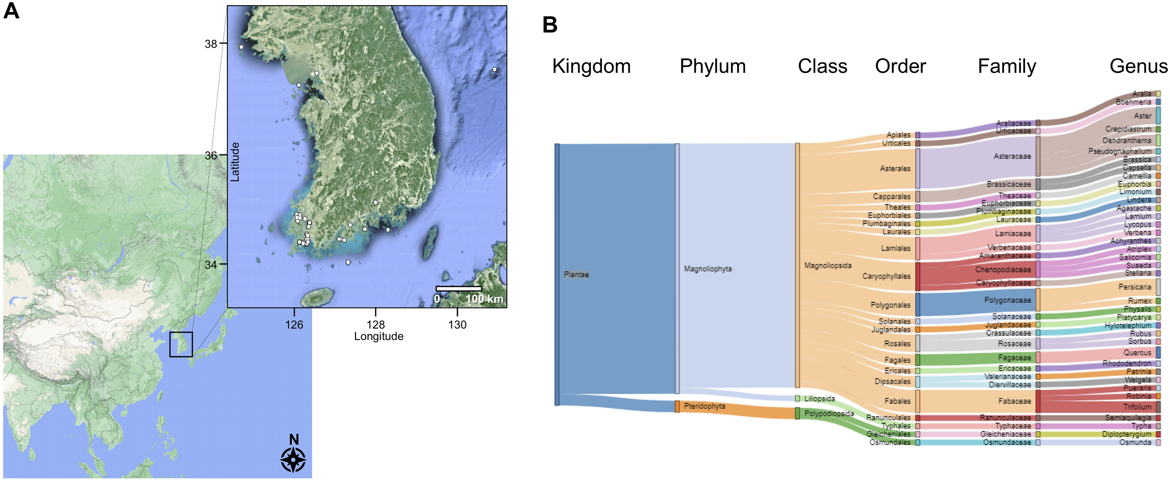
Plant collection and extract preparation. (A) Geographical locations of plant collection sites in Korean islands are visualized by an R package of ggmap with Google Maps. White dots represent the GPS coordinates of the sites of the 46 plants collected. (B) The 46 plants were classified based on kingdom, phylum, class, order, family, and genus.
Plant Extracts for Characterization
Preparation steps are crucial while exploring the pharmaceutical properties of the collected plants, such as their anti-inflammatory and immune-enhancing effects. The effective extraction of active chemicals is influenced by the selection of specific plant parts and the choice of solvent. 15 A total of 156 plant extracts were obtained by combining 5 different plant parts (flower, fruit, stem, leaf, and whole body) from the 46 species with 3 extraction solvents (water, 30% ethanol, and 70% ethanol). For Aralia cordata var. continentalis, 9 extracts were prepared from the leaves, stems, and flowers using water, 30% ethanol, and 70% ethanol, respectively. Additional information regarding the plant extracts used in this study is provided in Supplemental Table S1.
Anti-Inflammatory Effect of Plant Extracts
Inflammation serves as an essential defense mechanism pivotal to maintaining overall health. It functions as part of the immune system against deleterious stimuli, such as pathogens, cellular damage, toxic compounds, and ionizing radiation, to eliminate the sources of cellular damage. 16 This regulatory process plays a critical role in maintaining cellular and tissue homeostasis against acute actions that induce inflammation. However, inadequately regulated acute inflammation may transition into a chronic state, contributing to the development of various chronic inflammatory conditions. 17 To this end, we systematically investigated the anti-inflammatory effects of the 156 plant extracts derived from 46 indigenous plants in Korean islands. Notably, approximately 38% of the extracts (59 plant extracts from 31 species) exhibited statistically significant positive anti-inflammatory effects (P < 0.05) (Figure 2A). Of these, 18, 17, and 24 were subjected to extraction using water, 30% ethanol, and 70% ethanol, respectively (Figure 2B). Regarding plant parts, the fruits, leaves, stems, and whole body exhibited anti-inflammatory effects (Supplemental Table S2). Clustering based on the preparation solvents (water, 30% ethanol, and 70% ethanol) was conducted to identify which solvent yielded active chemicals with anti-inflammatory effects (Figure 2C). The anti-inflammatory effect of over 50% was achieved in high-concentration ethanol (70%) extracts from Osmunda japonica, Rumex crispus, and Aralia cordata var. continentalis; a similar anti-inflammatory effect was observed in both the high (70%) and low (30%) ethanol concentration extracts from the stems and whole bodies of Agastache rugosa, Rhododendron mucronulatum, Lindera obtusiloba, Trifolium pratense, and Aster ageratoides (Figure 2C, the first and second clades). Interestingly, Lamium amplexicaule and Physalis angulata exhibited specific anti-inflammatory activity when water was used for extraction; however, only high-concentration ethanol extracts from Aster yomena and Achyranthes bidentata var. japonica demonstrated anti-inflammatory activity (Figure 2C, the third and fifth clades). In addition to the active plant extracts exhibiting anti-inflammatory properties, a mild effect was observed in the case of 13 species (the fourth clade) subjected to extraction using either water or ethanol. Overall, this extensive screening involving 46 plants resulted in 59 functional plant extracts with anti-inflammatory properties. Notably, the anti-inflammatory efficacy varied not only with the extraction method but also with the specific plant part collected.

Anti-inflammatory effects of plant extracts. (A) The number of plant species demonstrating positive and negative anti-inflammatory effects. (B) The number of effective extracts depending on the extraction solvent. (C) Heat map representing clustering results of extracts produced using different solvents. Rows represent each plant species and part, labeled on the right side of the heat map. Columns indicate extraction solvents. The color key at the top left indicates the percentage of anti-inflammatory effects.
Immune Enhancement Effects of Plant Extracts
We explored and assessed the potential of the plant extracts to bolster immune response. Among the extracts assessed, approximately 22%, totaling 35 plant extracts from 24 species, significantly improved immune response (P < 0.05) (Figure 3A). Within this subset, 16, 12, and 7 plant derivatives were extracted using water, 30% ethanol, and 70% ethanol, respectively (Figure 3B). As opposed to the plant extracts exhibiting positive anti-inflammatory effects, no sample extracts from flowers enhanced immune function; however, extracts from fruits, leaves, stems, and whole bodies showed a positive effect on immune response (Supplemental Table S2). Regardless of the extraction method, extracts from 2 species—Lycopus maackianus and Quercus serrata—positively contributed to enhancing immune response (Figure 3C). Among these, immune responses were notably increased by the extract of Q. serrata, the only plant that provided its fruit (water, 25.9%; 30% ethanol, 35.5%; 70% ethanol, 32.8%). The most significant elevation in immune response enhancement effect (65%) was achieved by whole-body extracts (water extraction) of Typha orientalis (Figure 3C).
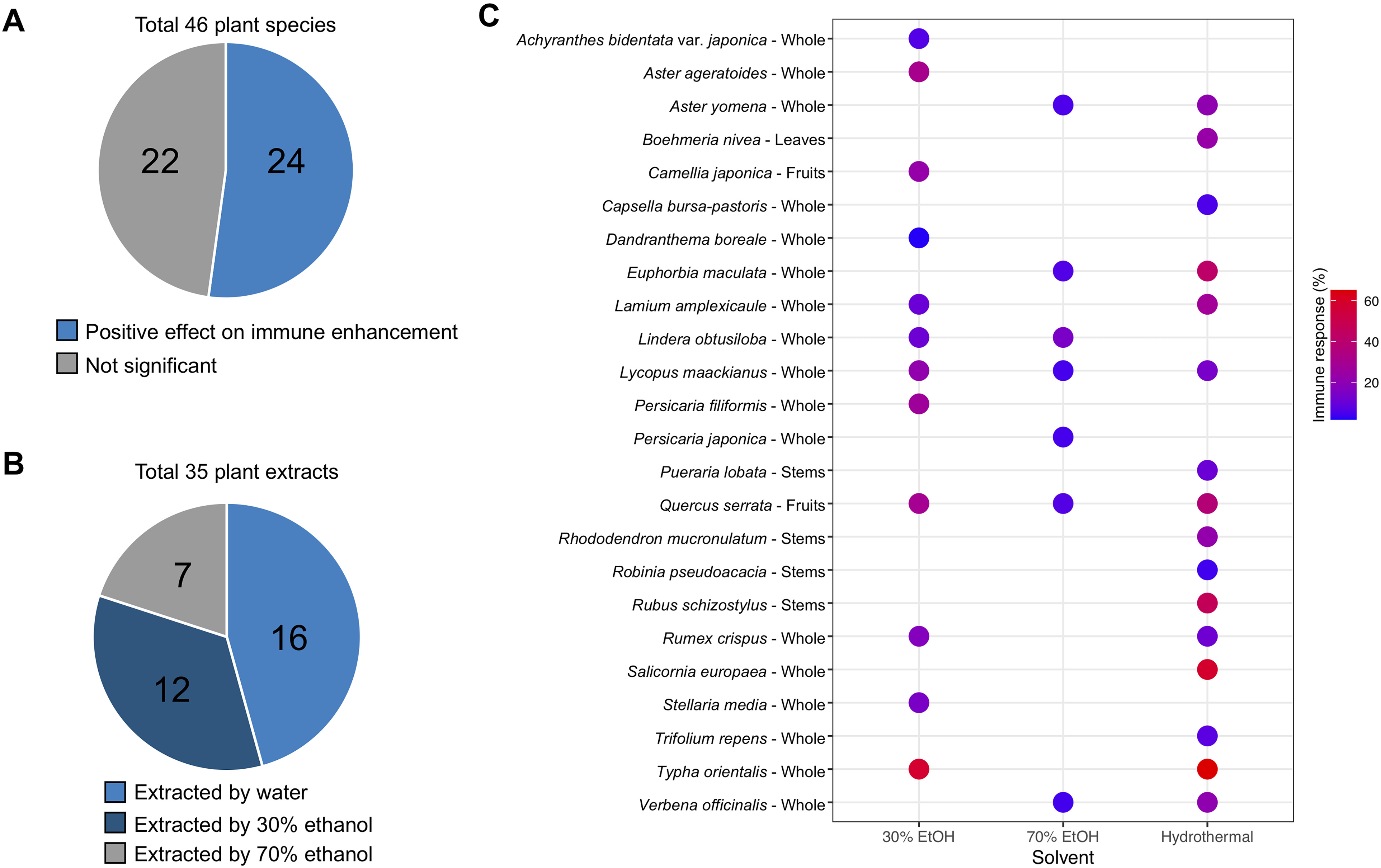
Immune response enhancement effects of extracts. (A) The number of plant species exhibiting positive and negative effects on immune response enhancement. (B) The number of effective extracts categorized based on the extraction solvent. (C) Dot plot illustrating the improvement of immune response by plant extracts and extraction methods. Rows represent each plant species and part, labeled on the left side of the dot plot. The color key in the middle right indicates the percentage of immune response enhancement.
Anti-Bacterial Effects of Plant Extracts
The increasing prevalence of infectious diseases poses a substantial threat to both human and animal health, leading to severe morbidity and mortality. This concern is exacerbated by several factors, such as the rapid emergence of multi-drug resistant pathogens, the limited anti-bacterial spectrum of available treatments, and the challenging quest for potential anti-microbial agents. 18 Consequently, we evaluated the potential of plant extracts in inhibiting the growth of representative pathogens Escherichia coli (E. coli) and Staphylococcus aureus (S. aureus). Seven and 11 plant extracts significantly inhibited the growth of E. coli and S. aureus, respectively (Table 1). Compared to the total number of plant extracts, the number of extracts that exhibited positive anti-bacterial activity was substantially lower (∼3-fold and ∼2-fold) than that for other functions, such as anti-inflammatory and immune response enhancement effects, respectively. Stem extracts from Rubus schizostylus, produced using water, 30% ethanol, and 70% ethanol, demonstrated the most effective inhibitory effect against E. coli. The growth assay also indicated that alcohol extracts from Quercus acuta inhibited approximately 25% of E. coli growth. In contrast to the inhibitory effect on E. coli, 11 plant extracts significantly reduced the growth of S. aureus. The growth of S. aureus was markedly suppressed when 30% ethanol extracts from the leaves of Pueraria lobata were introduced to the medium (Table 1). In addition, similar to the growth inhibition of the gram-negative bacteria, the growth of the representative gram-positive bacteria, S. aureus, was impeded by up to 42% with the addition of fruit extracts from Q. acuta. Taken together, these results suggest that these plant extracts can serve as alternatives to antibiotics.
Plant Extracts With Anti-Bacterial Activity.

EtOH, ethanol.
Multi-Functional Plant Extracts
Through the extensive characterization of 156 plant extracts, we observed positive results in each functional category: 59 extracts from 31 species exhibited anti-inflammatory effects, 35 extracts from 24 species enhanced immune responses, and 7 and 11 extracts demonstrated anti-bacterial activity against E. coli and S. aureus, respectively. Of the 156 extracts investigated, 90 remained functional, exhibiting at least 1 biological activity in the 3 experiments (Figure 4A, Supplemental Table S3). Specifically, 43, 20, and 9 extracts exhibited singular effects of anti-inflammation, immune response enhancement, and anti-bacterial activity, respectively. Additionally, 10, 3, and 2 extracts showed dual effects of anti-inflammation/immune response enhancement, anti-inflammation/anti-bacterial activity, and immune response enhancement/anti-bacterial activity, respectively. Of the 156 plant extracts, 3 exhibited all 3 biological effects. The whole-body extract of A. ageratoides prepared using 30% ethanol demonstrated all 3 biological effects; however, the growth inhibition of E. coli was limited to 1.3% (Figure 4B). Similarly, whole-body extracts of Persicaria filiformis prepared using 30% ethanol exhibited all 3 effects; however, the anti-inflammatory effect was less than 10%. Among the 156 extracts from 46 indigenous plants obtained from Korean islands, relatively high positive effects for all 3 biological examinations were obtained with the water extract of the stems of R. mucronulatum (Figure 4C).

Multi-functional plant extracts. (A) Venn diagram showing the number of single and multi-functional plant extracts regarding anti-inflammation, immune response enhancement, and anti-bacterial activity. 3D-plot showing the dual effect of anti-inflammation and immune response enhancement, as well as anti-bacterial activity against (B) gram-negative bacteria, Escherichia coli, and (C) against gram-positive bacteria, Staphylococcus aureus.
Discussion
The increasing utilization of plant-derived extracts in scientific research and commercial applications is driven by their rich bioactive constituents. These plant-based compounds serve as crucial alternatives to synthetic compounds, which may pose harm and exert carcinogenic effects. 19 In this study, we conducted a comprehensive exploration of the bioactive potential of plant-derived extracts. Over the course of a 1-year expedition in Korean islands, 46 indigenous plants were collected (Figure 1) and partitioned into 5 parts: flower, fruit, stem, leaf, and whole body. These parts were subjected to extraction using 3 different solvents—water, 30% ethanol, and 70% ethanol. The resulting 156 plant extracts were assessed to determine their potential anti-inflammatory (Figure 2), immune-enhancement (Figure 3), and anti-bacterial activities (Table 1). Even though comprehensive functional reports were carried out, this study, with its large number of plants and the separation of specific parts using 3 different solvents to produce extracts, is unique compared to other notable works, which examined fewer plants: 5 in Asia, 12 in Africa, and 19 medicinal plants.20–22
The increasing misuse of drugs, including prescription painkillers and over-the-counter medicines, in treating inflammation poses significant health risks.19,23 However, natural plant extracts have emerged as a promising and safer alternative. These extracts contain bioactive compounds that contribute to their effective anti-inflammatory properties. Plant derivatives play a crucial role in averting excessive or prolonged inflammation by modulating the activation of macrophages and subsequent increases in the secretions of NO, pro-inflammatory cytokines, such as interleukin-1β (IL-1β), IL-6, tumor necrosis factor-α (TNF-α), and cell adhesion molecules. 24 Our experimental results align with those of previous studies using the same plant but different extraction methods, where outstanding anti-inflammatory effects were observed. O. japonica has been reported to reduce pro-inflammatory cytokine levels in periodontal ligament fibroblasts upon stimulation with bacterial lipopolysaccharide (LPS). 25 Compared to the untreated control group, treatment with O. japonica extract resulted in a 63% and 81% reduction in IL-6 and IL-8 mRNA expression, respectively. The roots of R. crispus, a perennial plant that thrives in moist environments, have been traditionally used as a herbal medicine. 26 Root extracts from R. crispus with ethyl acetate and dichloromethane exhibited strong antioxidant activity and high flavonoid content. 27 The roots of R. crispus also demonstrated significant inhibition of NO production, suggesting the effectiveness of high flavonoid content on anti-inflammatory responses. 27 In addition to flavonoids, di- and tri-terpenes play a crucial role in exerting anti-inflammatory effects and inhibiting cyclooxygenase (COX)-1 and COX-2, serving as active constituents contributing to the pharmacological activity of Aralia cordata var. continentalis. 28
The immune system functions as a defense mechanism in various vertebrates, protecting against external harmful threats, including microbial invasions. This system is divided into 2 main categories: innate and adaptive immunosurveillance. 29 The innate immune system, comprising macrophages, natural killer cells, cytokines, and several types of white blood cells, rapidly responds to infections before the activation of the adaptive immune response, serving as the first line of defense. Among the contributors, macrophages play a key role in the innate immune system and host defense strategy. They are the first responders to external stimuli, and their activation induces changes, such as increased proliferation and dispersion capabilities, enhanced phagocytic function, and elevated production of NO and cytokines. 30 These changes may result in the effective suppression of the growth of cancer cells and other harmful microorganisms. In recent decades, extensive research has been conducted on numerous natural products to determine whether they improve immune response or restore diminished immune capabilities to a normal state through natural compounds. In a previous study with the 70% ethanol extract of Isodon japonicus, 31 a notable enhancement in the proliferation of T lymphocytes (up to 76.5%) and B lymphocytes (up to 15.6%) was observed compared to those of the mock control. The immunomodulatory potential of the extract was confirmed by assessing CD25 and CD69 markers on CD4+ and CD8+ T cells. In addition, the effect of methanol extracts of L. maackianus was demonstrated in brain immune cells, such as microglia and macrophages. 32 LPS-stimulated microglia induce pro-inflammatory agents, such as TNF-α, IL-6, prostaglandin E2, inducible nitric oxide synthase, and COX-2, potentially leading to a neurodegenerative process characterized by excessive inflammation and the generation of reactive oxygen species. 33 Heme oxygenase (HO) plays a role in cellular defense against oxidative stress. The methanol extract of L. maackianus stimulated the expression of HO-1, thereby enhancing antioxidant and anti-inflammatory properties. 32
The growing challenge posed by multi-drug-resistant bacterial strains has emerged as a significant global health concern, necessitating the search for alternative anti-bacterial treatments. 34 With traditional antibiotics facing diminishing efficacy against pathogens, plant extracts have emerged as a feasible solution. These natural extracts are rich in various bioactive compounds and exhibit a broad spectrum of anti-bacterial properties. Plant-derived compounds, such as alkaloids, flavonoids, and polyphenols, have demonstrated the efficacy to disrupt bacterial cell membranes, impeding bacterial proliferation. 35 Species of Rubus are recognized for their anti-bacterial activity. Ethanol-water extracts of R. idaeus and R. occidentalis were investigated in terms of their anti-microbial properties and active chemicals, such as sanguiin H-6, free ellagic acid, and anthocyanins. 36 Among 15 bacterial strains assessed, Corynebacterium diphtheriae and Moraxella catarrhalis, both human pathogens, showed significant growth reduction upon exposure to Rubus extracts. 36 This phenomenon is attributable to the presence of sanguiin H-6 and ellagic acid in Rubus; thus, these compounds can be developed into a narrow spectrum of antibiotics targeting Corynebacterium pathogens. Aligning with our findings on anti-bacterial activity, several studies and applications have explored the use of plant extracts for anti-bacterial purposes.37–40 Previous reports have demonstrated the efficacy of Pueraria lobate against inflammation and obesity through the modulation of the gut microbiota in a mouse model.41,42 In our study, S. aureus exhibited a higher growth inhibition rate than E. coli (Table 1), which may be attributable to the difference in cell wall thickness. The cell walls of gram-positive bacteria are thicker than those of gram-negative bacteria. 43 It is conceivable that substances, such as unique polysaccharides and puerarin extracted from P. lobate, may hinder the formation of thick cell membranes in gram-positive bacteria, causing material transport blockage or disrupting cell membrane formation, thereby impeding bacterial growth. However, further research is required to elucidate the anti-bacterial mechanisms of action of P. lobate extracts.
It is worth noting that this study has limitations. First, while the extracts from indigenous Korean plant species showed positive effects on anti-inflammatory, immune response-enhancing, and anti-bacterial activities, the specific active chemicals responsible for these effects were not identified. Without the identification and quantification of these bioactive compounds, it becomes difficult to find their specific targets and industrial applications. Second, the mechanisms by which these plant extracts show their observed biological activities remain unexplored. Detailed investigations into the cellular signaling pathways involved in mediating the anti-inflammatory, immune-modulatory, and anti-bacterial effects were beyond the scope of this study. However, understanding these mechanisms is crucial for elucidating the mode of action of compounds from the extracts and could provide valuable insights into their potential use in pharmaceutical and medical applications. With the comprehensive database provided in this study, future research should focus on isolating and characterizing active compounds, as well as examining molecular and cellular pathways, to provide strong evidence for the use and development of novel biomaterials derived from Korean indigenous plants.
Conclusion
This research evaluated the functional properties of 46 indigenous Korean plants collected from islands. Specimens were processed into 156 extracts and were revealed that 38% had anti-inflammatory effects, 22% enhanced immune responses, and some showed anti-bacterial activity. The study suggests these extracts could be valuable in pharmaceuticals and medicine, particularly for their anti-inflammatory, immune-boosting, and antibiotics alternatives.
Materials and Methods
Plant Collection
In 2022, we collected plants from 44 locations, encompassing both inhabited and uninhabited islands located between 34.0-37.9° latitude and 124.7-130.9° (Supplemental Table S1). For each species, a minimum of 3 individual plants were isolated from the area and reserved in the Bank of Bioresources from Island and Coast (BOBIC) at the Honam National Institute of Biological Resources (HNIBR), Korea.
Preparation of Plant Extracts
Prior to the extraction process, we separated the plant parts, washed them thoroughly in running water, and air-dried them at room temperature. Subsequently, the dried plant materials were ground into powder using a mechanical grinder. For extraction, 100 g of each plant parts were combined with 1 L of water, 30% and 70% ethanol (1:10, w/v, unless otherwise stated) and incubated for 24 h with centrifugation at 150 revolutions per minute (rpm). Following centrifugation at 4,500 rpm for 30 min, the residue was removed, and the supernatant was filtered using a Whatman filter (No. 2). The resulting solid extract was subjected to a decompression evaporation process, and the lyophilized extract (about 10-20 g), with a yield rate of about 10% to 20%, was resuspended in phosphate-buffered saline and used at a final concentration of 200 µg/mL for functional experiments.
Chemicals and Reagents
Fetal bovine serum (FBS) and penicillin/streptomycin were obtained from Thermo Fisher Scientific (MA, USA). Griess Reagent (1% sulfanilamide in 5% H3PO4, 0.1% N-(1-naphthyl)-ethylenediamine dihydrochloride), LPS from E. coli 0128:B18, and dexamethasone were purchased from Sigma-Aldrich (MO, USA).
Cell Cultures and Nitric Oxide Assay
Murine macrophage RAW264.7 cells were purchased from the American Type Culture Collection (ATCC; VA, USA). The cells were maintained in Dulbecco's modified Eagle's medium (Welgene, Daegu, South Korea) supplemented with 10% FBS and 1% penicillin/streptomycin and incubated at 5% CO2 and 37°C. The RAW264.7 cells were seeded into 48-well plates (SPL Life Sciences Co., Ltd, Pocheon-si, South Korea) at a density of 1 × 10⁵ cells/well overnight. The cells were treated with crude extracts at different concentrations with or without LPS (1 μg/mL) for an additional 28 h. The quantity of Nitric Oxide (NO) accumulated in the supernatants was measured following a previously described method. 44 Absorbance was measured at 570 nm using a microplate spectrophotometer (BioTek Instruments, Inc., VT, USA). The immune enhancement and anti-inflammatory effects were contingent on the presence or absence of LPS stimulation.
Bacterial Strains and Anti-Microbial Assay
Strains of E. coli ATCC 43895 and S. aureus KACC 14790 were obtained from American Type Culture Collection (ATCC; VA, USA) and Korean Agricultural Culture Collection (KACC; Jeollabuk-do, South Korea), respectively. S. aureus and E. coli strains were cultured in tryptic soy broth (TSB; Difco, MA, USA) at 37 °C in a shaking incubator. S. aureus and E. coli were diluted 1000-fold with fresh TSB, and the diluted cell culture was dispensed to every 90 μL per well in 96-well plates (SPL Life Sciences Co., Ltd, Pocheon-si, South Korea). Subsequently, the bacterial strains were treated with plant extracts at a concentration of 200 μg/mL overnight. The absorbance for optical density (OD) was measured at 630 nm using a microplate reader (BioTek Instruments, Inc., VT, USA). Compared to the ampicillin treatment at 100 μg/mL, the growth inhibition rate was calculated as follows:
Statistical Analysis
All experiments were repeated at least thrice. Statistical differences in the experimental data were calculated using GraphPad Prism version 5.01 (GraphPad Software, CA, USA). One-way analysis of variance was used for multiple-group comparison, with a P-value ˂ 0.05 indicating statistical significance.
Supplemental Material
sj-xlsx-2-npx-10.1177_1934578X241266706 - Supplemental material for Characterization of the Anti-Inflammatory, Immune Response, and Anti-Bacterial Properties of the Polar Extracts of Indigenous Korean Plants
Supplemental material, sj-xlsx-2-npx-10.1177_1934578X241266706 for Characterization of the Anti-Inflammatory, Immune Response, and Anti-Bacterial Properties of the Polar Extracts of Indigenous Korean Plants by Sungmin Hwang, Ji Soo Kim, Go Kyoung Na, Hyeon Jeong Lee, Bohyun Yun, Ji-Won Park, Seahee Han, Min-Ju Park, WonWoo Lee, Kyung-Min Choi and Jung Up Park in Natural Product Communications
Footnotes
Acknowledgments
The authors thank the members of the Division of Natural Products at Honam National Institute of Biological Resources for their preparations.
Author Contributions
Conceptualization, supervision: WWL, KMC. Investigation, methodology: JSK, GKN, HJL. Resources: SHH, MJP. Validation: BHY, JWP. Writing—Original, review & editing: SMH, JUP.
Declaration of Conflicting Interests
The authors declared no potential conflicts of interest with respect to the research, authorship, and/or publication of this research.
Funding
The authors disclosed receipt of the following financial support for the research, authorship, and/or publication of this article: This work was supported by the Honam National Institute of Biological Resources (HNIBR202302113), funded by the Korea Ministry of Environment (MOE). Additionally, this work was supported by the Korea Environment Industry & Technology Institute (KEITI) through a project to make multi-ministerial national biological research resources more advanced (RS-2023-00230403).
Ethical Approval
Ethical Approval is not applicable for this article.
Statement of Human and Animal Rights
This article does not contain any studies with human or animal subjects.
Statement of Informed Consent
There are no human subjects in this article and informed consent is not applicable.

Supplemental Material
Supplemental material for this article is available online.
References
Supplementary Material
Please find the following supplemental material available below.
For Open Access articles published under a Creative Commons License, all supplemental material carries the same license as the article it is associated with.
For non-Open Access articles published, all supplemental material carries a non-exclusive license, and permission requests for re-use of supplemental material or any part of supplemental material shall be sent directly to the copyright owner as specified in the copyright notice associated with the article.
