
Abstract
Introduction
Fibrosis is well-known chronic liver disease associated with an increasing death rate in the world. Statistical reports in 2020 revealed a significantly increased prevalence (13%) of hepatic injury and fibrosis since 2000, recording more than 1.5 billion cases. 1 According to 2019 data analysis, liver fibrosis is responsible for 2.4% of all mortality and 1.5% of all morbidity in all nations, making the 11th and 15th risk factors associated with mortality and morbidity, respectively. 2 Liver injury and hepatic fibrosis develop as a result of different exogenous and endogenous factors, including toxin exposure, viral infection, alcoholism, metabolic disorder, diabetes, biliary stones, and non-alcoholic fatty liver. 3
Liver fibrosis is an outcome of several biological modulations of the liver lobules, prolonged feast fibrosis, and micro-, and macro-nodules in parenchymal layers. These biological liver changes are followed by portal vein hypertension and hepatocyte proliferation, causing more oxidative and inflammatory damage. 4 The prognosis and the severity of liver disease can be different because of variability in the causative, area of damaged hepatic tissue, pro-fibrogenic paths, and pro-fibrogenic myofibroblast factors. 5 Liver fibrosis is well known for their increased accumulation of extracellular matrix proteins (collagen) commonly found in patients with chronic liver diseases. Fibrosis is considered an immune-responsive action of hepatic tissues toward several chronic inductions. At first, hepatic fibrosis starts with liver injury induced by numerous etiological factors, initiating inflammatory infiltration, and ignition of the inflammatory cascade. 6 Moreover, perisinusoidal cells (hepatic stellate cells) are converted into fibroblast-like cells capable of generating several types of collagen proteins along with laminin. Consequently, these liver protein alterations can have significant modulatory actions on the structure and function of hepatocytes subsequently affecting overall liver performance. 2
Thioacetamide (TAA) is a well-documented hepatotoxic agent capable of inducing liver damage and fibrosis in animal models, which were comparable to that of humans. 7 The TAA potentials in the stimulation of liver fibrosis have been correlated with its significant modulatory actions on the hepatic antioxidant enzymes and non-enzymatic, enhancing oxidative stress, lipid peroxidation, and initiation of hepatic necrosis. 8 Indeed, scientists declared a significant protective role of endogenous antioxidants against TAA-mediated liver injury. 9
Chronic inflammation and increased cytokine secretion trigger the gradual scaring damage of the liver subsequently causing liver fibrosis. The tumor necrosis factor (TNF-α), interleukin-1 β (IL-1 β), and interleukin-6 are pro-inflammatory cytokines that are well-known for their enhancing role in the progression of fibrosis through modulation of various cellular processes including lipid metabolism, biliary system obstruction, positive and negative acute phase proteins, and fibrosis progression. 5 While, anti-inflammation chemicals (IL-10) are major inhibitor of cellular processes involved in immune and inflammatory response, thereby reducing the activation of the glutaminase gene and lowering the incidence of hepatic encephalopathy. 10 The inflammatory cytokine production is thought to be regulated by the transcription nuclear factor-kappa B (NF-kB). Moreover, NF-κB serves as a central factor that can modulate inflammation, cell differentiation or proliferation, and oxidative stress-related disorders. It can be stimulated by a wide diverse factor and a complex network of cellular mechanisms including inflammation cascade, thus forming a cycle of auto-regulation that can amplify the inflammatory response for a prolonged time. 11 Therefore, the estimation of inflammatory cytokines can be a valuable tool for better monitoring inflammatory-related disorders including liver fibrosis. Moreover, the estimation of liver enzymes (AST, ALT, ALP) and liver proteins (albumin) are found to be very helpful in diagnosing the prognosis of acute and chronic liver diseases, as these parameters will drop significantly (30%-35%) in those cases. Despite the scientific revolutionary progress in figuring out initiating mechanisms of liver fibrosis in the past few decades, pharmaceutical drugs including silymarin (inhibitory rate of 30-35%) cannot exhibit significant protective action to halt the progression and cure these deleterious diseases. Therefore, the search for natural alternatives to antifibrotic therapy has become a continuous scientific mission in the last few decades.12,13
Medicinal plants have been utilized as therapeutic agents for liver disorders and liver disease, and in today's world, these natural sources are getting more popular due to their significant efficiency and lower side effects compared to chemical synthetics. 14 In the last few decades, numerous plant species were explored and reported as hepatoprotective products including those belonging to the genus Hypericum spp.13,15
Hypericum perforatum (Merr.) is a well-known therapeutic traditional Islamic medicine, Chinese medicine, and Greek medicine 15 that has been utilized for the attenuation of liver-related disorders, including hepatic dysfunctionality, steatosis, cholestasis, and liver fibrosis. 16 This plant species belongs to Hypericaceae (Clusiacaeae) family commonly known as St. John's wort (SJW) (Hypericum or Millepertuis). It is a herbaceous perennial native plant in Western Asia, Europe, and Northern Africa. 17 Traditionally, the flowers, leaves, and other aerial parts of Hypericum perforatum have been consumed for medicinal purposes. 18 Previous biological studies on various parts of H. perforatum reported several bioactivities including, antioxidant and anticancer, 19 anti-inflammatory, 20 anti-depressant, 21 anti-microbial actions, 22 and for skin diseases.23,24 Most of their pharmacological actions were assigned to their phytochemical (photosensitive naphthodianthrone; hypericin, hyperforin, quercitrin, p-coumaric acid, rutin, and quercetin) contents.25-27
In today's Pharmaceutical industry, dry powder of aerial parts of Hypericum is available in the form of capsules, tablets, tea, and tinctures for various health issues.18,28 The increased demand for the utilization of Hypericum products as a therapeutic agent could be related to its advantages, including high tolerability, high efficacy, inexpensiveness, good accessibility, and lower side effects compared to conventional drugs. 29
Despite previous reports on the therapeutic efficacy of H. perforatum against liver disorders, cholestasis, 15 and hepatitis B virus, 30 the literature data lacks a concise study detailing the possible molecular mechanisms behind hepatoprotection action of H. perforatum. Therefore, the present study investigates the hepatoprotective potentials of H. perforatum in TAA-stimulated liver injury in rats.
Materials and Methods
Plant Extract
The methanolic (95% concentration) extract of Hypericum perforatum leaves (yield 4.3%) was a gift from Erbil Medical Laboratory, Erbil-Iraq.
Ethical Approval
The research was approved by the Ethical Committee of the Faculty of Pharmacy, King Abdel-Aziz University, Jeddah, Saudi Arabia (approval # PH-1445-1, 12/9/2023).
Chemicals
The hepatotoxic inducer (TAA) and (silymarin) were from Sigma-Aldrich (Merk, Germany). TAA was dissolved in a flask containing 10% Tween 80, liquefied, and used as a stock solution. TAA solution was given by intraperitoneal injection (3 doses weekly) to rats for 2 months. The reference drug (silymarin) was prepared in sterilized distilled water, which was given in 50 mg/kg to rats. 8
Acute Toxicity Experiment
Sprague Dawley male and female rats (7-8 weeks) weighing about 170 to 180 g were obtained from the Faculty of Pharmacy, King Abdel-Aziz University, Jeddah, Saudi Arabia. Thirty rats (equal number from both genders) were distributed into 3 wide-mesh wire cages (avoiding coprophagia) and they had a standard diet and tap water for 7 days of adaptation. After that, the 3 rat clusters underwent overnight fasting and they were treated as follows the next day:
A, Normal rats received 10% Tween 80 (5 mL/kg): B, rats supplemented with 2000 mg/kg of MEHP; C, rats ingested with 5000 mg/kg of MEHP.
Rats were not allowed to have food following 3 to 4 h after supplementation. The observation started immediately and continued for the next 48 h for any potential toxic incidence or abnormalities. After 14 days, all rats received overdose of anesthesia (ketamine and xylazine) and sacrificed. The blood samples from intracardial blood puncture were analyzed for biochemical contents and various organs were examined by histological assays using hematoxylin and eosin stain.
31
Hepatoprotective Trial
Experimental Design
Thirty mature male rats were obtained from the Faculty of Pharmacy, King Abdulaziz University, Jeddah, Saudi Arabia. The animals were housed in plastic cages (7 animals each) at a temperature of 25 °C and 70% humidity, with 12 h light/ dark cycle, and had free access to standard rodent diet and water.
The rats were clustered into 5 wire-mesh cages (6 rats in each group) and were given the following treatments (Figure 1):
A, normal control rats were administered a daily dosage of 10% Tween 80 (5 mL/kg) by oral gavage and received 3 injections (5 mL/kg) of sterile distilled water each week for 2 months. B, fibrosis rats were administered a daily dosage of 10% Tween 80 (5 mL/kg) by oral gavage and injected with 3 intraperitoneal injections of 200 mg/kg of TAA each week for 2 months. C, reference rats were administered a daily dosage of 50 mg/kg silymarin by oral gavage and injected with 3 intraperitoneal injections of 200 mg/kg of TAA each week for 2 months. D and E, rats were administered daily dosages of 250 and 500 mg/kg of MEHP by oral gavage and injected with 3 intraperitoneal injections of 200 mg/kg of TAA each week for 2 months.

Experimental design of hepatoprotective procedure.
After that, food was removed from rats overnight and they were given 100 m/kg anesthesia (Ketamine and Xylazine) and sacrificed. The blood specimens collected from the jugular vein were analyzed for different biochemical parameters and the dissected livers were evaluated by histopathologic techniques. 32
Macroscopic Viewing of the Liver
The dissected liver organs were cleaned with normal saline (cold), and dried by the filter papers for the estimation of gross weight and gross morphological studies.
33

Histology of Liver
The liver lobes were kept in the flask containing 10% phosphate buffered formalin for 1 day as a tissue fixation technique. After that, the liver tissues were parafinized in paraffin blocks through a well-developed tissue machine (Leica, Germany). Small slices (5 μm) of liver tissues were cover slipped on slides and investigated histopathologically by staining with hematoxylin, eosin, and Masson Trichrome stains. 5
Immunohistochemistry
The rat monoclonal Anti-PCNA (proliferating cell nuclear antigen) antibody [PC10] and monoclonal anti-actin (anti-α-Sm-1) were purchased for PCNA and (α-SMA) detections, respectively. Briefly, by utilizing poly-L-lysine-coated slides, liver sections were kept in an oven (DON-HE Series, Infitek, Shandong, China) for 25 min at 60 °C. After heating, xylene was used for deparaffinization, and alcohol (series-graded alcohol) was used for the hydration. The antigen recovery technique was possible via 10 mM sodium citrate buffer after boiling in a microwave. The slides were colored following the instructions of the producer's company (Sigma Aldrich, Merck, Germany). Briefly, 0.03% hydrogen peroxide sodium azide was utilized for blocking the endogenous peroxidase for 5 min. Liver sections were buffer washed, cleaned, and incubated for 15 min with anti-bodies (PCNA) (α-SMA) (1: 200). After careful washing and reincubation for 15 min with streptavidin HRP, the liver sections were incubated for 7 min with diaminobenzidine substrate chromogen (7 min) and buffer washed again for the coloring with counterstain hematoxylin (5 s). Finally, tissue sections dipped in 0.037 mol/L ammonia (5 times), cleaned, and cover-slipped for the microscopic detection of cytoplasmic brown granules and brown-colored nuclei for the positive appearance of α-SMA and PCN antigens, respectively. The staining intensity of PCN was found by estimating the number of colored cells divided by 1000 hepatocytes. 34
Liver Antioxidant
The liver samples (1 g) from both left liver lobes were transferred into a flask of 10 mL containing PBS solution (10%, pH 7.2) for normalization and homogenization (5000 rpm, 15 min at −4 °C). The supernatant part was separated and stored in a freezer (−80 °C) for later use. Different antioxidant kits of CAT, SOD, GPx, and MDA were bought from Sigma Ald-rich (Merck, Germany). 35
Serum Inflammatory Cytokines
The present estimation of TNF-α, IL-6, and IL-10 cytokines in serum samples obtained from experimental rats followed the instructions provided in the ELISA kit (Cusabio Biotech Co. China).
Biochemistry of Liver Functions
The serum samples were analyzed for the liver enzymes (ALT, AST, and ALP), liver synthetic functions (total protein and albumin), and the excretory (bilirubin) functions of the liver. 36
Statistical Analysis
The values are available as mean ± SEM. This was archived by GraphPad Prism 9 software. The statistical procedure also included 1-way ANOVA, post hoc, and a significance level setup at P < .05.3
Results
Acute Toxicity
The toxicity results showed non-observable changes in the behavioral or physiology of rats exposed to oral ingestion of 2 and 5 g/kg of MEHP. Furthermore, there was no significant variance in the food intake and body weight (BW) between normal and MEHP-treated rats. The present results did not detect any toxicity effects on fur, eyes mucous membrane, diarrhea, skin, tremors, or salivation) in MEHP-ingested rats. The present biochemical evaluations showed comparable results of liver and kidney parameters between normal and MEHP-treated rats. The gross evaluation at autopsy and histopathological investigations of the different organs colored with hematoxylin and eosin showed comparable tissue structure arrangements for normal and MEHP-treated rats (Tables 1 and 2, and Figure 2A-C). The outcomes expect the toxic dose of MEHP to exceed 5 g/kg.

Microscopic presentation of the liver and kidney tissues of different experimental rats. A, normal controls fed on 10%tween 80; B, rats ingested 2000 mg/kg MEHP; C, rats ingested 5000 mg/kg MEHP. Non-significant changes are observed in the alignment of the kidney and livers histological layers of normal control and MEHP-treated rats. The kidney tissues appeared as normal Bowman ’s capsule with glomeruli (yellow arrow) and adequate interlobular blood vessels (black arrow); green star, distal convoluted tubule; black star, proximal convoluted tubules. The hepatic tissues appeared with central vein (blue star), sinusoidal capillaries (green arrow), kupffer cell (pink arrow), and normal liver cell with circular nucleus (blue arrow) for all tested rats (hematoxylin and eosin stain, 40×).
Effect of MEHP Supplementation on Liver Function Tests.

A, normal cluster received 10%tween 80; B and C, rats ingested 2 and 5 g/kg MEHP, respectively.
Effects of MEHP Administration on Kidney Functional Parameters.
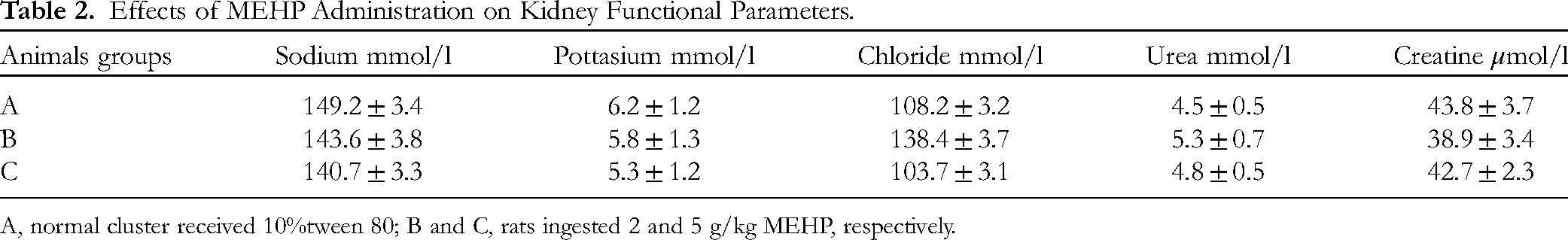
A, normal cluster received 10%tween 80; B and C, rats ingested 2 and 5 g/kg MEHP, respectively.
The data analysis of the liver (Table 1) and kidney (Table 2) parameters revealed non-significant changes between supplemented (2000 and 5000 g/kg MEHP) rats and normal controls.
The oral supplementation of 2 and 5 g/kg to rats did not alter the kidney functional parameters depending on the comparison to control rats (Table 2 and Figure 2).
Hepatoprotectives of MEHP
Body and Liver Masses
The outcomes revealed significant variation in the BW of different treated rats compared to fibrosis control rats. Results from fibrosis control rats (10%tween80 + TAA) showed significantly lower BW (183.4 g) compared to that (325.8, 292.8, 229.2, 259.3 g) of normal control, silymarin or 250 and 500 mg/kg MEHP-treated rats, respectively. Moreover, rats fed on 500 mg/kg of MEHP revealed similar data results in their body when compared to reference drug (silymarin)-treated rats (Table 3).
Effect of MEHP on Body Parameters of Rats Administered TAA Hepatotoxic Compound.

Values with same letters within column denotes non-significant variance at P < .05.
The liver weightiness was significantly modulated following different oral and intraperitoneal injections. The normal controls (A) showed usual liver weight (7.8 g) and liver index (2.42%), which were significantly lower than that of all other experimental rats. Rats in group B presented highest liver weight (12.8 g) and liver index (6.97%), which significantly higher than in the normal control (7.9 g, 2.42%, respectively), silymarin (9.3 g, 3.27%, respectively), or 250 mg/kg (10.9 g, 4.75%, respectively) and 500 mg/kg (10.1 g, 3.89%, respectively) MEHP-treated rats. Moreover, rats nourished with 500 mg/kg of MEHP showed statically non-significant changes in the liver weight and liver index compared to reference drug (silymarin)-treated rats (Table 3).
Morphology of Liver
The gross morphology of livers dissected from different experimental rats showed different levels of tissue surface alterations due to delivery of TAA and various treatments. Normal controls showed usual criteria of smooth surface tissues with regular tissue arrangements. Fibrotic control rats had livers covered with numerous micro and macronodules with rough surface area on their epithelial layers. Reference drug (silymarin)-rats had much smoother surface liver and less micronodules compared to group B. MEHP treatment (250 and 500 mg/kg) lead significant liver protection represented by reduced liver tissue injury, more arranged tissue layers, fewer miconodules compared to fibrotic rats (Figure 3).

Histology of liver stained with H&E, hematoxylin and eosin; MT, Masson trichrome stains; GA, gross appearance. A, normal rats showed usual tissue structure without any collagen deposition or tissue inflammation; B, fibrotic rats showed increased micronodules and elongated bile duct, large size fibrous septum, and increased collagen deposition about central vein (green area); C, rats received TAA + 50 mg/kg silymarin had reduced hepatic injury shown by fewer micronodules, fewer fibrous septa, and the lowest collagen deposition (green area); D, rats had TAA + 250 mg/kg MEHP showed moderate hepatic damage represented by reduced micronodules, less inflammation, and lower collagen deposition compared to group B; E rats received TAA + 500 mg/kg MEHP revealed similar hepatic structural arrangement compared to that of silymarin-treated, which were shown with fewer surface micronodules, lower inflammatory infiltrations, and fewer collagen depositions compared to group B. Nikon microscope (Y-THS, Japan). 20× magnification.
Microscopic Results
The present histological results showed significant modulation of hepatic structural arrangement due to the delivery of TAA and different treatments to rats. Normal controls showed the usual criteria of liver tissues with the absence of any inflammation signs or necrosis.
In the present experiment, fibrotic rats experienced significant liver tissue disruption shown by endothelial tissue injury, ambiguous nucleus, and elevated cytoplasmic vacuoles, all of these denoting severe inflammation and tissue necrotic condition. Moreover, the parenchymal layers were seriously altered as a result of fibrous septa that adjust the collagen bond in the hepatic triangles, denoting various micro- and macro-nodules in the liver cells. These nodules were found inside bundles of connective tissues that separate the liver into several lobules, accompanied by increased inflammation rate and hepatic necrosis. The histological results of silymarin-treated rats revealed noticeable recovery from TAA-hepatotoxicity damage represented by inflammatory cell infiltration, fewer centrilobular tissue necrosis, and clearer round nucleus compared to fibrosis controls. Moreover, hepatic lobules were more organized, and capillary veins were distributed across connective tissues. The present supplementation (250 and 500 mg/kg) to rats caused retained the hepatic tissues from TAA toxic damage shown by reduced tissue necrosis in centrilobular, fewer fibrosis scores, less nucleus and tissue fibrosis, and reduced vacuolization, which were significant compared to group B (fibrosis rats) but not as significant as rats received silymarin. Moreover, microscopically evaluation revealed less tissue penetration, less necrotic zones, and higher parenchymal cell regenerations in their tissue layers (endothelial and sub-endothelial layers) (Figure 3, H&E).
The fibrosis incidence rate was evaluated by Masson's trichrome staining procedure for all dissected liver sections from different treated rats as shown in Figure 3. Liver tissue sections obtained from normal controls appeared in a normal state without any obvious signs of collagen depositions (Figure 3A, MT). Fibrotic rats experienced increased collagen deposition around the central vein, denoting increased tissue fibrosis (Figure 3B, MT). Rats treated with silymarin had very reduced collagen deposition, denoting the lowest hepatic fibrosis (Figure 3C, MT). Rats ingested 250 mg/kg of MEHP showed moderate collaged deposition and moderate congestion about the central vein (Figure 3D, MT). Finally, rats that received 500 mg/kg of MEHP experienced mild collagen deposition and mild congestion in the hepatic central vein (Figure 3E, MT). The outcomes revealed a significant protective effect of MEHP against hepatotoxic injury in rats represented by significantly lower collagen deposition compared to fibrotic rats.
Immunohistochemical Protein Expression
The current results showed different expression rates of immunohistochemical proteins in liver tissues obtained from experimental rats as a result of various treatments. Normal controls revealed the lowest intensity of α-SMA in their hepatic parenchymal layers (Figure 4A and F). While, fibrotic rats (TAA + 10%tween 80) showed elevated expression of α-SMA about central veins, which were significantly more abundant compared to all treated rats and denoted severe hepatic injury and tissue proliferation (Figure 4B and F). Silymarin-treated rats revealed reduced α-SMA expression in their hepatocytes, denoting the hepatoprotective action of this drug against TAA cytotoxicity in rats (Figure 4B and F). Rats received 250 mg/kg of MEHP showed moderate intensity of α-SMA concentration in their hepatocytes. Moreover, rats administered with 500 mg/kg of MEHP demonstrated a mild appearance of α-SMA in their liver tissues, indicating the significant resistant effect of these plant extracts against TAA hepatotoxic damage.

Influence of MEHP on α-SMA expression in the hepatocytes. A, rats received 10%tween 80 + distilled water; B, fibrotic rats treated with 10%tween 80 + TAA; C, rats received 50 mg/kg silymarin + TAA; D, rats treated with 250 mg/kg MEHP + TAA; E, rats ingested 500 mg/kg of MEHP + TAA injection. The TAA injections caused significant liver injury and tissue fibrosis. While, MEHP ingestion resisted this toxic effect of TAA and retained the structured arrangement of hepatic tissues. Nikon microscope (Y-THS, Japan). 100× magnification.
The present TAA-induced liver injury followed by various treatments caused significant modulation in the expression of PCNA expression in hepatic tissues. As expected, normal controls revealed the absence of PCNA stains in their liver tissues, denoting a lack of cell renewal. Fibrotic rats revealed significantly increased intensity of PCNA proteins in their hepatic tissues, denoting severe hepatic damage with elevated necrotic cells. While, silymarin treatment caused noticeably reduced expression of PCNA stain in the liver tissues, indicating lower proliferation incidence in the necrotized hepatocytes and higher apoptotic actions. MEHP-treated rats showed significantly decreased necrotized hepatocyte proliferation represented by reduced PCNA stains in their hepatic tissues. These outcomes suggest that one of the hepaprotective actions of MEHP could be through up-regulation of apoptotic actions in damaged hepatic cells and attenuation effects on the cellular proliferations (Figure 5).

Influence of MEHP on the intensity of PCNA appeared in hepatic tissues. A, rats received 10%tween 80 + distilled water had no signs of PCNA stains; B, fibrotic rats treated with distal 10%tween 80 + TAA showed severe fibrosis with increased intensity of PCNA stain (arrow); C, rats received 50 mg/kg silymarin + TAA revealed less PCNA-stained liver cells (arrow) denoting less hepatocyte regeneration; D, rats treated with 250 mg/kg MEHP + TAA had moderate regeneration of liver cells shown by moderate PCNA expression (arrow) in the hepatocyte; E, rats ingested 500 mg/kg of MEHP + TAA injection showed minor regeneration of liver cells shown by minor PCNA expression (arrow) in the hepatocyte. Nikon microscope (Y-THS, Japan). 20× magnification.
MEHP Effects on Liver Antioxidants
The present data showed significant modulation of endogenous antioxidants as a result of TAA and different treatments in the experimental rats. Normal clusters had normal concentrations of antioxidant enzymes and non-enzymatic in their liver tissue homogenates represented by SOD (27.30 U/mg), CAD (144 CAT nmol/min/ml), GPx (19.20 CAT nmol/min/ml), and lower MDA content (1.78 U/mg) compared to other experimental rats. While, fibrotic rats showed the lowest antioxidant enzymes (SOD, 7.23 U/mg; GPx, 4.82; CAT, 91.2 nmol/min/ml) and the highest MDA (6.93 U/mg) levels in their hepatic tissue homogenates. MEHP (250 and 500 mg/kg) treatment caused significant positive modulation of tissue antioxidant and negative regulation of lipid peroxide indicator (MDA) in the hepatic tissues shown by higher SOD (11.83, 15.20 U/mg), GPX (17.28, 22.73 nmol/min/ml), and CAT (122.8, 135 nmol/min/ml), and lower MDA values (3.47, 2.73 U/mg), but not as significant as silymarin-treated rats (18.30 U/mg, 27.39, 142.1 nmol/min/ml, 2.8 U/mg, respectively) (Figure 6).

Effect of MEHP on the hepatic antioxidant enzyme and non-enzymatic in TAA-mediated fibrosis in rats. A, rats received 10%tween 80 + distilled water; B, fibrotic rats treated with 10%tween 80 + TAA; C, rats received 50 mg/kg silymarin + TAA; D, rats treated with 250 mg/kg MEHP + TAA; E, rats ingested 500 mg/kg of MEHP + TAA injection.
Inflammatory Cytokines
The present data revealed a noticeable difference in the serum inflammatory cytokines between different experimental rats receiving TAA + treatments. Normal control rats had significantly the lowest serum anti-inflammatory chemicals and the maximum anti-inflammatory chemicals in their serum compared to other experimental rats. Fibrotic rats suffered from significant inflammatory conditions represented by increased TNF-α (632 pg/ml) and IL-6 (429 pg/ml) and the highest IL-10 cytokine levels (99.2 pg/ml) than in the other treated groups. The levels of TNF-α, IL-6, and IL-10 were significantly modulated positively in silymarin (143.2, 151, 214.1 pg/ml, respectively) or 250 mg/kg (211.3, 182.3, 173 pg/ml, respectively) and 500 mg/kg (181.5, 174, 188.2 pg/ml, respectively) MEHP-treated rats, which statistically far away from that of fibrotic rats. The outcomes suggest significant anti-inflammatory potentials of MEHP that may contribute with its hepatoprotective action against TAA-mediated liver damage (Figure 7A-E).

Effect of MEHP on the serum inflammatory markers in rats exposed to hepatotoxicity. A, rats had 10%tween 80 + distilled water; B, fibrotic rats treated with 10%tween 80 + TAA; C, rats received 50 mg/kg silymarin + TAA; D, rats treated with 250 mg/kg MEHP + TAA; E, rats ingested 500 mg/kg of MEHP + TAA injection. The TAA induction significantly up-regulated the inflammatory status of fibrotic rats, while silymarin or MEHP treatment resisted these inflammatory cytokine modulations and significantly reduced anti-inflammatory cytokine release into the serum.
Effect of MEHP on Liver Biochemistry
The present biochemical evaluation showed significant differences due to TAA injection and various treatments in the experimental rats. Normal clusters had normal values of liver synthetics. While rats in group B (10%tween 80 + TAA) showed significant enzyme leakage and reduced protein production (albumin) as a result of heavy hepatic injury mediated by TAA. The liver synthetic and excretory function were more improved following silymarin or MEHP treatments shown by higher liver protein (albumin) and lower AST, ALT, bilirubin, and ALP levels in their serum samples compared to that of fibrotic rats (Table 4). The present TAA induction (group B) caused significant structural hepatic damage and liver dysfunctionality that lowered protein production in rats and led to the significant leakage of liver enzymes and bilirubin into the circulation, which was more significant in fibrotic rats in group B than in the silymarin or MEHP-treated rats. The present MEHP supplementation evidenced significant toxic resistant action of these plant extracts against TAA-hepatotoxicity to the point that almost normalized the liver synthetic and excretory functions (Table 4).
Effects of MEHP Supplementation on the Liver Biochemical Parameters in Rats.

A, rats received 10%tween 80 + distilled water; B, fibrotic rats treated with 10%tween 80 + TAA; C, rats received 50 mg/kg silymarin + TAA; D, rats treated with 250 mg/kg MEHP + TAA; E, rats ingested 500 mg/kg of MEHP + TAA injection. Values with same letters within column denotes non-significant at P < .05.
Discussion
The estimation of the toxic effect of herbal medicine is indispensable to ensure the safety utilization of any plant with therapeutic interest. Toxicity trial enables the intrinsic toxic effect of various physiological processes. Laboratory animals (rats) are sensitive to toxic materials present in plants and their extracts, therefore, oral ingestion of increased dosage of these extracts provides a scientific record for its toxicity limits. The toxicity test is usually applied in 2 doses, on both sexes considering various factors, age, weight, diet, species, and environmental conditions. 37 The present MEHP supplementation (2000 and 5000 g/kg) to rats did not show any toxic damage even after a 14-day toxicity trial.
The histological evaluation of various organs showed comparable tissue structural arrangement compared to normal controls. Moreover, the serum biochemical evaluations showed a non-significant difference between normal control and MEHP-treated rats. Accordingly, researchers have shown non-toxic effects (alertness, convulsion, touch response, irritability, fearfulness, restlessness, and gait) of H. perforatum extracts, which were given orally in different doses (500, 1000, 3000, and 5000 mg/kg) to rats. 38
The hepatotoxic trial showed significant potential of TAA in the induction of hepatotoxicity after delivery of 2 intraperitoneal injections weekly for 3 weeks to adult rats. In the present study, fibrotic rats showed significantly lower BW and higher liver index compared to other treated rats. Moreover, TAA controls had noticeable hepatomegaly conditions, which may have been linked to the increased aggregation of fats that could deteriorate the hepatocytes (alcoholic steatosis). This TAA cytotoxicity action is found parallel with the previous findings regarding the negative modulation of TAA on the liver weight/BW ratio in hepatotoxic rats. 39 In contrast, rats that ingested silymarin or MEHP (250 and 500 mg/kg) showed significantly retained BW and liver weight almost near to normal values; this resistant effect of MEHP against TAA could be correlated with its phytochemical profile (hypericin, hyperforin, quercitrin, p-coumaric acid), which were repeatedly labeled as hepatoprotective agents.15,40,41 Moreover, the present protective effect of MEHP on body and liver weight might be due to the antioxidant and anti-inflammatory potentials of their chemical contents, as previously detailed.42,43
Liver fibrosis can be an outcome of persistent inflammatory action where the normal lobular structure of the liver parenchyma is converted into a parenchymal nodule circled by the fibrous tissue. The gross morphologic characterization of liver fibrosis is recognized by the size of the parenchymal nodules: micro-, macro-nodules, or both. Micronodular fibrosis is easily identified by regenerative nodules of almost similar and small size, while macronodules more appear in various shapes and very large. 44 The present study found an increased incidence of micro- and macro-nodules on the surface of liver epithelium with obvious inferior liver margins in TAA control rats. These were parallel to the previous studies that declared the TAA potentials in the alteration of liver surface layers (irregularities), denoting cellular proliferation and severe liver tissue damage. MEHP treatment resisted this toxic damage of TAA shown by a smoother surface layer of the livers obtained from rats ingested with oral dosages of 250 and 500 mg/kg of MEHP compared to fibrotic rats. This bioactivity of MEHP could be linked with its phytochemicals, which were repeatedly evidenced as hepatoprotective agents against several chemical-induced liver damages in rats.45-48
The histological evaluation of hepatic sections dissected from TAA control rats revealed extensive damage, recognized by fatty degeneration, severe necrosis, sinusoidal dilatation and congestion, proliferation of bile duct, centrilobular necrosis, and collagen bundles about the lobules, subsequently thickened fibrotic septae causing further disruption of the cellular architecture.
Moreover, the TAA control group exhibited increased collagen deposition around central veins (using Masson's trichrome stain), denoting significant modulation of the membrane permeability of the liver cells. While, rats ingested silymarin or MEHP (250 and 500 mg/kg) had significant liver recovery from TAA hepatotoxicity and the hepatic architecture was noticeably improvement with fewer hepatocyte injuries shown by narrow fibrotic septae, less necrotized tissues, elevated bile ductless, and increased amount of Kupffer cells and fat-storing cells. Moreover, the collagen deposition was significantly lower in silymarin or MEHP-treated rats compared to fibrotic rats. Similarly, researchers have shown significant preventive actions of hypericin and quercetin (the main chemical content of MEHP) against TAA-induced liver fibrosis in rats.40,49,50
The present study evaluated the hepatic tissues based on their expression of different immunohistochemical proteins, which were noticeably found to vary between different treated rats. Smooth muscle α-actin (SM α-actin) protein is a well-documented contractile properties protein and a key component of hepatic stellate cells, which were increased during liver injury due to their transition action into myofibroblasts. Therefore, α-SMA can serve as a good indicator of hepatocellular injury. 5 The present results showed increased expression of α-SMA and PCNA proteins in the hepatic tissues obtained from TAA controls, denoting an increasing rate of fibrosis, cellular proliferation, and reduced tissue regenerations. While, rats who received silymarin or MEHP (250 and 500 mg/kg) showed significantly reduced appearance of these 2 proteins in their liver tissues, subsequently indicating less fibroblast formation and cellular proliferation. Accordingly, previous studies showed positive modulatory actions of TAA on the hepatic α-SMA and PCNA proteins, which were significantly down-regulated following ingestion of hypericin and quercetin (main chemical content of MEHP).51,52
The endogenous antioxidants (SOD, CAT, and GPx) in the liver homogenates are well-known indicators to determine the rate of oxidative stress related to the increasing incidence of liver damage. 53 The present results revealed elevated rates of oxidative damage in fibrotic rats shown by decreased levels of antioxidants (SOD, CAT, and GPx) and increased amount of endogenous MDA contents in the hepatic tissue, which stimulates inflammation cascade and further liver tissue damage, as researchers declared. 54 Accordingly, scientists have found significant stimulatory potentials of TAA in the initiation of ROS formation in the hepatocytes, which could be one of the mechanisms by which TAA leads to tissue necrosis and liver fibrosis. 55 Silymarin or MEHP treatment was very effective in the positive modulation of tissue antioxidants and negative regulation of MAD levels, denoting lower oxidative tissue damage which could be correlated with lower TAA-mediated hepatotoxicity in those groups compared to fibrotic rats. The present anti-radical actions of MEHP could be linked with its chemical contents, which were found very active in lowering free radicals in different experimental studies.56,57
The immune system can be seriously altered following delivery of TAA either by route of intraperitoneal or intravenous injections. 58 The immune defense factors are another physiological regulator that is modulated as a result of chemical administration, alcohol, diet, stress, etc. The TNF-α and IL-6 as pro-inflammatory cytokines are usually increased and IL-10 as anti-inflammatory cytokines are significantly down-regulated during TAA administration, subsequently leading to free radical aggregation and oxidative tissue damage. 59 In the present study, TAA control rats had significantly elevated serum inflammatory cytokines and reduced anti-inflammatory cytokines compared to all experimental rats, which consequently have a negative outcome on the hepatotoxic damage mediated by TAA administrations. While silymarin or MEHP supplementation caused positive alteration in the inflammatory markers, shown by lower TNF-α and IL-6 and higher IL-10 cytokines compared to fibrotic rats. Similarly, numerous previous publications revealed significant anti-inflammatory actions of H. perforatum extracts, which were mainly correlated with its phytoconstituents (hypericin, hyperforin, quercitrin, and p-coumaric acid).60,61
The serum biochemical parameters including liver enzymes (ALP, ALT, AST, GGT) waste product (bilirubin), and liver proteins (total protein, and albumin) considered valuable indicators that will be estimated during liver toxicity. 62 The leakage of liver enzymes into the circulated blood can give clear indications of the severity level of the liver injury. Moreover, a significant drop in the liver proteins (albumin or hypoalbuminemia and total protein) may result from malnutrition or liver tissue injury. The present data showed significant modulation of liver biomarkers following TAA administrations. TAA control rats had significantly increased liver enzymes and reduced liver proteins in their serum, denoting increased liver dysfunctionality due to TAA intoxication-related liver injury. Accordingly, scientists have correlated the TAA stimulatory effects of liver enzymes with its interaction with genetic materials (DNA and RNA), which further injured hepatic tissues and up-regulated the enzyme synthesis and leakage into blood. 63 The TAA administration also down-regulated the total protein and albumin levels, which were mostly linked with its inhibitory action on the transcriptional pathways (mRNA) and enhancement of the nuclei acid leakage from the nucleus to the cytoplasm, developing severe cellular injury and enzyme leakages. The sustained liver enzyme leakage subsequently caused diminished essential proteins in the intracellular and extracellular fluids. 64 While MEHP treatment showed significant hepatoprotective actions that restored the modulated serum liver biomarkers parallel to that of silymarin. These results were also parallel to the gross and histopathological evaluations. Similarly, previous studies declared the positive modulatory effects of hypericin, hyperforin, quercitrin, and p-coumaric acid (main chemical contents of H. perforatum) on the liver proteins (albumin) and its restoration action on liver enzymes (ALT, AST, and ALP) against chemical-induced liver injury in rats.65,66 Moreover, oral ingestion of H. perforatum extracts (25, 50, and 100 mg/kg) caused significant normalization of liver enzymes and liver proteins in CCl4-mediated liver damage in rats, which were similar to that of silymarin (reference)-treated rats. 67 Similar protective effects of H. perforatum extract (27 and 81.0 mg/kg/day) were found against Olanzapine (OLZ)-mediated oxidative stress associated with liver damage. 68
Conclusions
The toxic evaluation of MEHP (up-to 5 g/kg) did not reveal any observable undesired changes in rats even after 14-days trial. In the hepatoprotective experiment, MEHP treatment caused significant restoration of hepatic dysfunctionality (reduced total protein and albumin levels), hepatotoxicity, and necrotized cell proliferation mediated by TAA injection in rats. Hepatoprotective action of MEHP could be explained by its modulatory effect on different molecular mechanisms including liver synthetics, immunohistochemical (α-SMA and PCNA) proteins, tissue antioxidants, and inflammatory cytokines, altogether caused reduced hepatocyte proliferation and reduced mitotic index. Because of the current study limitations (small sample size, poor facility, and lack of laboratory equipment), further investigation must follow to fully explain the exact molecular mechanisms responsible for the hepatoprotective actions exhibited by phytochemicals (hypericin, hyperforin, quercitrin, and p-coumaric acid) of MEHP.
Footnotes
Author Contribution
A.A.J. and Z.Z.A. were contributed equally in conceptualization and investigation; A.A.J. writing and editing of the manuscript.
Declaration of Conflicting Interests
The authors declared no potential conflicts of interest with respect to the research, authorship, and/or publication of this article.
Funding
The authors received no financial support for the research, authorship, and/or publication of this article.
Ethical Approval
The research was approved by the Ethical Committee of the Faculty of Pharmacy, King Abdel-Aziz University, Jeddah, Saudi Arabia (approval # PH-1445-1, 12/9/2023).
Statement of Animal Rights
Rats were handled according to international regulation for animal use in laboratory. The animal handling procedure was in accordance with international guidelines. 69
Statement of Informed Consent
Not applicable.