Objectives: The aim of the project was the isolation, structure elucidation, and anti-inflammatory evaluation of compounds from the leaves of Ligustrum sinense Lour. Methods: Chromatographic techniques were used to isolate anti-inflammatory compounds from the methanol extract of L sinense leaves. The structures of compounds were elucidated by analyses of mass spectra, nuclear magnetic resonance data, and Circular dichroism spectra. Compounds were evaluated using anti-inflammatory assays. Results: One new neolignane glucoside, neoligustrume A (1), along with eleven known compounds, ligujaponoside A (2), ligujaponoside B (3), ligustroside (4), oleuropein (5), ligustaloside A dimethyl acetal (6), ligustaloside B dimethyl acetal (7), olivil (8), (+)-cycloolivil (9), kaempferol 3-O-β-D-glucopyranoside-7-O-α-L-rhamnopyranoside (10), oleanoic acid (11), and hydroxytyrosol (12) were isolated from the methanol extract of L sinense leaves. Compounds 2, 3, and 12 showed significant inhibitory NO production with IC50 values of 16.3 ± 0.5, 18.2 ± 1.1, and 15.7 ± 1.9 μM, respectively. Compounds 4-7 and 11 showed moderate inhibitory NO production with IC50 values ranging from 21.4 to 45.0 µM. Moreover, compound 12 showed the most TNF-α inhibition with 58.1 ± 5.7% at a concentration of 50 μM. Conclusions: One new and 11 known compounds were isolated from L sinense. This is the first report of compounds 2-9 from L sinense. Compounds 2, 3, and 12 showed significant inhibitory NO production with IC50 values of 16.3 ± 0.5, 18.2 ± 1.1, and 15.7 ± 1.9 μM, respectively. Compound 12 could be the anti-inflammatory source.
Ligustrum genus (Oleaceae) contains about 80 accepted names, including erect, deciduous, or evergreen shrubs, small, and medium-sized trees. They are native species in Europe, North Africa, Asia, and Australia.1 The most Ligustrum species are important ingredients in traditional Chinese medicine. The chemical components of Ligustrum showed the presence of phenylethanoid glycosides, terpenoids, iridoids, and flavonoids.1 However, limited chemical and biological studies on L sinense have been carried out.2 In our screening project for anti-inflammatory effect from the herbal plants, we found that the methanol extract of L sinense leaves possess nitric oxide inhibitory effect with 50.4 ± 2.3% at a concentration of 50 μg/mL. As a part of our continuing research to find bioactive compounds of this plant, we report here the isolation, structural elucidation, and evaluation of the nitric oxide inhibitory effect of one new neolignan glucoside and 11 known compounds from the leaves of L sinense (Figure 1).
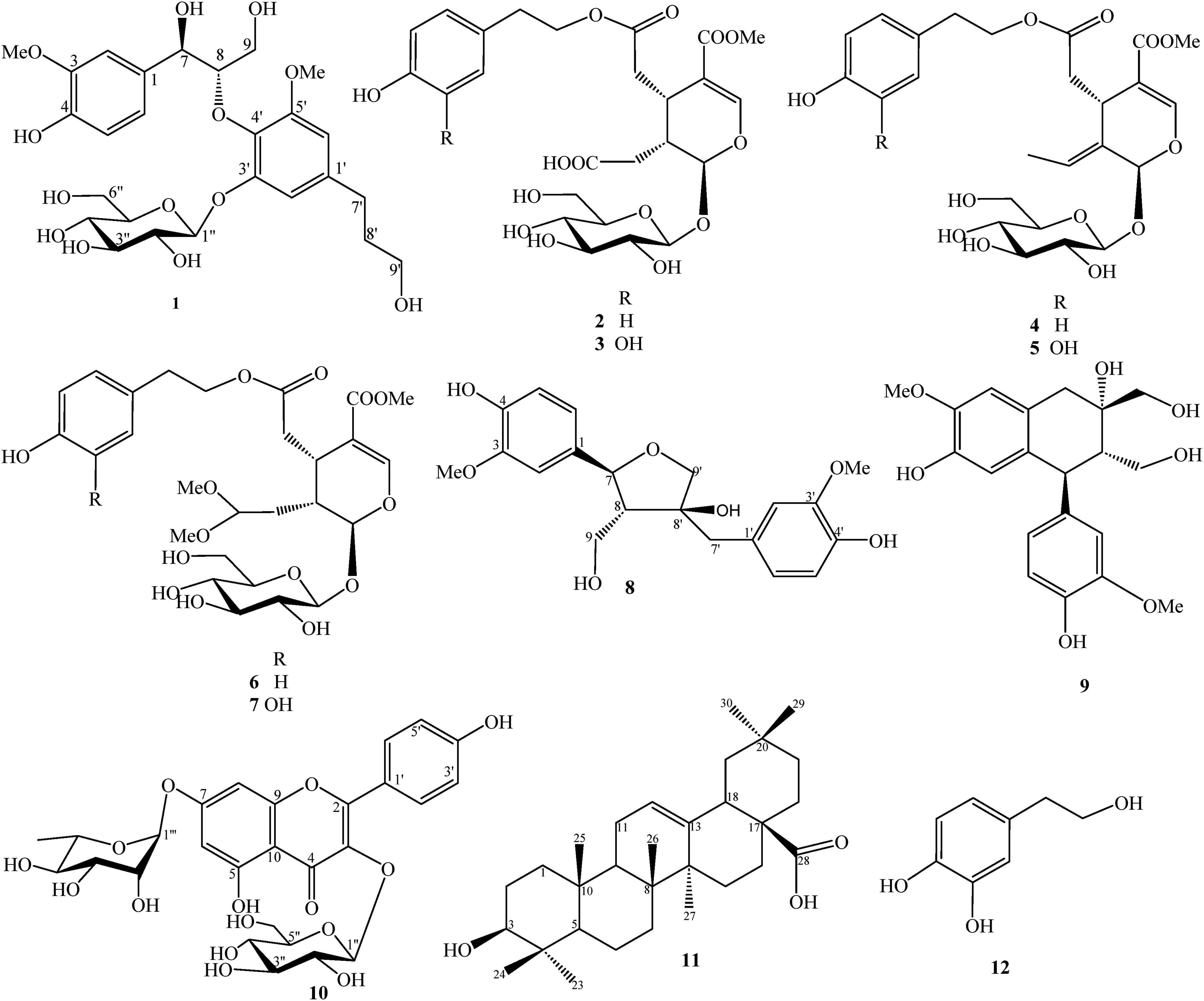
Chemical structures of compounds 1-12.
Results and Discussion
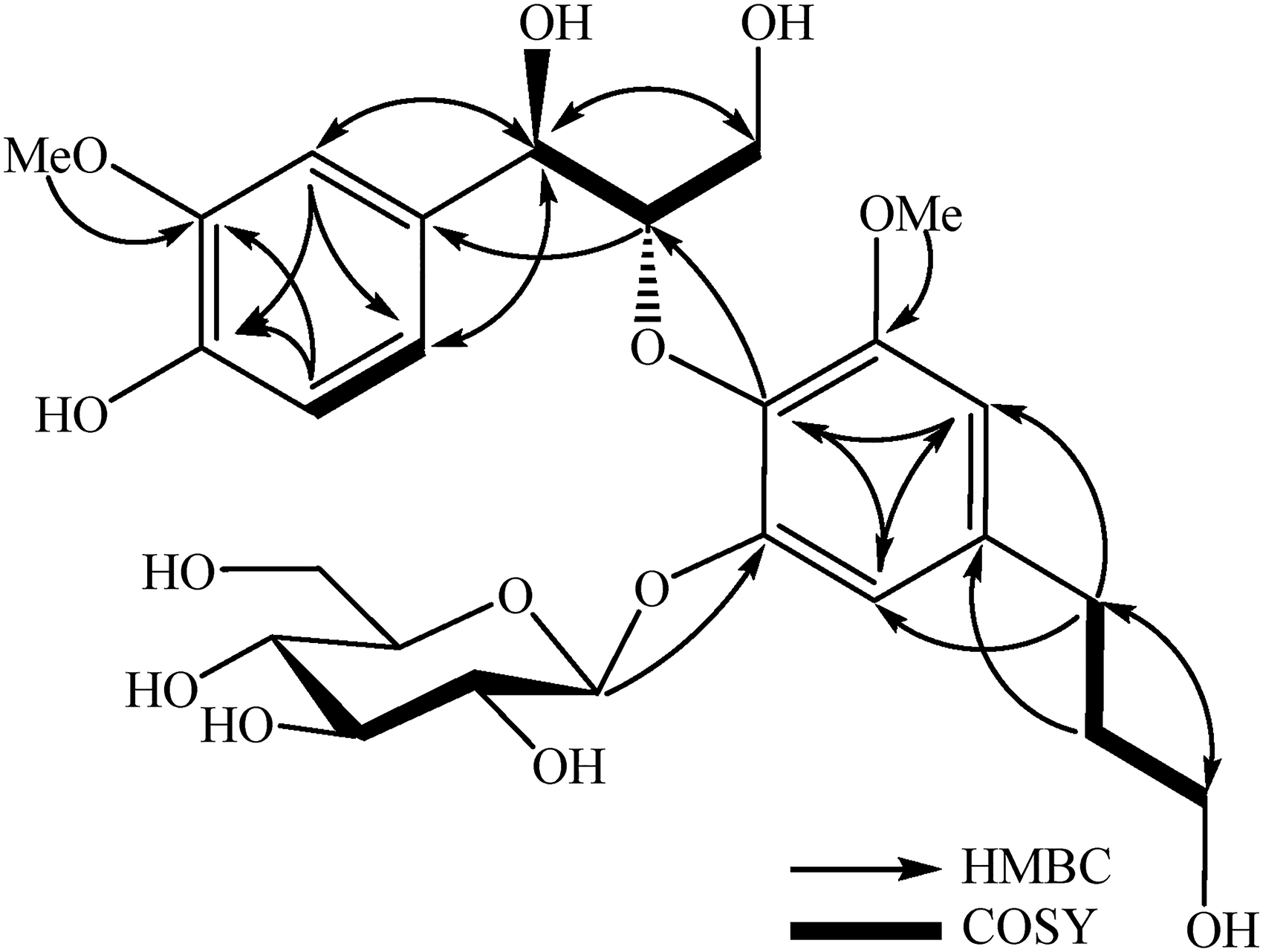
The molecular formula of 1 was determined as C26H36O13 on the basic of HR-ESI-MS ion at m/z 557.2204 [M + H]+ (Calcd. for [C26H37O13]+, 557.2229). The 1H-NMR spectrum of 1 showed proton signals of one ABX aromatic ring system at δH 6.63 (1H, d, J = 2.0 Hz) and 6.81 (1H, d, J = 2.0 Hz), 2 methoxy groups at δH 3.83 and 3.86 (each 3H, s), and one anomeric proton at δH 4.90 (1H, d, J = 7.5 Hz). The 13C-NMR and HSQC spectra of 1 revealed signals of 26 carbons, of which 18 carbons were assigned to an 8-O-4ʹ-neolignan moiety, 6 carbons to a monosaccharide moiety, and 2 methoxy carbons. Analysis of 1H and 13C-NMR data of 1 indicated its structure was similar to that of pinnatifidanin B VII3 except for adding a glucopyranosyl moiety at C-3′. HMBC correlations from H-7 (δH 4.92) to C-1 (δC 133.5)/C-2 (δC 111.3)/C-6 (δC 120.3)/C-8 (δC 87.9)/C-9 (δC 61.5), from methoxy proton (δH 3.86) to C-3 (δC 148.8), as well as COSY correlations of H-5 (δH 6.77)/H-6 (δH 6.79) and H-7 (δH 4.92)/H-8 (δH 4.34)/H-9 (δH 3.61 and 3.91) indicated the presence of 3-methoxy-4-hydroxyphenylpropane-12,3-triol (Figure 2). Additionally, HMBC correlations from H-2′ (δH 6.81) to C-1′ (δC 139.9)/C-3′ (δC 152.4)/C-4′ (δC 135.6)/C-6′ (δC 108.5), from H-6′ (δH 6.63) to C-1′ (δC 139.9)/C-2′ (δC 111.7)/C-4′ (δC 135.6)/C-5′ (δC 154.6), from H-7′ (δH 2.66) to C-1′ (δC 139.9)/C-2′ (δC 111.7)/C-6′ (δC 108.5)/C-8′ (δC 35.2)/C-9′ (δC 62.2), and from the methoxy proton (δH 3.83) to C-5 (δC 154.6) suggested the presence of 3,4-dihyhydroxy-5-methoxyphenylpropane-3-ol. The bond between 2 phenyl propanoids via oxygen atom (8-O-4′) was confirmed by HMBC correlation from H-8 (δH 4.34) to C-4′ (δC 135.6). The large coupling constant between H-1′′ and H-2′′ (J = 7.5 Hz) and 13C-NMR chemical shifts of a monosaccharide unit [δC 103.5 (C-1′′), 75.0 (C-2′′), 77.8 (C-3′′), 71.5 (C-4′′), 78.4 (C-5′′), and 62.6 (C-6′′)] indicated the typical signals of a β-glucopyranosyl moiety. The position of the β-glucopyranosyl at C-3′ was confirmed by HMBC correlation from H-1″ (δH 4.90) to C-3′ (δC 152.4). The small coupling constant of H-7 and H-8 (J = 4.5 Hz) suggested an erythro configuration of 3-methoxy-4-hydroxyphenylpropane-12,3-triol by comparing the coupling constant of erythro configuration: pinnatifidanin B VII (J = 3.9 Hz) and threo configuration: pinnatifidanin B IX (J = 7.0 Hz).3 Moreover, the 7R,8S configuration was confirmed based on Cotton effects ([θ]25 (rel, nm) ‒1.00 (209), +0.82 (240), and +0.22 (278) in the CD spectrum (Figure 3).3 Thus, the structure 1 was established and named neoligustrume A.
The key HMBC and COSY correlations of compound 1.
Experimental CD spectrum of compound 1.
The known compounds were identified to be ligujaponoside A (2),4 ligujaponoside B (3),4 ligustroside (4),5 oleuropein (5),5 ligustaloside A dimethyl acetal (6),6 ligustaloside B dimethyl acetal (7),6 olivil (8),7 (+)-cycloolivil (9),8 kaempferol 3-O-β-D-glucopyranoside-7-O-α-L-rhamnopyranoside (10),9 oleanoic acid (11),10 and hydroxytyrosol (12)11 (Figure 1). Their structures were identified by comparing their spectroscopic data with those reported in the literature.
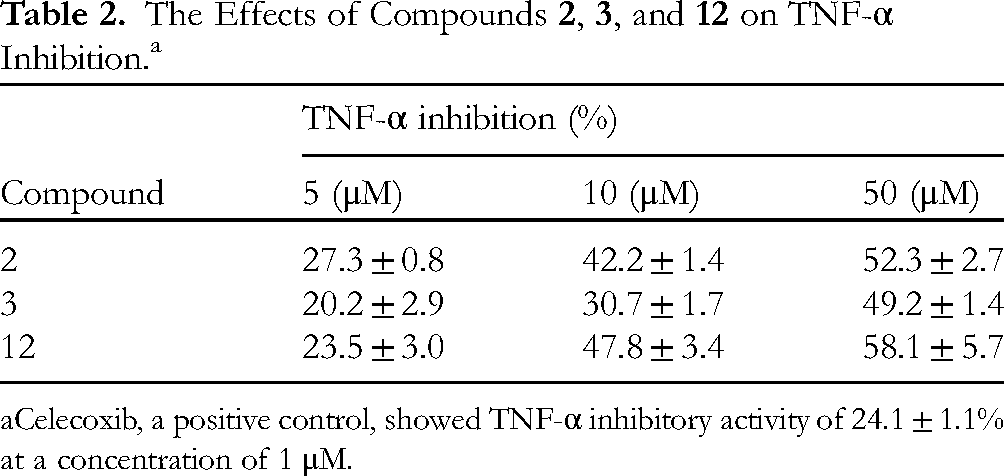
All isolated compounds were evaluated for their inhibitory effect against NO production in LPS-stimulated RAW 264.7 macrophages.12 Firstly, the cytotoxic activity of each compound was evaluated at a concentration of 50 µM. All compounds did not show cytotoxic activities (cell viability > 80%). Thus, these compounds were evaluated for inhibitory NO production in LPS-stimulated RAW264.7 macrophages at concentrations of 1, 5, 10, and 50 µM. L-NMMA was used as a positive control with IC50 value of 12.4 ± 1.6 µM. Compounds 2, 3, and 12 showed significant inhibitory NO production with IC50 values of 16.3 ± 0.5, 18.2 ± 1.1, and 15.7 ± 1.9 μM, respectively (Table 1). Compounds 4-7 and 11 showed moderate inhibitory NO production with IC50 values ranging from 21.4 to 45.0 µM. Our findings were consistent with previous reports of secoiridoids and anti-inflammatory components from L lucidum and L obtusifolium.13,14 Tumor necrosis factor-α (TNF-α) is a well-characterized pro-inflammatory cytokine primarily released from monocytes and macrophages upon invasion of the host by a wide variety of pathogens. Thus, compounds 2, 3, and 12 were further evaluated for their inhibition of TNF-α at concentrations of 5, 10, and 50 μM (Table 2). Among them, compound 12 showed the most TNF-α inhibition with 58.1 ± 5.7% at a concentration of 50 μM. Compound 12 showed strong anti-inflammatory activity (diminished secretion of cytokines, and chemokines and reduced the expression of genes of inducible nitric oxide synthase).15
Inhibitory Effect of NO Production of Compounds 1-12 and the MeOH Extract in LPS-Stimulated RAW264.7 Macrophages.
The Effects of Compounds 2, 3, and 12 on TNF-α Inhibition.a
Compound
TNF-α inhibition (%)
5 (μM)
10 (μM)
50 (μM)
2
27.3 ± 0.8
42.2 ± 1.4
52.3 ± 2.7
3
20.2 ± 2.9
30.7 ± 1.7
49.2 ± 1.4
12
23.5 ± 3.0
47.8 ± 3.4
58.1 ± 5.7
Celecoxib, a positive control, showed TNF-α inhibitory activity of 24.1 ± 1.1% at a concentration of 1 μM.
Material and Methods
General
See Supplemental Material.
Plant Material
The leaves of Ligustrum sinense Lour. (Oleaceae) were collected at Thai Nguyen city, Thai Nguyen province, Vietnam, in January 2020 and identified by Dr Bui Hong Quang, Institute of Ecology and Biological Resources, VAST. A voucher specimen (AT03) was deposited at the Institute of Ecology and Biological Resources, VAST.
Extraction and Purification of Compounds
The dried powder of L sinense leaves (5.0 kg) was sonicated 3 times (each 2 h) with hot methanol (15 L MeOH at 45 °C) and then filtered through filter paper, removed solvent under reduced pressure to yield 500 g of MeOH extract. The MeOH extract was suspended in water and successively partitioned with n-hexane and ethyl acetate, giving n-hexane (LS1, 66.0 g), ethyl acetate extracts (LS2, 53.3 g), and water layer (LS3). LS2 was subjected to a silica gel column chromatography (CC) eluting with gradient solvents of CH2Cl2/acetone (20/1, 10/1, 5/1, 2.5/1, vol/vol, each 1 L) and then MeOH (1 L) to give 5 fractions, LS2A-LS2E. LS2A was chromatographed on a silica gel CC eluting with n-hexane/CH2Cl2/EtOAc (4/1/1, vol/vol/vol) to yield 3 fractions, LS2A1-LS2A3. LS2A2 was chromatographed on an RP-18 CC eluting with acetone/MeOH/water (3/2/1, vol/vol/vol) to yield 2 fractions, LS2A2A and LS2A2B. LS2A2B was chromatographed on a silica gel CC eluting with n-hexane/acetone (15/1, vol/vol) to yield compound 11 (6.9 mg). LS2D was chromatographed on a silica gel CC eluting with CH2Cl2/MeOH (15/1, vol/vol) to obtain 3 fractions, LS2D1-LS2D3. LS2D1 was chromatographed on an RP-18 CC eluting with MeOH/water (1/2.5, vol/vol) to obtain 2 fractions, LS2D1A and LS2D1B. LS2D1A was purified by an HPLC using 10% MeCN in water to yield compound 12 (29.0 mg). LS2D1B was purified by an HPLC using 20% MeCN in water to yield compound 8 (24.0 mg). LS2D3 was chromatographed on a silica gel CC eluting with CH2Cl2/acetone (3/1, vol/vol) to obtain 2 fractions, LS2D3A and LS2D3B. LS2D3B was purified by an HPLC using 20% MeCN in water to yield 9 (3.5 mg). LS2E was chromatographed on a silica gel CC eluting with CH2Cl2/MeOH/water (3/1/0.1, vol/vol/vol) to obtain 4 fractions, LS2E1-LS2E4 was chromatographed on an RP-18 CC eluting with MeOH/water (1/1.3, vol/vol) to obtain 2 fractions, LS2E3A and LS2E3B. LS2E3A was purified by an HPLC using 18% MeCN in water to yield 10 (9.0 mg).
The water layer (LS3) was chromatographed on a Diaion HP-20 CC eluting with water to remove polar components, then increase the concentration of MeOH in water (25%, 50%, 75%, and 100% MeOH, each 1 L) to obtain 4 fractions, LS3A-LS3D. LS3A was chromatographed on a silica gel CC eluting with gradient solvents of dichloromethane/MeOH (20/1, 10/1, 5/1, 2.5/1, vol/vol) to give 4 fractions, LS3A1-LS3A4. LS3A3 was chromatographed on an RP-18 CC eluting with MeOH/water (1/2, vol/vol) to obtain 2 fractions, LS3A3A and LS3A3B. LS3A3A was purified by an HPLC using 23% MeCN in water yield 3 (30.0 mg). LS3A3B was purified by an HPLC using 25% MeCN in water yield 2 (4.6 mg). LS3C was chromatographed on a silica gel CC eluting with gradient solvents of dichloromethane/MeOH (20/1, 10/1, 5/1, 2.5/1, vol/vol) to give 4 fractions, LS3C1-LS3C4. LS3C3 was chromatographed on a silica gel CC eluting with CH2Cl2/MeOH (7/1, vol/vol) to obtain 3 fractions, LS3C3A-LS3C3C. LS3C3A was chromatographed on an RP-18 CC eluting with MeOH/water (1/1.5, vol/vol) to obtain 2 fractions, LS3C3A1 and LS3C3A2. LS3C3A2 was purified by an HPLC using 30% MeCN in water to yield compounds 4 (5.0 mg) and 7 (4.0 mg). LS3C3C was chromatographed on an RP-18 CC eluting with MeOH/water (1/1.5, vol/vol) to obtain 2 fractions, LS3C3C1 and LS3C3C2. Compound 1 (13.2 mg) was obtained from LS3C3C1; compounds 5 (14.9 mg) and 6 (13.0 mg) were obtained from LS3C3C2 on an HPLC system using 25% MeCN in water.
Neoligustrume A (1)
White amorphous powder; : −15.0 (c 0.01, MeOH); CD (c = 1.0 × 10‒3, MeOH): [θ]25 (rel, nm): see Figure 3; HR-ESI-MS: m/z 557.2204 [M + H]+ (Calcd. for C26H36O13: 557.2229); 1H- and 13C-NMR data (CD3OD): see Table 3.
The 1H- and 13C-NMR Spectral Data for Compound 1 in CD3OD.
One new neolignane glucoside, neoligustrume A (1), and 11 known compounds (2-12) were isolated from the leaves of L sinense. Their structures were elucidated by analyses of mass spectra, nuclear magnetic resonance data, and Circular dichroism spectrum. Compounds 2, 3, and 12 showed significant inhibitory NO production with IC50 values of 16.3 ± 0.5, 18.2 ± 1.1, and 15.7 ± 1.9 μM, respectively. Compound 12 showed the most TNF-α inhibition with 58.1 ± 5.7% at a concentration of 50 μM. Thus, compound 12 could be anti-inflammatory source.
Supplemental Material
sj-docx-1-npx-10.1177_1934578X241226825 - Supplemental material for Neolignan Glycoside and Other Constituents From the Leaves of Ligustrum sinense and Their Anti-Inflammatory Activity
Supplemental material, sj-docx-1-npx-10.1177_1934578X241226825 for Neolignan Glycoside and Other Constituents From the Leaves of Ligustrum sinense and Their Anti-Inflammatory Activity by Nong Thi Anh Thu, Lo Huyen Linh, Dao Anh Hoang, Nguyen Tu Oanh, Vu Mai Thao, Nguyen Thi Minh Hang and Nguyen Xuan Nhiem in Natural Product Communications
Footnotes
Acknowledgments
The authors would like to thank Mr Dang Vu Luong, Institute of Chemistry, VAST, for recording NMR and the Korea Basic Science Institute (KBSI, Chuncheon Center) for recording HR-ESI-MS spectra.
Declaration of Conflicting Interests
The author(s) declared no potential conflicts of interest with respect to the research, authorship, and/or publication of this article.
Funding
The author(s) disclosed receipt of the following financial support for the research, authorship, and/or publication of this article: This research was supported by Vietnam Ministry of Education and Training under grant number B2022-TNA-28.
ORCID iD
Nguyen Xuan Nhiem
Supplemental Material
Supplemental material for this article is available online.
References
1.
GaoBBSheGMSheDM. Chemical constituents and biological activities of plants from the genus Ligustrum. Chem Biodiversity.2013;10(1):96-128.
2.
OuyangMAHeZDWuCL. Anti-oxidative activity of glycosides from Ligustrum sinense. Nat Prod Res.2003;17(6):381-387.
3.
HuangXXZhouCCLiLZ, et al.The cytotoxicity of 8-O-4′ neolignans from the seeds of Crataegus pinnatifida. Bioorg Med Chem Lett.2013;23(20):5599-5604.
4.
NgoQMTLeeHSNguyenVTKimJAWooMHMinBS. Chemical constituents from the fruits of Ligustrum japonicum and their inhibitory effects on T cell activation. Phytochemistry. 2017;141:147-155.
5.
HeZDDongHXuHXYeWCSunHDButPPH. Secoiridoid constituents from the fruits of Ligustrum lucidum. Phytochemistry. 2001;56(4):327-330.
6.
Mohammed H, Ehab AR, Abd El-salam IM, Usama YS. New secoiridoids from Ligustrum ovalifolium and their hypotensive activity. Pharmacognosy Res.2009;1(2):91-97.
7.
YeoHChinYWParkSYKimJ. Lignans of Rosa multiflora roots. Arch Pharm Res. 2004;27(3):287-290.
8.
LiHZLuoGJLiHMLiXLLiRT. A new aryltetrahydronaphthalene lignan from Epimedium brevicornum. Chin Chem Lett.2011;22(1):85-87.
9.
ÖzdenSDürüstNTokiKSaitoNHondaT. Acylated kaempferol glycosides from the flowers of Delphinium formosum. Phytochemistry. 1998;49(1):241-245.
10.
ThuNTHHoaPTHDatNTTuyenPNK. Triterpenoids, steroid, and aromatic compounds from Combretum indicum leaves. Vietnam J Chem. 2022;60(5):629-635.
11.
LeeSYChoiSULeeJHLeeDULeeKR. A new phenylpropane glycoside from the rhizome of Sparganium stoloniferum. Arch Pharm Res. 2010;33(4):515-521.
12.
NhiNPKPhuong HaTAnhLTChiVTQCuongLCV. Secondary metabolites from the leaves of Solanum melongena and their nitric oxide production inhibitory activity. Vietnam J Sci Technol. 2022;60(3):362-370.
13.
PengYSLiuJXJiaoJ, et al.Secoiridoid glycosides from the fruits of Ligustrum lucidum and their in vitro anti-inflammatory activity. Fitoterapia. 2023;171:105705.
14.
SuhWSKwonOKLeeTHSubediLKimSYLeeKR. Secoiridoid glycosides from the twigs of Ligustrum obtusifolium possess anti-inflammatory and neuroprotective effects. Chem Pharm Bull.2018;66(1):78-83.
15.
RichardNArnoldSHoellerUKilpertCWertzKSchwagerJ. Hydroxytyrosol is the major anti-inflammatory compound in aqueous olive extracts and impairs cytokine and chemokine production in macrophages. Planta Med.2011;77(17):1890-1897.
16.
HuyenLTOanhLT. Diterpenoids from Rosmarinus officinalis L. and their nitric oxide inhibitory activity. Vietnam J Chem. 2021;59(2):229-234.
Supplementary Material
Please find the following supplemental material available below.
For Open Access articles published under a Creative Commons License, all supplemental material carries the same license as the article it is associated with.
For non-Open Access articles published, all supplemental material carries a non-exclusive license, and permission requests for re-use of supplemental material or any part of supplemental material shall be sent directly to the copyright owner as specified in the copyright notice associated with the article.