
Abstract
Chinese herbal medicine (CHM) represents a potent, safe, and efficacious reservoir of treatment options against an array of microbial infections and inflammatory diseases. It has a long history of positive clinical outcomes with minimal or no side effects while enhancing and bolstering the host's protection against infections. With its unique ability to prevent, treat, and manage a wide range of diseased conditions, CHM has been successfully practiced in China for thousands of years. In the modern medical era, where harsh therapeutic drugs and antimicrobial resistance (AMR) present a significant challenge, CHM warrants further exploration. The present review highlights and focuses on 4 major CHM-based herbs, that is, (Lonicerae flos [LF], Lonicerae japonicae flos [LJF], Scutellaria baicalensis Georgi [SBG], and Forsythia suspensa [FS]) in terms of their antibacterial and anti-inflammatory efficacies. A detailed literature survey was done by the team using a systematic electronic search from PubMed, Science Direct, Google Scholar, Research Gate, books, etc. This was followed by data collecting, pertinent data extraction, in-depth analysis, and composing the final review. Each herb has been discussed in detail describing its mechanism adopted and the bioactive components involved in alleviating bacterial infections and inflammatory damage. Further, proof of efficacy by detailing the major past studies and major findings has been discussed for each of the 4 herbs. This review will give the scientific community the opportunity to update their knowledge on the subject, which is crucial for heralding the process of bringing CHM-based medicines closer to clinical development given the area of alternative medicine's rapid advancements.
Introduction
A traditional method of healthcare, traditional Chinese medicine (TCM) aims to prevent or treat a variety of illnesses, including cancer, diabetes, depression, Alzheimer's, cardiovascular disorders, infectious diseases, and others. The fundamental tenet of TCM is the preservation of the ying-yang balance for a healthy trend while eliminating the pathogenic elements.1,2 The use of Chinese herbal medicine (CHM) is the most crucial component of TCM. CHM represents one of the most abundant sources of medicinal herbs that have been used for centuries to treat and cure a wide range of infections and inflammatory conditions. The bioactive herbal components and their therapeutic potencies are the key to CHM's success as a common complementary and alternative medicine (CAM) against various medical conditions.3–5 Owing to the positive therapeutic results noted in past studies and the minimal adverse effects reported, there has been a recent resurgence in interest and renewed focus on further researching these herbal medicines.
With the ongoing challenge of rising antimicrobial resistance (AMR), there is an imperative need to focus on nonantibiotic approaches and alternative solutions to combat bacterial infections not responding to antibiotics.6,7 Furthermore, the use of gentler yet more effective substitutes is justified because high doses of antibiotics are frequently linked to significant toxicity in patients.CHM has been shown to inhibit the growth of several pathogens via different mechanisms adopted at the molecular level while posing minimal side effects postadministration.8,9 Additionally, it has been demonstrated that Chinese herbs have synergistic effects when used with conventional antibiotics, making them a promising adjunct therapy strategy that should be used to enhance the clinical outcomes.10,11
Four important CHMs that have been frequently used and referred to as “heat-clearing herbs” (Lonicerae flos,[LF] Lonicerae japonicae flos [LJF], Scutellaria baicalensis Georgi [SBG], and Forsythia suspensa [FS]) have been identified in the current review. These herbs are known to have potent antibacterial and anti-inflammatory properties. The present review aims to discuss in detail each of these 4 herbs and summarize the major studies of the past and their findings in order to understand the possible mechanisms toward antibacterial (via direct cell damage, reversal of resistance, quorum sensing inhibition, synergistic action, etc.) and/or anti-inflammatory activity. Both the antibacterial and anti-inflammatory potential of each medicinal herb has been discussed under separate headings. This aims to provide quantifiable data on these alternative, safe antibacterial and anti-inflammatory compounds, updating the scientific community with recent developments and serving as a reference for the application and clinical success of these medicinal plants in the future.
Materials and Methods
PubMed, as well as internet searches (Google search engine), were used without time restriction following the use of related keywords that included names of the 4 major herbs, that is, LF, LJF, SBG, and FS and search terms like Chinese herbs, CHM, Chinese medicinal plants, and Chinese herbal extracts applying a combination of one or more keywords in relevance to antibacterial, antimicrobial, anti-biofilm, in vitro and/or in vivo efficacy, synergy, antibiotic resistance, and anti-inflammatory activity. Articles were considered if they contained information on 1 or more of the 4 herbs that were the subject of this review. Studies that did not cover 1 of the 4 plants but instead focused on the antibacterial or anti-inflammatory characteristics of other Chinese medicinal herbs were not taken further and were excluded. All 3 authors (XM, MY, WL) screened the titles and abstracts of articles obtained from the initial search while excluding articles that did not fit into the context for this review. Further, the full text of the shortlisted articles was read and relevant information was abstracted. Information from the source documents was organized into various categories and subsections as per the flow of the review article.
LF and LJF: Antibacterial potential
LF: LF, also known as Shanyinhua in Chinese, is one of the significant TCM herbs listed in the Chinese Pharmacopoeia (2015 edition). The dried flower buds belonging to 4 main species of the genus Lonicera (Caprifoliaceae), that is, Lonicera hypoglauca, L. confusa, L. fulvotomentosa, and L. macranthoides are all referred to as LF. Numerous pharmacological benefits of LF include its hepatoprotective, anti-tumor, anti-angiogenic, anti-diabetic, and anti-tumor properties.12,13 The herb has demonstrated effectiveness against a range of pathogens as it possesses significant antibacterial and anti-inflammatory potential. However, the studies of LF based on its antibacterial activity are limited and thus few major studies will be discussed.
The studies of the antibacterial effects of LF mostly focus on L. macranthoides Hand.-Mazz. The antibacterial effects tend to vary as per the method of preparation used. For example, L. macranthoides Hand.-Mazz. contains phenolic acids (total chlorogenic acids), glycosides, flavonoids (total flavones), and volatile oils. These ingredients have varying degrees of inhibitory effects on bacteria including Escherichia coli, Staphylococcus aureus, and Pseudomonas aeruginosa, with S aureus being more inhibited than E coli. The Sichuan province's L. macranthoides Hand.-Mazz has inhibitory effects on S aureus, Streptococcus haemolyticus, E coli, and Shigella flexneri. The same herb, nevertheless, had no effect on Salmonella typhimurium.14–16 In another in vitro study by Du et al. 17 the authors cultured Helicobacter pylori using a tomato juice culture medium, and LF exhibited antibacterial activity against this bacterium, one of the main reasons for peptic ulcers and gastrointestinal problems. In a recent study, Tao et al. 18 examined the antibacterial impact of the essential oils isolated from LF in a recent study. The oils significantly inhibited the growth of E coli, S aureus, and P aeruginosa, as demonstrated by the minimum inhibitory concentration (MIC) test and the disc diffusion assay. Essential oil tested at (25%, 50%, 75%, and 100% (v/v)) against E coli showed inhibition diameter ranging from 11.5mm to 16.5 mm. For S aureus, the inhibition diameters ranged from 13.7 mm to 21.4 mm. However, the diameter for P aeruginosa was less ranging from 8.7 mm (25%) to 11.8 mm (100%). The MIC values for S aureus (20.0 μL/mL) were comparatively lower than the values seen for E coli and P aeruginosa (37.0 and 71.0 μL/mL, respectively) suggesting superior results against Gram-positive pathogens. Additionally, research has shown its effectiveness in treating oral pathogens like Streptococcus mutans, Actinomyces viscosus, and Bacteroides melaninogenicus. 19
The possibility of using this herb in combination therapy with antibiotics would be advantageous as it may offer synergistic activity improving the treatment outcome and reducing the dose of the antibiotic involved thus reducing the associated side effects and frequency of antibiotic drug resistance. This is demonstrated by a study that was preprinted but not peer reviewed. Using the checkerboard dilution method and the fractional inhibitory concentration index (FICI) to check the synergy, Cui et al. 20 conducted an in vitro study to assess the synergistic and dose–effect relationship of Trimethoprim (TMP) and LF. The MIC of LF alone against S aureus, E. coli, and Salmonella was 125 mg/mL, 250 mg/mL, and 125 mg/mL, respectively. When LF (62.5 mg/mL) was used in combination with TMP (20µg/mL), it showed an additive effect with a FICI = 0.75. Further, the bactericidal rates were fitted with a least square method. The 95% optimized additive concentration of TMP to enhance the bactericidal activity of LF on test organisms were 231 to 249μg/mL (S aureus); 237 to 259μg/mL (E coli); and 235 to 259μg/mL (Salmonella). In another study , 21 the team reported synergistic effects of LF with vancomycin and linezolid against both planktonic and biofilm-forming strains of methicillin-resistant S aureus (MRSA). In this study, the authors used Tanreqing (TRQ) injection which is a commercial TCH formula. The ingredient TRQ, which is derived from Scutellariae radix, LF, Forsythia fructus (FF), and Naemorhedi cornu, is effective against pneumonia, MRSA, and upper respiratory tract infections. Results showed that TRQ exhibited a MIC value of 4125 μg/mL for MRSA, while the MIC values for both tested antibiotics (vancomycin and linezolid) were 2.5 μg/mL. When the combination was used, MIC values of both antibiotics were reduced, that is, vancomycin and TRQ showed a 2-fold reduction in MIC (from 2.5 µg/mL to 1.25 µg/mL) while in the case of linezolid, it was an 8-fold reduction seen by checkerboard analysis assay. One possible explanation for this synergic effect seen with TRQ might be its ability to inhibit the efflux pump system. The efflux pump represents a key resistance mechanism adopted by bacteria allowing the antibiotics to be extruded before they reach their intended targets .23,24 In a separate study, the authors showed that TRQ was able to exhibit a synergistic antibacterial effect with amoxicillin, aztreonam, meropenem, ceftazidime, and cefoperazone against 2 multi-drug resistance P aeruginosa efflux pump-positive strains. Also, TRQ was able to downregulate the expression levels of bacterial efflux pump genes. 24 These encouraging results point to a viable strategy for battling antibiotic resistance and enhancing treatment results: combining traditional medicine with conventional medicines. Table 1 shows a summary of the main studies that concentrated on the antibacterial properties of Lonicerae flos.
Summary Table of Major Studies Focussing on Antibacterial Effects of Lonicerae flos (LF).
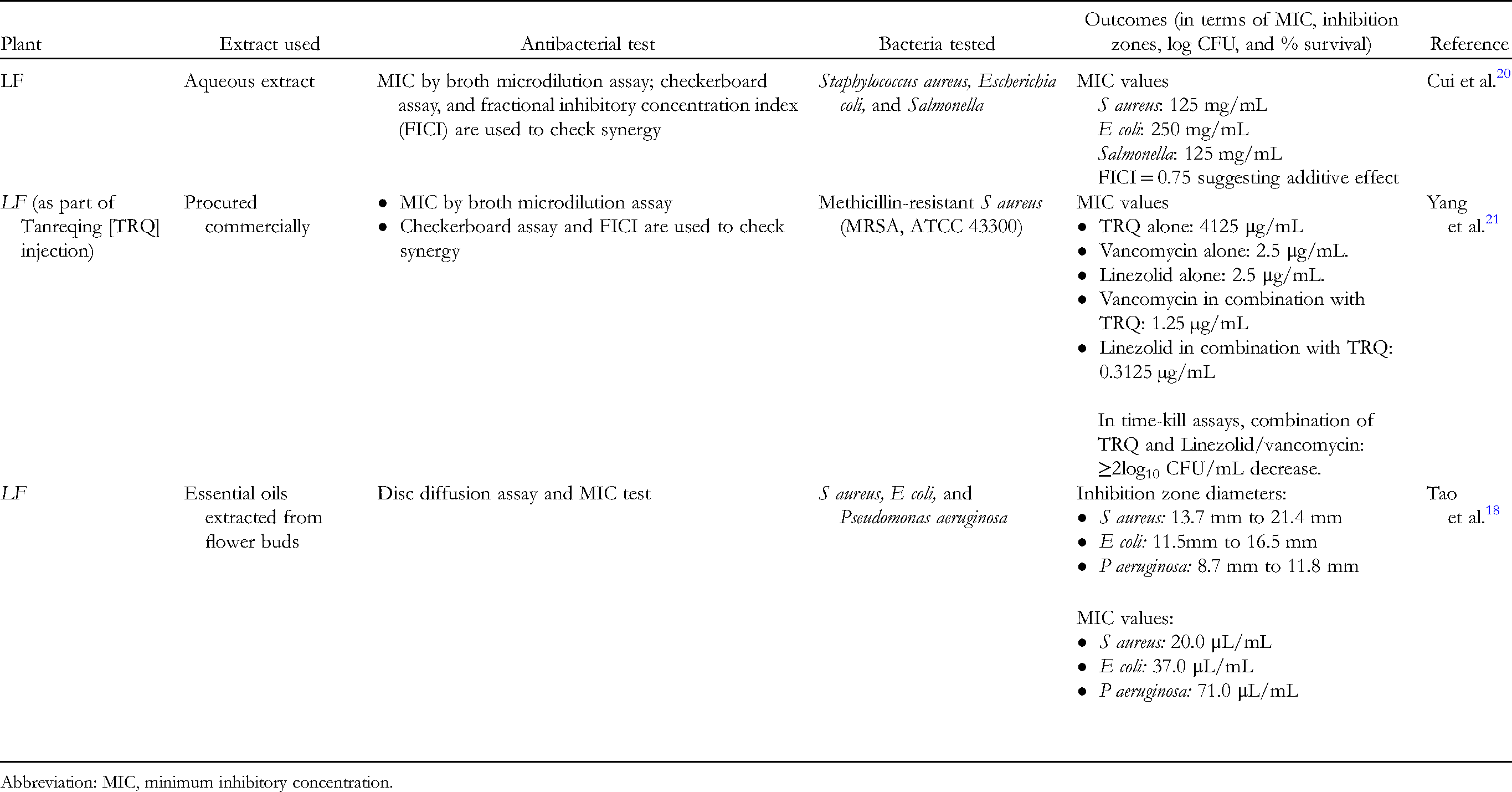
Abbreviation: MIC, minimum inhibitory concentration.
LJF: LJF, also known as Japanese honeysuckle, Jin YinHua, or Ren Dong, is a common Chinese medicinal herb and a member of the Caprifoliaceae family. According to the Chinese Pharmacopeia (2005 edition), LJF and LF are distinct herbs with vastly different morphologies, therapeutic benefits, and bioactive component concentrations. Furthermore, Lonicera japonica Thunb is the sole source of LJF in plants.13,25,26 LJF exhibits a broader antimicrobial spectrum, and more powerful antibacterial activity acting even against drug-resistant bacteria which is the need of the hour. 15
The list of bioactive compounds present in the extract is complicated and so far, more than 300 chemical compounds have been isolated and identified. The major components include phenolic acids, organic acids, essential oils, flavones, iridoids, and saponins.13,18,27 Going into the details, there are 49 different kinds of phenolic acids which include: CGA derivatives and cinnamic acid derivatives. The CGA and its derivatives are neochlorogenic acid (NGC), isochlorogenic acid A, isochlorogenic acid B, isochlorogenic acid C, etc. The cinnamic acid derivatives isolated and identified are caffeic acid (CA), 1-O-caffeoylquinic acid, trans-cinnamic acid, trans-ferulic acid, caffeic acid methyl ester, etc. 28–30 A total of 52 flavonoids have been isolated from LJF. The flavonols mainly include rutin, quercetin, isoquercitrin, astragalin, quercetin 3-O-hexoside and the flavones are primarily cynaroside, luteolin, chrysoeriol 7-O-neohesperidoside, chrysoeriol 7-O-glucoside, lonicerin, tricin, etc. Among the essential oils, the main constituents in flower buds are (Z, Z)-farnesole (16.2%) and linalool (11.0%), the main constituents in leaf fraction include hexadecanoic acid (16.0%) and linalool (8.7%), and the main constituents in the stem are hexadecanoic acids (31.4%). Among the iridoids, the major ones are loganin, sweroside, secologanoside, ethyl secologanoside, centauroside, etc., that exhibit strong anti-inflammatory and antioxidant activity. In addition, these bioactive substances found in LJF have been shown by Xu et al. to effectively block DNA gyrase activity, which in turn inhibits bacterial growth. 31 The team studied the effect of 15 Chinese herbal drugs (CHDs) in form of their methanolic extracts against the supercoiling activity of bacterial DNA gyrase using a DNA gyrase kit. When compared to other herbs, the results showed that LJF extracts (LFFE) (0.4 g/mL) were highly effective at inhibiting the supercoiling activity of gyrase, and the inhibitory effect was concentration dependent. This suggests that LJF, a gyrase inhibitor, may operate as an effective substitute due to rising resistance to currently available DNA gyrase inhibitors (ie, quinolone drugs).
The different components f LJF tested by researches include water extract, alcoholic extract, extracted oil, polysaccharides, etc., and each extract varies in its final activity and effect. Most of the water extracts of LJF are more powerful against S aureus and E coli and show weak activity against P aeruginosa and Shigella spp.32–36 In 2009, Rahman et al. 37 evaluated the effect of extracted volatile oil with significant antibacterial potential against Listeria monocytogenes, Bacillus spp., S aureus, E coli, Salmonella enteritidis, and S typhimurium as tested by MIC as well as disc diffusion assay. In another study by Song et al. 38 the team reported marked antibacterial activity of LJF against 14 strains, including S aureus, S haemolyticus, E coli, Bacillus dysenteriae, Bacillus comma, Bacillus typhosus, Bacillus paratyphosus, P aeruginosa, Klebsiella pneumoniae, Mycobacterium tuberculosis, S mutans, Bacillus adhaerens, Haemophilus actinomycetemcomitans. The extract was able to inhibit 87.5% of the test strains with an average MIC value of 25 µg/mL.
Instead of testing a volatile oil extract, Kang et al. 39 instead investigated aqueous and alcoholic extracts of LJF. The MIC and MBC values (expressed as percent inhibition) for the water extract on S aureus were 19.25 and 38.50%, respectively, and for the alcohol extract on S aureus, it was 19.60 and 39.20%, respectively. The major bioactive component of all these extracts prepared from LJF is CHA. MIC of CHA to Shigella and Salmonella was 0.125 mg/mL, in comparison to the antibiotic kanamycin. Also, the MIC values against E coli, Sarcina luteus, Bacillus subtilis, and S aureus were 0.025 g/mL, 0.025 g/mL, 0.1 g/mL, and 0.8 g/mL as reported by Wu et al., 40 LJF demonstrates to be a promising alternative option in light of the developing antibiotic resistance. According to Tang et al, 16 one of the bioactive components of LJF, flavonoids, was able to significantly inhibit MRSA with MIC ≤ 5 mg/mL. The agar dilution method was used to evaluate the butanol LJFE on a very large group of 104 clinical isolates of Gram-positive and Gram-negative anaerobes. 41 LJF and imipenem (among the set of 5 antibiotics) were highly active against Bacteroides fragilis, Bacteroides ovatus, C. difficile, C. perfringenes, and Propionibacterium acnes, with MIC90 (where 90% of isolates get inhibited) ranging from 0.032 mg/L to 0.5 mg/L. The extract was active against all anaerobic groups, but not B ovatus and Peptostreptococci. These results reinforce LJF's ability to combat clinical strains, the majority of which show a high level of resistance. Additionally, they emphasize that LJF used in combination with other conventional herbs or antibacterial medicines will improve the effectiveness of the treatment. In one such study, LJFE and Magnolia obovata extract were used in combination against strains of S aureus, B subtilis, and C. albicans. When examined using an in vitro time-kill assay, the 2 herbs had a synergistic impact with greater killing efficacy against all test strains. 42
A similar study by Yang et al. 43 reported the enhanced killing effect of silver nanoparticles (AgNPs) when they were used in combination with LJF water extract. The researchers first evaluated the survivability of pathogenic E coli CMCC44113 using the antibacterial properties of AgNPs and LJFE alone. AgNPs demonstrated a concentration-dependent drop in viable population, with only 8% and 4% of the population able to form colonies at concentrations of 0.5 mg/mL and 1 mg/mL, respectively. The LJFE at 1 × LJFE, showed a 50% decrease in the E coli population and at 5 × LJF strength, complete eradication was evident. Further, when the combination of AgNPs and LJFE was used (the mixture of 3 × LJF and 0.1 mg/mL AgNPs), a 4.24% survival rate was seen in contrast to a 42.57% survival rate seen when 3 × LJF was used alone or 22.75% survival rate when 0.1 mg/mL of AgNP's was used singly. This indicates that in combination with 3 × LJF, the concentration of AgNPs e required to eradicate the bacteria was reduced to one-tenth (0.1 mg/mL). This is equally essential as silver may pose adverse effects at high concentrations.45,46 and thus a combination approach can be used to achieve a similar level of killing at low concentrations. Further, upon studying the possible mechanism of the synergistic effect seen, it was delineated that AgNPs caused cell wall lysis and damage to the cell membrane integrity of E coli cells. This physical damage increased the penetration and easy entry of LJF into the bacterial cell allowing the active constituents of the extract, that is, CHA to exhibit its damage resulting in enhanced cell death. These findings open the possibility of the development of promising nanoparticle-enhanced herbal pharmaceuticals. However, more such studies focussing on the use of combination approaches involving LJF along with other antibacterial or antibiotics are required as data is still limited.
Animal studies on the antibacterial efficacy of LJF are also scarce but those that have been done so far support its antibacterial potential. In a study by Miami and Makino, 46 the researchers evaluated the LJFE on Citrobacter rodentium-induced digestive tract infection in a mouse model. For this, mice were infected with bacterial suspension orally followed by LJFE. 24 h after infection, the majority of mice with C rodentium perished. However, the mice that received LJFE showed enhanced survival till 5 days and beyond. In the histological investigation, control untreated mouse tissue displayed bacterial mass in colonic mucosa, whereas LJFE (2g/kg)-treated mouse tissue did not. In the LJFE-treated group, bacterial counts in the large intestine were fewer than 2 log CFU/g, compared to 3.5 log CFU/g in the control group that was not treated. Similarly, for control animals, bacterial load in mesenteric lymph nodes and blood was 2 log CFU/mL whereas mice treated with 2 g/kg of LJFE, the bacterial load was minimal (less than 0.5 log CFU/mL in lymph nodes and close to negligible in blood). In the ex vivo experiments, LJFE-treated mice showed increased levels of IL-1β, IFN-γ, and as TNF-α in both serum and culture supernatant of intraperitoneal macrophages. This supports the immunostimulatory effects of LJFE leading to accelerated clearance of bacteria with faster resolution and higher survival rates. Table 2 includes details of this and other significant research on the type of extract, a thorough technique, and outcomes in terms of bacterial load decrease.
Summary Table of Major Studies Focussing on Antibacterial Effects of Lonicerae Japonicae Flos (LJF).

Abbreviation: MIC, minimum inhibitory concentration.
The ethanolic extract of LJF (LJEE) was examined for its antibacterial activity as well as its wound repair and healing efficacy in a rat excision wound model in a different animal investigation. 47 Wound excision model was established in male Wistar rats with 500-mm 2 full thickness wound inflicted on their backs. This was followed by treating their wounds with simple ointment, LFJ extract (10%) and nitrofurazone (10%), respectively, as per different groups of 8 animals each. Results of this study depicted that the CHA content LJFE was 64.2 ± 0.18 μg/g of the dry extract. The extract exhibited significant antimicrobial activity against S aureus (10.96 ± 0.08 mm), Staphylococcus epidermidis (10.67 ± 0.11 mm), and E coli (8.17 ± 0.09 mm). The ointment formulation containing 10% (w/w) LFJ exhibited high healing ability and significant wound contraction (21.6% on day 3 to 85.6% by day 12) with complete healing was seen by day 15 whereas simple ointment group showed slow wound contraction (13.2% on day 3 and only 52.2% by day 12). In comparison to the group treated with ordinary ointment, the wound treated with LFJ extract also showed significantly improved fibroblast proliferation, angiogenesis, and reepithelialization. These promising results advocate the use of purified LJEE in wound and skin infections owing to its excellent antibacterial, wound healing and reepithelization properties. More in vivo research is needed, though, on parameters other than efficacy, such as the impact of co-treatment, dose–response studies, the impact of various administration routes, and safety considerations against a range of bacterial infections, particularly drug-resistant strains in different animal models.
LF and LJF: Anti-inflammatory potential
Inflammatory dysregulation can cause consistent tissue damage further worsening the disease outcomes. Evidence suggests that both LF and LJF have significant potential to alleviate inflammation and related damage as seen in animal studies involving various models of inflammation. These include studies done in carrageen- and croton oil-induced paw edema and ear edema models 39,48 cotton ball granulomatous hyperplasia, 49 egg white induced localized acute inflammation model. 32 Both the water and alcohol extracts of Lonicerae japonicae flos have shown significant anti-inflammatory potential.15Similarly, LF water extracts were found to decrease inflammation as seen in the acetic acid-induced capillary permeability increase model, paw swelling model, cotton ball granulomatous hyperplasia mode, carrageenan-induced paw swelling model, etc,16,50,51
Few studies that have focused on understanding how the extracts of LJF work in regulating the inflammatory pathways are notable as they examine the bioactive components and associated processes. Inami et al. 52 used a three-dimensional human keratinocyte culture to investigate the inhibitory efficacy of water extract from LJF flower buds and its primary ingredient CHA against histamine synthesis. They also looked at the levels of L-histidine decarboxylase (HDC). According to the results of ELISA and western blotting experiments, both CHA and the whole extract significantly reduced the release of histamine by keratinocytes and the expression of the 53kDa HDC. The effect was greater for the extract than for a single CHA, indicating that more components may cooperate to reduce histamine release. In addition, as studied in the activated macrophage-like cell line RAW 264.7 cells, the water LJEE demonstrated potent inhibition of both NO production and TNF-α secretion in a dose-dependent manner. It also prevented mast cell activation induced by trypsin by interfering with the phosphorylation of the extracellular signal-regulated kinase. 53
During inflammation, macrophages get activated and start releasing proinflammatory mediators such as tumor necrosis factor α (TNF)-α, cyclooxygenase-2 (COX-2), and inducible nitric oxide synthase (iNOS). 54 Further, macrophage activation leads to triggering the activation of transforming growth factor-beta (TGF-β-) activated kinase 1 (TAK1) which causes phosphorylation of mitogen-activated protein kinases (MAPKs) and inhibitor of nuclear factor kappa-B kinase (i.e., IκB kinase). This leads to the final activation of the most important regulator of inflammation, that is, nuclear factor-κB (NF-κB), the principal transcriptional activator.55,56 Park et al. 57 studied the detailed mechanism involved in the role of polyphenols isolated from LJF in exerting anti-inflammatory effects. A total of 18 polyphenols consisting of 11 hydroxycinnamic acids and 7 flavonoids were isolated by HPLC and then an array of experiments were done to see the effects of polyphenols on (a) cell viability of RAW 264.7 cells, (b) COX-2 mRNA and its protein expression in lipopolysaccharide (LPS)-stimulated RAW 264.7 cells, (c) on iNOS mRNA and related protein expression in LPS-stimulated RAW 264.7 cells, (d) degradation and phosphorylation of IκB-α and expression levels of NF-κB p65 protein, and (e) LPS-induced phosphorylation of MAPKs. iNOS and COX-2 are important regulators of inflammation and hence they represent key target proteins for anti-inflammatory therapies. Our results showed that pretreatment with 200 μg/mL polyphenols decreased the mRNA expression of LPS-induced COX-2. Polyphenols also markedly suppressed COX-2 protein induction at concentrations of 100 and 200 μg/mL. Similarly, the iNOS mRNA expression was also inhibited with 100 and 200 μg/mL of polyphenols in the LPS-stimulated RAW 264.7 cells along with reduced iNOS protein levels. Further, mRNA expressions of TNF-α, IL-1β, and IL-6 were all significantly decreased posttreatment with polyphenols at 200 μg/mL and 100 to 200 μg/mL. Next, polyphenols inhibit the translocation of NF-κB p65 protein from the cytosol to the nucleus and polyphenols prevented the phosphorylation of IκB-α in a dose-dependent manner. This may be crucial as degradation and phosphorylation of IκB-α are important for NF-κB activation. Phosphorylation of MAPK is essential for further triggering the inflammatory cascade. However, co-treatment with 100 and 200 μg/mL polyphenols reduced the LPS-induced phosphorylation of p38 MAPK. In a similar study, the team 39 reported that Luteolin (3′,4′,5,7-tetrahydroxylflavone), a flavonoid isolated from flower buds of LJF was also able to suppress NF-κB activation, IκB degradation and reduce the expression of TNF-α, IL-8, IL-6, GM-CSF, and COX-2 through a decrease in the intracellular calcium levels. Lin et al 58 investigated the pathways through which water extract of LJF (FLJWE) reduced virus-induced inflammation. Results indicated that FLJWE suppressed the levels of TNF-α, IL-6, RANTES, and monocyte chemoattractant protein-1 (MCP-1). All these are inflammatory cytokines that are released postactivation of NF-κB. Further, FLJWE dose-dependently inhibited the expression of NF-κB, COX-2, and iNOS as determined by the western blotting technique. Pretreatment with the FLJWE showed reduced phosphorylation of STAT1 and STAT3 in a dose-dependent manner while it upregulated the protein levels of both pNrf2 and heme oxygenase 1 (HO-1) when compared to control. HO-1 is an inducible anti-inflammatory enzyme that via its antioxidant and anti-inflammatory role helps to protect against oxidative stress.59,60 The authors also compared the effect of the whole extract, luteolin (a flavonoid from flower buds), and CHA and found that FLJWE and luteolin inhibited the levels of TNF-α, IL-6, MCP-1, and RANTES in infected RAW264.7 cells unlike seen with CHA. Also, FLJWE and luteolin, but not CHA, strongly inhibited the expression of NF-κB, iNOS, and COX-2 in virus-infected RAW264.7 cells and upregulated levels inducible HO-1 expression According to these results, luteolin is a significant bioactive ingredient that helps to reduce the inflammatory reactions brought on by viruses. These findings provide the pharmacological basis advocating further use of FLJWE, in the treatment of inflammatory damage caused by bacterial or viral agents. Apart from flavonoids, polyphenols, and CHA, Yang et al. 61 also reported the presence of a new lignan, loniceralanside A, together with 3 known compounds, isolated from L. Japonicae Flos. The bioassays showed that all 4 compounds showed moderate to high anti-inflammatory activities with IC50 values ranging from 3.05 to 27.80 mM with the lignan showing similar anti-inflammatory activity as the control sample.
This detailed insight clears the mechanism adopted by the various polyphenols and bio constituents present and how they act at multiple levels to alleviate the exaggerated inflammatory response.
SBG: Antibacterial potential
SBG, often known as Chinese Skullcap, is a perennial Lamiaceae herbaceous plant species. It is a plant indigenous to the countries of East Asia. The Chinese have utilized its dried roots as traditional medicine for centuries, referring to it as Huang-Qin. 12,27,62
Due to the many bioactive components present, SBG extract possesses an array of pharmacological activities. The principal ingredients are flavones, such as baicalin, wognoside, and their aglycones baicalein and wogonin. Baicalin is a monomer with the chemical formula C12H18O5 and a molecular mass of 446.3, while baicalein is its glucuronide form (C15H10O5; molecular mass of 270.24). In addition to flavones, SBG creates a number of other natural substances, such as amino acids, essential oils, flavonoids, phenylethanoids, and sterols 63 Additionally, SBG extract inhibits the growth of bacteria, fungus, viruses, mycoplasma, and spirochetes. SBG extract has been shown to have strong antibacterial activity against Bacillus cereus, E coli, Listeria monocytogenes, Salmonella anatum, and S aureus, among the 46 herbs and spices.17,64,65 Past studies have demonstrated that SGB utilizes multiple mechanisms and multiple targets to exert bactericidal actions. In an intriguing study conducted by Zhao et al, 66 the inhibitory effect of baicalin on isolated E coli strains obtained from bovine mastitis milk was investigated. Further, the resistance rates of the E coli isolates to 13 antimicrobial agents before and after the baicalin (2mg/mL for 8 h) incubation were also examined. The team identified 56 strains of E coli from 341 milk samples, and the MIC of baicalin against all E coli strains was found to be 4 mg/mL. The resistance rates of all lactams decreased from 1.7% to 14.3% after incubation with baicalin. The antibiotics with the greatest decreases were streptomycin (25.0%), ciprofloxacin (19.6%), and ampicillin (14.3%). In addition, after incubation with baicalin, the isolates were more susceptible to specific antimicrobial drugs compared to their preincubation state. The second observation was that after incubation with baicalin, the surface of E coli became concave and shriveled as seen under scanning electron microscopy. These findings suggest that baicalin influences the structural integrity and stability of bacterial cell walls.
In addition to these effects, SBG and its active component baicalein can diminish the pathogenicity of bacteria by inhibiting enzyme activity, interfering with normal nucleotide synthesis, and interfering with the bacteria's energy consumption. 67 According to a recent study by Jia et al. 68 baicalin can also inhibit the bacterial toxin-induced death of healthy cells. The researchers evaluated the effects of matrine and baicalin on preventing the death of bovine mammary epithelial cells caused by Panton-Valentine leucocidin (PVL). PVL is an extracellular toxin produced by community-associated strains of MRSA composed of 2 components, namely LukS-PV and LukF-PV. This toxin forms dimers into an octameric ring structure with pores that cause significant harm to bovine tissue cells, macrophages, neutrophils, etc. Results showed that both matrine and baicalin significantly decreased PVL apoptosis induced by PVL-producing S aureus strains. Baicalin exhibited a concentration-dependent protective effect between 2.5 and 10 g/mL. Baicalin lowered the expression of RNA III in S. aureus, despite the fact that it had no effect on the expression of LukS-PV or LukF-PV. RNAIII regulates the expression of this 2-component system in S aureus. Additionally, baicalin inhibited the expression of cleaved caspase-9, an essential component of the endogenous death process. This unique mechanism of baicalin clearly supports its application in the treatment of infections caused by PVL-producing community strains of S aureus, including CA-MRSA.
Variable effects of SGB on Gram-negative bacteria have been documented. In one study by Trinh and co-workers, 69 no activity of SGB was observed for E coli and P aeruginosa. The researchers examined the antimicrobial activity of alcoholic extracts of SBG and Artemesia apiacea H. (ART), an annual grass weed, against a variety of Gram-positive, Gram-negative, and fungal strains. The antibacterial activity of the 3:5 mixture of ART and SBG was greater than that of ART or SBG alone, showing that the combined usage of plant extracts provides greater synergistic advantages. MIC values for ART and SBG against B subtilis and S aureus ranged from 0.03125 to 1 mg/mL, although SBG was inactive against E coli and P aeruginosa.
The anti-biofilm ability of SBG to represents a crucial success mechanism that can be leveraged in the treatment of chronic difficult-to-treat biofilm infections. Luo et al. studied the in vitro and in vivo anti-biofilm effects of baicalin, the active component of SGB extract, against P aeruginosa, as well as the anti-quorum mechanism involved. At sub-MIC doses of baicalin (64, 128, and 256 μg /mL), cell attachment and biofilm biomass formation were inhibited in a dose-dependent manner. At 32 μg /mL, baicalin inhibited the production of P aeruginosa PAO1 biomass, whereas values below 64 μg /mL showed no discernible influence on biofilm bacterial counts. In addition, biofilm mass production was inhibited more effectively after longer (24 or 96 h) baicalin treatment. Baicalin was able to show minor biofilm dispersion activity even at 256 μg/mL.
However, a synergistic effect was detected when baicalin was used with antibiotics (levofloxacin, amikacin, or ceftazidime). Exposure to baicalin (256 µg/mL), levofloxacin (1 µg/mL), tobramycin (8 µg/mL), and ceftazidime (2 µg/mL) resulted in significant decreases in biofilm biomass and bacterial counts when compared to the respective single antibiotic treatment groups. The combined effect was time-dependent, and the combination [baicalin (256 µg/mL)/antibiotic] treatment was able to disrupt 96-hour P aeruginosa PAO1 biofilms by reducing biofilm mass and viable cell counts significantly. Scanning electron microscopy (SEM) revealed that baicalin or antibiotics alone were ineffective at eradicating preformed biofilms; however, the combination of baicalin and antibiotics was efficient at disrupting biofilms. Baicalin demonstrated dose-dependent inhibitory effects on quorum-regulated virulence phenotypes (LasA protease, LasB elastase, pyocyanin, rhamnolipid, motility, and exotoxin A) of P aeruginosa. Sub-MIC levels of baicalin effectively repressed the expression of QS-regulatory genes, such as lasI, lasR, rhlI, rhlR, pqsR, and pqsA, resulting in considerable reductions in QS signaling molecules. In the C. elegans worm model, sub-MIC baicalin treatment (256 µg/mL) significantly increased LT90 life to 96 h, compared to 24 h in the control group with no treatment. In the mouse model of P aeruginosa-induced foreign body infection, baicalin therapy greatly increased the removal of adherent P aeruginosa cells from the mice's implants. When baicalin was used in conjunction with antibiotics, the reduction in bacterial load adhered to implants was greater than in the antibiotic treatment group alone or the baicalin treatment group alone. The same group also reported a similar past study with baicalein, the aglycone derivative of baicalin. Baicalein also showed anti-QS activity in P aeruginosa PAO1 by inhibiting the expression of QS genes and inhibiting acyl homoserine lactone (AHL) production. 11 With baicalin and baicalein exhibiting superior ability to interrupt bacterial quorum sensing, reducing virulence, and reducing or preventing biofilm-formation, this opens a new window worth exploring in the fight against difficult-to-treat biofilm infections.
Baicalin has been shown to exhibit a strong synergistic effect against resistant S aureus strains when used in combination with β-lactams, tetracycline, oxytetracycline, and ciprofloxacin.71,72 Using 16 µg/mL of baicalin, the MIC values of benzylpenicillin against MRSA and penicillin-resistant S aureus decreased significantly from 125g/mL and 250 µg/mL to 4 and 16 µg/mL, respectively, indicating that baicalin has the potential to restore the efficacy of beta-lactam antibiotics against MRSA and other resistant S aureus strains. 71 It is possible that the synergistic action of baicalin/baicalein on MRSA is due to its capacity to block NorA efflux pumps. Chan et al. 72 demonstrated that baicalin could overcome ciprofloxacin resistance in MRSA by blocking the NorA efflux pump. In their study, the ciprofloxacin-resistant MRSA bacterium SA-1199B that overexpressed the Nor A efflux pump was employed. Baicalein's antibacterial activity against ciprofloxacin-resistant organisms was modest, with MICs ranging from 64 to 256 µg/mL. However, when coupled with ciprofloxacin, the MICs of both drugs were dramatically lowered (MIC range for ciprofloxacin: 4-128 µg/mL; MIC range for baicalein: 2-64 µg/mL). Baicalein with ciprofloxacin exhibited synergism (fractional inhibitory concentration index × 0.5) against 12 of the 20 tested resistant strains, as evaluated by the checkerboard experiment. The synergy was further confirmed by in vitro time-kill curves for SA-1199B. After 24 h of co-incubation with baicalin and ciprofloxacin, bacterial counts reduced considerably (4 log cycles) from an initial inoculum of 5.93 ± 0.66 to 2.19 ± 0.27 log10 CFU/mL. Additionally, the hydrophobic quinolone pefloxacin, a poor substrate of the NorA efflux pump, was utilized as a negative control to evaluate whether baicalein is unique to the NorA efflux pump. The FICI of pefloxacin in combination with baicalein was 1.75 (range: 1.5-2.0), and no synergy was identified. Fujita et al. 73 observed a similar mechanism of inhibition of the TetK efflux pump (the multidrug efflux pump) in which baicalein strongly inhibited the transport of tetracycline in a TetK-positive strain of E coli. The usage of an efflux pump inhibitor explains the synergy observed when such medicinal herbs are combined with conventional antibiotics, since they enhance the efficacy of the drug (preventing its extrusion by inhibiting efflux pump action) and reverse the antibiotic-induced resistance of bacterial strains. 74
In a similar study, Huang et al. 75 treated 4 multidrug-resistant strains of H pylori with emodin, baicalin, schizandra, or berberine at half the MIC for 48 h. The changes in MIC levels pre and posttreatment were determined. Baicalin was able to significantly decrease the MIC of amoxicillin and tetracycline against some H pylori strains. Further, those strains showed decreased hefA mRNA expression. hefA gene codes for an active efflux system and MDR strains show high expression of this gene (Liu et al.). In addition, past studies have shown that baicalin has a strong inhibitory effect on H pylori including the MDR strains.77–79 This provides a basis for investigating the potential of these traditional herbs to reverse MDR in H pylori strains via their action on efflux pumps.
In addition to these processes, baicalin may also inhibit penicillinase, an enzyme associated with penicillin resistance in penicillinase-producing MRSA strains. 80 SBG extract and injectables decrease the action of -lactamases in drug-resistant E. coli strains, according to studies.81,82 Chen et al. 83 report that SBG eradicates the resistance plasmids (which create -lactamase) as stated in their study. Thus, owing to the advantages of abundant resources, moderate price, and multicomponent and multi-target mechanisms, SBG represents a potential approach to address issues of antibiotic resistance.
In light of the reduced efficacy of antibiotics, the use of combination therapy including SGB along with antibiotics seems a promising approach. Qiu et al. 84 investigated the in vitro activities of 5 major flavones, that is, baicalein, baicalin, wogonin, wogonoside, and oroxylin A in combination with cefazolin against MRSA strain and the antibacterial process was presented by time-kill curves. It was found that out of these 5 flavones from SGB, baicalein, and baicalin exhibited a stronger effect and this effect was further improved when used with cefazolin. This study showed these flavones could restore the activity of cefazolin against MRSA. A similar observation was reported by Brackman et al. 85 who studied both in vitro and in vivo efficacy of baicalin. MICs of antibiotics (tobramycin, vancomycin, and clindamycin) in the presence as well as an absence of baicalin hydrate (used here as Quorum-sensing inhibitor, ie, QSI), were determined and a checkerboard assay was used to find FICI [synergistic if FICI≤ 0.5) or indifferent (FICI > 0.5)] followed by in vitro biofilm formation. MIC values of antibiotics in the absence or presence of QSI exhibited minor differences, and the FIC index >0.5 demonstrated that there was no synergistic interaction between any antibiotic and QSI, namely baicalin hydrate. Treatment of 24-hour-old B. cenocepacia and B. multivorans biofilms with tobramycin and baicalin hydrate resulted in considerable biofilm eradication, with biofilm adherent counts reducing from log 8 CFU/mL to log 5 CFU/mL. During the in vivo studies, he C. elegans survival model revealed that just 25% of untreated nematode worms artificially infected with test strains survived after 48 h. However, after treatment with baicalin and tobramycin, survival rates were greater than 85%. BH was not toxic for G. mellonella. There was significantly higher larvae survival from infection with B. multivorans and B. cenocepacia after treatment with tobramycin or BH compared to no treatment. But, when BH and tobramycin were used as a combination, the G. mellonella survival rate showed a highly significant increase not seen with either agent alone. In the mouse lung infection model, tobramycin was used singly at a concentration of 10, 20, and 30 mg/kg. The reduction in pulmonary bacterial counts was 47.5%, 88%, and 91%, respectively. When tobramycin was used at a low concentration, that is, 10 or 20 mg/kg along with baicalin hydrate (2mg/kg), the addition of baicalin showed no improvement in bacterial load reduction. However, the combination of a higher dose of tobramycin (30 mg/kg) with baicalin (2 mg/kg) resulted in a significantly (P < .05) reduced pulmonary bacterial load reaching as high as 99%. These findings (Table 3) indicate a prospective role for SBG extract alone or in combination with conventional antibiotics in the treatment of infections caused by resistant bacteria. However, in-vivo efficacy investigations in animal infection models will provide additional justification for advancing herbal formulations toward clinical phase trials.
Summary of the Major Studies Focusing on the Antibacterial Effect of Scutellaria Baicalensis Georgi (SBG) and its Active Constituents.
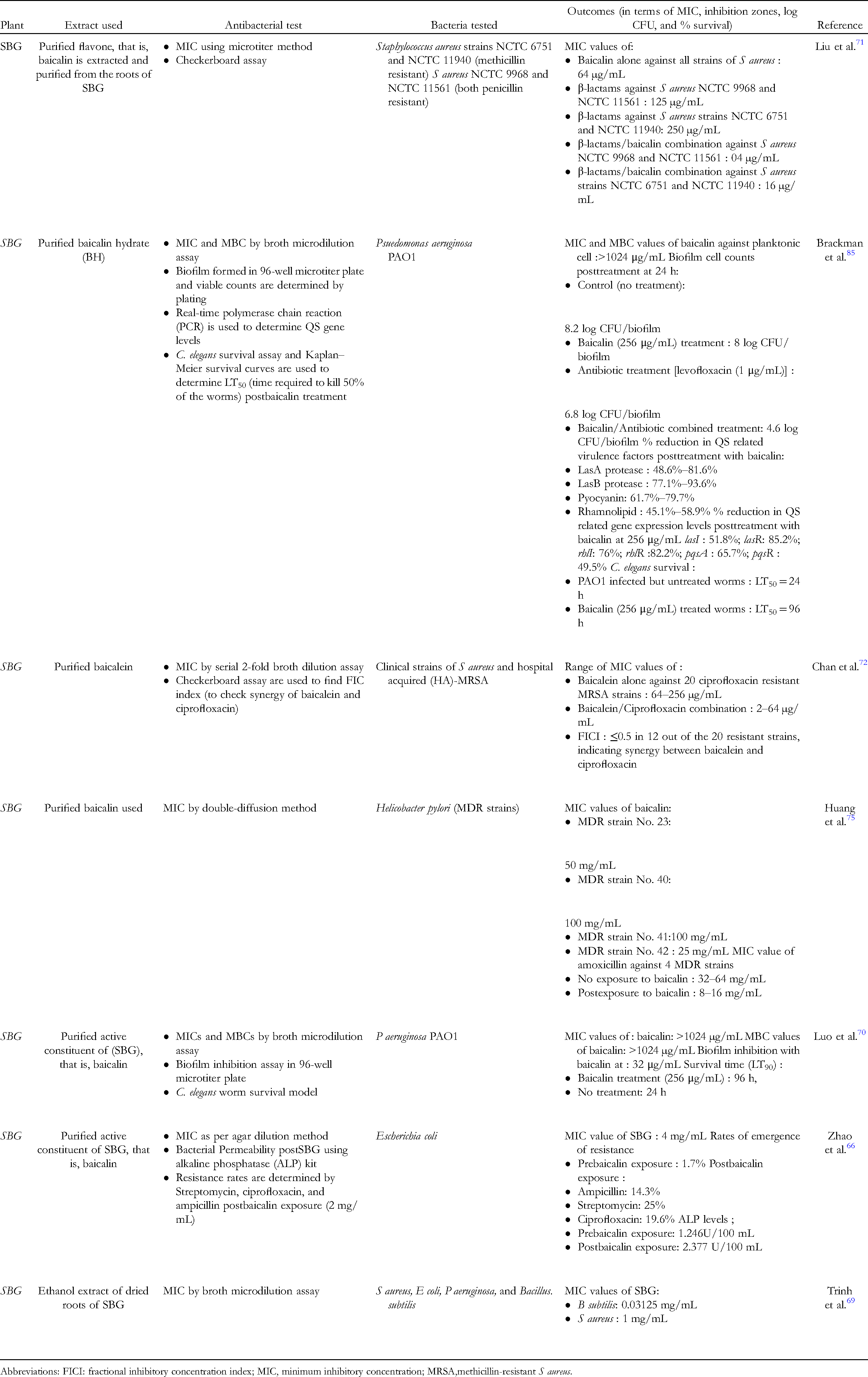
Abbreviations: FICI: fractional inhibitory concentration index; MIC, minimum inhibitory concentration; MRSA,methicillin-resistant S aureus.
SBG: Anti-inflammatory potential
SBG is also equipped with anti-inflammatory properties. The flavonoids baicalin, baicalein, wogonin, and wogonoside, which are abundant in the dried roots known as Scutellariae radix (SR), are what give the plant its medicinal properties and are responsible for the pharmacological effects.12,62
Chronic obstructive pulmonary disease (COPD) is one of the common chronic inflammatory illnesses wherein airway blockage and gradual lung damage are its major hallmarks. Cigarette smoke (CS) serves as a major risk factor 86 for COPD. Apoptosis of epithelial and endothelial cells, excessive nuclear factor-B (NF-B) and activator protein-1 (AP-1) activity, and high levels of pro-inflammatory cytokines (IL-6, IL-8, and TNF-α) are the results of oxidative stress brought on by CS, which also results in massive recruitment of neutrophils and macrophages to the lung tissues. Also, COPD-associated inflammation responds poorly to corticosteroids.87,88 This calls for investigating natural products as an adjunct therapy. In one such study 89 mice were exposed to the smoke of 15 cigarettes (1.0 mg of nicotine and 13 mg of tar per cigarette) for 1 h/day and 6 days/week for a total period of 3 months, and later dosed with baicalin (25 mg/kg, 50 mg/kg, and 100 mg/kg) or dexamethasone (1 mg/kg). Results showed that baicalin-dosed mice had significantly protected pulmonary function with a significant decrease in inflammatory cell recruitment and lower levels of TNF-α, IL-8, and MMP-9, unlike the dexamethasone-treated group.
CS has been shown to inhibit the enzyme histone deacetylase (HDAC2), that act as a cofactor for the glucocorticoids to exert their anti-inflammatory effect. This impaired HDAC2 activity leads to increased inflammation and corticoid insensitivity. Results showed that baicalin blocked CS-induced HDAC2 degradation, and 2 doses of baicalin (50 and 100 mg/kg) were able to restore HDAC2 protein expression. In addition, research showed that baicalin at high doses, that is, 100 mg/kg reduced the phosphorylation of HDAC2. This supports the critical role and mechanism adopted by baicalin in enhancing levels of HDAC2 via upregulating HDAC2 protein expression and inhibiting its phosphorylation leading to anti-inflammatory effects seen in test mice.
In a similar recent study by Zhang et al. 90 the protective role of baicalin was reconfirmed in rats wherein rats were exposed to CS for 1 h/day, 6 days/week, for 24 weeks and then treated with baicalin (40, 80, and 160 mg/kg). The team reported that baicalin was able to increase the HDAC2 protein expression and inhibit the expression of NF-κB. Also, it reduced the expression of plasminogen activator inhibitor-1 (PAI-1) in both rats and HBE cells. PAI-1 is an important regulator of fibrosis and is involved in COPD progression with high levels seen in patients with acute COPD and lung damage. 91 Consequently, baicalin is a key component of the roots of SBG and has the ability to reduce airway inflammation in COPD patients by effectively attacking at multiple levels leading to downregulating the exaggerated inflammatory damage seen.
Rheumatoid arthritis (RA), which is accompanied by synovitis and joint degeneration, is another common chronic condition. Over time, joint degeneration leads to the gradual breakdown of bones and cartilage due to the ongoing infiltration of inflammatory cells, the release of cytokines, and other factors. 92 Interleukin-17 (IL-17) has been linked to both the onset and severity of RA. Proinflammatory cytokine IL-17 is mostly raised in the synovial fluid of RA patients and is primarily produced by T-helper-17 (Th17) cells.93,94 Therefore, IL-17 inhibition is a key goal in stopping the joint degeneration seen by RA patients. In a mouse adjuvant-induced arthritis model, baicalin was found to exert its anti-inflammatory activity by reducing splenic Th17 cell population expansion and suppressing IL-17 levels. 96 Baicalin administered at a dose of 100 mg/kg significantly improved the joint injury scores and considerably reduced inflammatory cell infiltration, synovial associated hyperplasia, cartilage erosion, and bone erosion in arthritic mice ankles. Furthermore, baicalin IL-17-mediated lymphocyte adherence to cultured synoviocytes and demonstrated a considerable inhibition of splenic Th17 cell population increase in vivo. A similar study employing rat model of adjuvant-induced arthritis 92 confirmed that baicalin is a promising agent against RA joint destruction. The maximum dose of 200 mg/kg resulted in significantly lower arthritic indices in the rats. Further, the histopathological analysis also supported that baicalin improved chronic inflammation of synovial tissue rats with decreased cartilage destruction pannus formation, bone erosion, and low cellular infiltration and this effect were dose-dependent. TNF-a, IL-6, IL-1, IL-17, COX2, and COX1 levels were lower in the synovial tissue of rats given baicalin as tested by real time PCR. Further, the western blot test revealed decreased expression of the autophagy-related proteins Atg5, Atg7, Atg12, LC3-II, Beclin1, and Bcl-2 after baicalin therapy. Autophagy (a process of removing dead organelles, cells) aids to reduce the inflammatory responses through apoptosis. 98 Results showed that postbaicalin intervention, the expression levels of anti-apoptotic factor Bcl-2 protein and inflammatory factors were decreased, while those of pro-apoptotic factor Bax protein were increased. Therefore, baicalin inhibits splenic Th17 cell population growth, inhibits IL-17 levels, and restrains and induces autophagy in order to exert its anti-arthritis and anti-inflammatory activities. In addition to baicalin, other polyhydroxyflavonoids also exhibit significant anti-inflammatory and antioxidative activities. Among all, wogonin showed superior potency in inhibiting NO levels with IC50 = 45:3 ± 0.2 µM. Further, the highest inhibition of superoxide formation and free radical scavenging was seen with baicalein followed by oroxylin A and then wogonin. However, wogonin proved to be the most potent (82.9% inhibition) in its anti-inflammatory activity against carrageenan-induced rat hind paw edema. Thus, the polyhydroxyflavonoids possess anti-inflammatory, cytoprotective effects as well as strongly radical-scavenging effects advocating their promising role in the treatment of various chronic inflammatory diseases.
FS: Antibacterial potential
Forsythia suspensa (Thunb.) Vahl. belonging to the family Oleaceae is a flowering plant with unique healing properties. This plant is native to China, Korea, Japan, and numerous European countries. The dried fruit of FS which is also called FF has also received much attention. Both FS and FF have been widely used as part of TCM by Chinese and Japanese people in the treatment of skin disorders, inflammation, carbuncles, pyrexia, ulcers, etc.12,99 According to the 2015 edition of the Chinese Pharmacopoeia, 114 Chinese medicinal formulations contained FS. Modern pharmacological research confirms that this plant possesses anti-inflammatory, antioxidant, antibacterial, anticancer, neuroprotective, hepatoprotective, and diuretic properties, among others.100–103 The leaves of FS also have medicinal properties.
More than 100 compounds have been identified from FS include including alkaloids, flavonoids, phenylethanoid glycosides, pentacyclic triterpenoids, and lignans. The major constituents of FF include quercetin, rutaecarpine, forsythiaside A, betulinic acid, and forsythialan A. Forsythoside A possesses potent antioxidant, antibacterial, and antiviral properties, whereas forsythin and rutin exhibit potent antioxidant properties.104–106 In addition, phillygenin and 8-hydroxypinoresinol isolated from the fruit of FS have antioxidant and antimicrobial properties. Kuo et al. 105 purified and identified the chemical constituents of the methanolic extracts of fruits of F. suspensa. Among the 34 compounds extracted and identified, a new triterpene, that is, 3β-acetoxy-20α-hydroxyursan-28-oic acid was reported for the first time. The MIC values of purified compounds (compound Nos. 1, 2, 6, 10, 11, 12, and 16) was in the range of 1.20 to 5.00 mg/mL. Among the tested compounds, triterpenoids betulinic acid (6) and ψ-taraxasterol (10) exhibited the most significant inhibition against E coli with MIC values of 1.20 mg/mL. In another investigation, 107 FS fruit samples were extracted with 70% (v/v) ethanol using a percolator. The extract was subsequently filtered, dried, and the volume was adjusted using 70% ethanol (v/v). Results showed that fruit extract of FS possessed significant inhibition against E coli (MIC of 3.13 µg/mL), B subtilis (MIC of 12.5 µg/mL), and S aureus (MIC of 3.13µg/mL). As per disc diffusion assay, the extract of FS fruits showed the highest inhibition against S aureus (21.9 ± 1.1 mm) and B subtilis (16.6 ± 0.7 mm) but weak inhibition against E coli.
When compared to the fruit of FS, the leaves of FS have received less attention and there are few studies related to them. In one study by Endo et al. 108 antibacterial effects of FS leaves were studied, and found that forsythosides A, B, C, and D showed antibacterial activity against S aureus at <2 μM. The majority of FS leaves are abandoned during harvesting; however, the leaves are a significant source of bioactive compounds. In a recent study, Zhou et al. 11 utilized microporous adsorption resin to concentrate the active component (phenethyl alcohol glycosides) in FS leaf. There were a total of 31 chemicals found including glycosides, flavonoids, organic acids, phenolic, lignans, phenylpropanoid, and terpenoids. In addition, the MIC values for FS extract were 7.81 mg/mL for S aureus and 3.91 mg/mL for E coli, whereas the MBC values were 7.81 and 15.63 mg/mL. SEM analysis showed that after incubation with FS extract, both E coli cells and S aureus cells exhibited a change in surface morphology with cell membrane appearing pitted, shrunk, with holes evident. The possible mechanism is that FS extract disrupts the membrane's integrity, allowing nucleic acids and proteins to leak out and triggering bacterial cell death. Additionally, it may enter the cell membrane and cause cell death and bacterial growth suppression.11,109 Similarly, high antibacterial potential against food pathogens of FS fruit extract loaded green nanoparticles have been reported by Du et al. 110
The potential use of FS extract as a substitute for antibiotics, represents an intriguing application area. Antibiotics are excessively administered to agricultural animals to increase muscle mass and enhance growth performance. However, this has resulted in animals and animal farms serving as a reservoir for drug-resistant bacteria that easily enter the human food chain.111,112 Han et al. 101 investigated the potential of FS extract as a substitute for antibiotics in suppressing the growth of the 3 most prevalent broiler pathogens (E coli, Salmonella, and S aureus) The feeding program comprised of a starting diet from day 1 to day 21 and a finisher diet from day 22 to day 42. Three dietary supplement groups included (i) NC (negative control diet with no extract or antibiotic), (ii) FC (test group fed with 100 mg FS extract/kg diet), and (iii) PC (positive control diet) with Chlortetracycline (80 mg/kg). After a 12-hour fast on days 21 and 42, body weight (BW) and feed intake (FI) were assessed to calculate the average daily gain (ADG), average daily feed intake (ADFI), and feed conversion ratio (FCR). Additionally, caecal segments were examined for microbiological counts. The FS extract exhibited inhibition on E coli K88, S. aureus, and Salmonella 34R99 that disappeared after 25, 12.5, and 1.56 mg/mL, respectively. In the finisher phase, ADG of FC and PC was greater (16.2% and 12.2%, respectively) than negative control diet with no supplementation. ADFI of FC and PC was also higher ((9.6% and 11.5%, respectively) than NC. Histological measurements revealed that the FS-supplemented group had increased villus height and villus height-to-crypt depth ratios, hence enhancing digestibility. At the end of the finisher phase, the apparent digestibility of energy and calcium was enhanced in FC and PC compared to NC. Both FS and chlortetracycline supplementation lowered E coli levels in the cecum from an initial 6.31 log CFU to 5.51 log CFU on days 21 and 42 compared to the NC group. Long et al. 113 demonstrated that dietary FS extract supplementation increased nutritional digestibility and intestinal architecture in addition to providing anti-inflammatory and antioxidant effects in treated broilers. These findings suggest a potential shift away from the use of antibiotics in farm animals in favor of diets enriched with natural medicinal herbal extracts for growth enhancement and other benefits.
In addition to this, FS has shown synergistic antibacterial effects when used along with other medicinal herbs and antibacterial agents. Gu et al. 114 showed a synergistic antibacterial activity of FS and Origanum vulgare L (perennial Lamiaceae Origanum plant) against Staphylococcus cremoris, B subtilis, and E coli. Using the supercritical carbon dioxide extraction (SCDE) method, the team isolated essential oils from FS and Origanum vulgare L. Individual MICs for Origanum vulgare L essential oil ranged from 0.0025 to 0.005 mg/mL, whereas MICs for FS essential oil ranged from 0.01 to 0.05 mg/mL for the 3 test bacterial strains. In contrast, Origanum vulgare L and FS essential oil had a synergistic impact against all 3 bacteria (FICI = 0.2-0.4) when used in combination. Similar synergistic inhibition was seen when Patrinia scabiosaefolia ethyl acetate extract was combined with FF extract against E. coli. 115 These results encourage the combined use of herbal plant extracts and more studies focus on adding animal data in this direction which is presently limited. Table 4 summarizes the major studies focusing on the antibacterial effect of FS extracts.
Summary of the Major Studies Focusing on the Antibacterial Effect of Forsythia Suspensa (FS).

Abbreviations: FICI: fractional inhibitory concentration index; MIC, minimum inhibitory concentration.
FS: Anti-inflammatory potential
FF, also known as Lianqiao in Chinese, is a common ingredient in traditional CHM remedies. This herb is ranked in the top 10 most effective anti-inflammatory herbs. Its ethanolic extract and forsythosides A and B display strong anti-inflammatory activities, according to pharmacological research.116,117 FS helps to alleviate inflammation by acting on multiple targets. In one animal-based investigation, 118 the researchers used a mouse model of compound 48/80-induced paw edema to examine the effect of FF in reducing the associated inflammation. It is well known that mast cells play an important role in developing allergic inflammatory reactions and are involved in diverse pathologies related to hypersensitive reactions such as asthma, arthritis, and fibrosis. Results showed that when FF extract (100 µg/g) was orally supplied to mice, it significantly decreased edema scores and vascular permeability, with a greater effect than ketotifen (known mast cell stabilizer). Also, fructus showed a dose-dependent inhibition of histamine release (with the highest inhibition of 72% at 1 mg/mL) and a dose-dependent decrease in TNF-α secretion from human mast cells. The downregulation of mast cell activation by FF may make it a useful herb for treating inflammatory illnesses, according to this study.
In a similar investigation, it was discovered that Rengyolone, a cyclohexylethanoid isolated from the fruits of Forsythia koreana, inhibited NO and TNF-α levels effectively. In addition, the component suppressed the gene expression of inflammatory cascade genes iNOS and COX-2, which were stimulated by LPS. On further evaluation of the underlying mechanism, it became evident that Rengyolone blocked the phosphorylation of the p38 MAP kinase pathway and prevented LPS-induced DNA binding of NF-kB. 119 Another bioactive component, arctigenin, isolated from the ethyl acetate fraction of FF extract, demonstrates strong anti-inflammatory activity at several levels, as assessed in mouse models of paw and ear edema and the RAW 264.7 cell line. Results showed that arctigenin at a dose of 100 mg/kg decreased paw edema in carrageenan-induced mice. In the arachidonic acid (AA)-induced ear edema, the reduction was seen at a dose of 0.1 to 1.0 mg/ear. Further, the activities of 2 enzymes, that is, myeloperoxidase (MPO) and eosinophil peroxidase (EPO) in the edematous tissue homogenate showed a significant decrease at a painting dose of 0.1 to 1.0 mg/ear. These enzymes are indicators of neutrophils and eosinophils’ recruitment into the inflamed tissue. Arctigenin showed a dose-dependent decline in silica-induced reactive oxygen species (ROS) production indicating its ability to inhibit the process of neutrophil recruitments, exudation, and release of high levels of oxygen radicals at the inflamed tissues site. 120 Forsythiaside is an active component of FS that possesses anti-inflammatory and antioxidant properties. Cheng et al. 121 examined the protective effects of forsythiaside against CS-induced lung inflammation in a COPD mouse model in which Forsythiaside was delivered 2 h prior to CS exposure for 5 days. Forsythiaside inhibited the infiltration of inflammatory cells and the release of NO and inflammatory cytokines (production of TNF-α, IL-6, and interleukin 1 [IL-1]). Also, CS-induced NF-κB activation was reduced by forsythiaside, as determined by western blotting. Forsythiaside increased the expression of nuclear factor-erythroid 2-related factor 2 (Nrf2) in a dose-dependent manner . Nrf2 is a transcription factor that maintains cellular redox equilibrium and protects against oxidative damage. 122 Thus, this bioactive component of FS protects against lung damage and inflammation caused by CS. In a similar study 123 FS extract showed considerable protection against LPS-induced liver damage. LPS depletes cytosolic Nrf2 and inhibits nuclear translocation of Nrf2 in the liver, resulting in the production of ROS and malondialdehyde (MDA) in serum and liver. Results showed that FS extract elevated the Nrf2-mediated activation of HO-1 in the liver, as well as the levels of superoxide dismutase (SOD) and glutathione peroxidase. In addition, FS extract was able to suppress NF-κB activation by reducing serum and living rats’ levels of TNF-α, IL-1, and IL-6. Thus, FS demonstrates its involvement in treating and regulating chronic inflammatory disorders by interfering with NF-κB activation pathways and promoting the production of Nrf2, which counteracts the negative consequences of excessive inflammatory reactions.
Neuroinflammation is now considered an important underlying reason associated with many of the prevalent neurodegenerative diseases such as Alzheimer's disease, frontotemporal dementia, Parkinson's disease, amyotrophic lateral sclerosis, and Huntington's disease.124,125 The use of natural products from TCM with both neuroprotective and anti-inflammatory activities offers an excellent option for treating and managing such conditions. According to a recent study by Shao et al,. 126 FS and its fruit extract have a protective effect against neuroinflammation. Twelve new and bioactive phenylethanoid glycosides were extracted and discovered from the fruits of FS for this purpose. Three novel C6–C7′/C6–C16′-linked phenylethanoid glycoside dimers (1-3), 3 novel phenylethanoid glycosides (4-6), and 6 recognized compounds (7-12) were found and further investigated. Compounds 1 and 10 demonstrated strong neuroprotective properties, as assessed by serum deprivation and rotenone-induced PC12 cell line damage. In addition, compound 1 demonstrated a significant decrease in LPS-induced TNF-α production in RAW 264.7 cells, suggesting that bioactive phenylethanoid glycosides are involved in reducing neuroinflammation.
Another recent study by Chao et a.l 127 focussed on studying the role of FS extract against a common inflammatory disease, that is, ulcerative colitis (UC) employing a mice model of UC induced by dextran sodium sulfate (DSS). The team discovered that FS extract reduced serum IL-1 levels and alleviated colon shortening in mice. Additionally, colon tissue MDA levels and MPO activity were also significantly lower in mice given FS extract doses. To delineate the underlying mechanism, in vivo mechanistic studies revealed that FS extract showed inhibition of pyroptosis (a type of programmed cell death that is involved in experimental colitis in mice). Western blot analysis and quantitative real-time PCR indicated that FS extract was able to suppress the expression of pyroptosis-related genes and the levels of their encoding proteins, unlike the control group. Additionally, FS extract decreased the amount of ROS in the J774A.1 cell line. FS extract revealed its anti-inflammatory actions via the Nrf-NLRP3 pathway, as inhibition of pyroptosis was not observed when NLRP3 expression was decreased, demonstrating the important function of NLRP3 in pyroptosis. In addition, comprehensive metabolomics demonstrated that FS extract restored the aberrant levels of 9 metabolites (including glutathione metabolism, aminoacyl-tRNA biosynthesis, and linoleic acid metabolism) in mice with colitis, indicating a considerable improvement in metabolic function. A similar past study 128 employing the DSS-induced colitis model also showed that FS significantly decreased BW loss, colon length shortening, and reduced TNF-α and interleukin-6 elevations unlike in the control group (not administered with FS extract).
The results of various past studies support that FS extract and its fruit are rich in bioactive components which have significant potential to ameliorate inflammatory damage via acting on different pathways and molecules involved thus downregulating the elevated inflammatory reaction.
Conclusion
It is imperative to research and use innovative antibacterial medicines with minimal side effects and maximal potency in view of the rise in antibiotic resistance and the number of fatalities caused by life-threatening-resistant bacterial infections. Chinese medicinal herbs and their recently discovered bioactive components/phytochemicals are a rich source of novel antibacterials, and represent a potential solution in reducing the problem of rising antibiotic resistance. As demonstrated by multiple studies from the past and cited in this review, these medicinal herbs and their bioactive constituents are equipped with the ability to reverse resistance (via multiple mechanisms including anti-QS, anti-biofilm, resistant plasmid elimination, efflux pump inhibitors, etc.) and, when combined with antibiotics, can restore the effectiveness of conventional antibiotics in killing resistant populations. Consequently, they are great candidates for adjunct therapy, as they can be used in conjunction with antibiotics as part of a standard treatment plan. For clinical approval and success of these medicinal herbs and their constituents, future studies, particularly in vivo studies, are required to detail the efficacy, dosage, and safety against different resistant bacterial strains and diseased conditions using animal models of infection and inflammation. These can be administered as pure forms or extracts and represent an important complementary alternative medicine to improve the disease outcomes.
Footnotes
Availability of Data and Material
All the data pertaining to the above study is available in the manuscript.
Declaration of Conflicting Interests
The author(s) declared no potential conflicts of interest with respect to the research, authorship, and/or publication of this article.
Funding
The author(s) received no financial support for the research and/or authorship of this article.
Author’s Contribution
XM, MY, and WL: Literature search, data collection and extraction, draft writing, and editing and approval of the final manuscript.