A new sesquiterpenoid, chaenomelesterpenoid A (1), and a new norisoprenoid, chaenomelesterpenoid B (2), were isolated from the fruits of Chaenomeles sinensis (Thouin) Koehne. Their structures were determined by NMR spectroscopy and MS. In addition, the protective effects of the compounds were tested against corticosterone-induced damage in PC-12 cells using real-time cellular analysis (RTCA). Compounds 1 and 2 significantly improved cell viability and corticosterone-induced damage in PC-12 cells with EC50 values of 15.7 and 12.6 µM, respectively.
Chaenomeles sinensis (Thouin) Koehn, commonly known as Chinese quince or “Guang Pi Mu Gua,” is a deciduous or semi-evergreen tree of the family Rosaceae and native to China.1 Its fruits are of high economic value and reported to be rich in dietary fiber, organic acids, and vitamins,2 while its sugar content is relatively low. After appropriate dilution and supplementation with a sweetener, the fruits can be made into a delicious food with a unique flavor. Chaenomeles fruits are used as medicinal herbsin Korea and China for the treatment of throat diseases, anaphylaxis, viral infection, and neurodegenerative diseases.3 Modern pharmacological investigations have demonstrated significant biological properties such as anti-hyperuricemic, antiacetylcholinesterase, and antidiabetic effects.4 The fruits of C. sinensis contain pentacyclic triterpene acids, flavonoids, lignans, and simple phenolic compounds.5 As a continuation of our recent research on the neuroprotective constituents of C. sinensisfruits,6 we further isolated a new sesquiterpenoid, chaenomelesterpenoid A (1), and a new norisoprenoid, chaenomelesterpenoid B (2) (Figure 1). Here, we report the isolation, structure elucidation, and evaluation ofthe protective effects of compounds 1 and 2 on corticosterone-induced pheochromocytoma cell damage in rats (PC-12 cells).
Structures of compounds 1 and 2.
Results and Discussion
Compound 1 was isolated as a white powder. The molecular formula was determined to be C21H30O9 based on the HR-ESI-MS ion at m/z 449.1786 [M + Na]+(calculated for 449.1782, C21H30O9Na) (Supplemental Figure S8), combined with 1H and 13C NMR spectroscopic data. The UV spectrum of 1 displayed absorption of a conjugated double bond at 266 nm (Supplemental Figure S10). The IR spectrum displayed absorption bands at 3385 (OH), 1654 (conjugated C = O), and 1030 (ether bond) cm-1 (Supplemental Figure S9). The 1H NMR spectrum of 1 showed a pair of trans-olefinic proton signals [δH 6.48 (1H, d, J = 15.7 Hz, H-8) and 6.29 (1H, d, J = 15.7 Hz, H-7)], two olefinic proton signals [δH 5.90 (1H, s, H-4) and 5.86 (1H, s, H-10)], and four methyl groups [δH 2.28 (3H, s, H-15), 1.90 (3H, s, H-14), 1.05(3H, s, H-13), and 1.00 (3H, s, H-12)]. In addition, a pair of free anomeric protons of glucose were observed at δH5.08 (0.5H, d, J = 3.7 Hz, Hα−1′) and 4.47 (0.5H, d, J = 7.7 Hz, Hβ−1′) (Supplemental Figure S1). The 13C NMR and DEPT spectra indicated the presence of 27 carbons, of which 12 were assigned to the pair of free glucopyranose moieties [δC 98.3 (Cα−1′)/94.0 (Cβ−1′), 72.0 (Cα−2′)/75.4 (Cβ−2′), 73.8 (Cα−3′)/74.8 (Cβ−3′), 70.7 (Cα−4′)/71.8 (Cβ−4′), 76.2 (Cα−5′)/77.9 (Cβ−5′), 64.4(Cα−6′)/64.5 (Cβ−6′)] (Supplemental Figure S2 and S3). According to the glycosylation shift, the aglycone unit was assumed to be connected to C-6′ of glucose.7,8 The other carbons were observed as two carbonyl groups [δC 200.9 (C-3) and 168.3 (C-11)], four quaternary carbons [δC 166.2 (C-5), 153.2 (C-9), 80.3 (C-6), and 42.7 (C-1)], four methine groups [δC 127.5 (C-4), 137.4(C-7), 135.0(C-8), and 120.5(C-10)], a methylene group [δC 50.7 (C-2)] and four methyl groups [δC 24.7(C-12), 23.6(C-13), 19.4(C-14), and 14.3(C-15)], which suggested that the aglycon of 1 might be (2-trans, 4-trans)-abscisic acid,9 except for the additional signals associated with the free glucopyranosyl unit. Acid hydrolysis of 1 with 2 M HCl afforded glucose, revealing the occurrence of a D-glucopyranosyl moiety in 1.10 The cross peaks between 4.21 (Hα−6′) and 4.25 (Hβ−6′) of glucose and C-11 in the HMBC spectrum indicated that C-6′ of the glucose unit is connected to C-11 (Figure 2 and Supplemental Figure S6), which was confirmed by the glycosidation shift.11 Thus, compound 1 was identified as (7E, 9E)-abscisic acid-(α and β)-glucopyranoside and named chaenomelesterpenoid A (Figure 1).
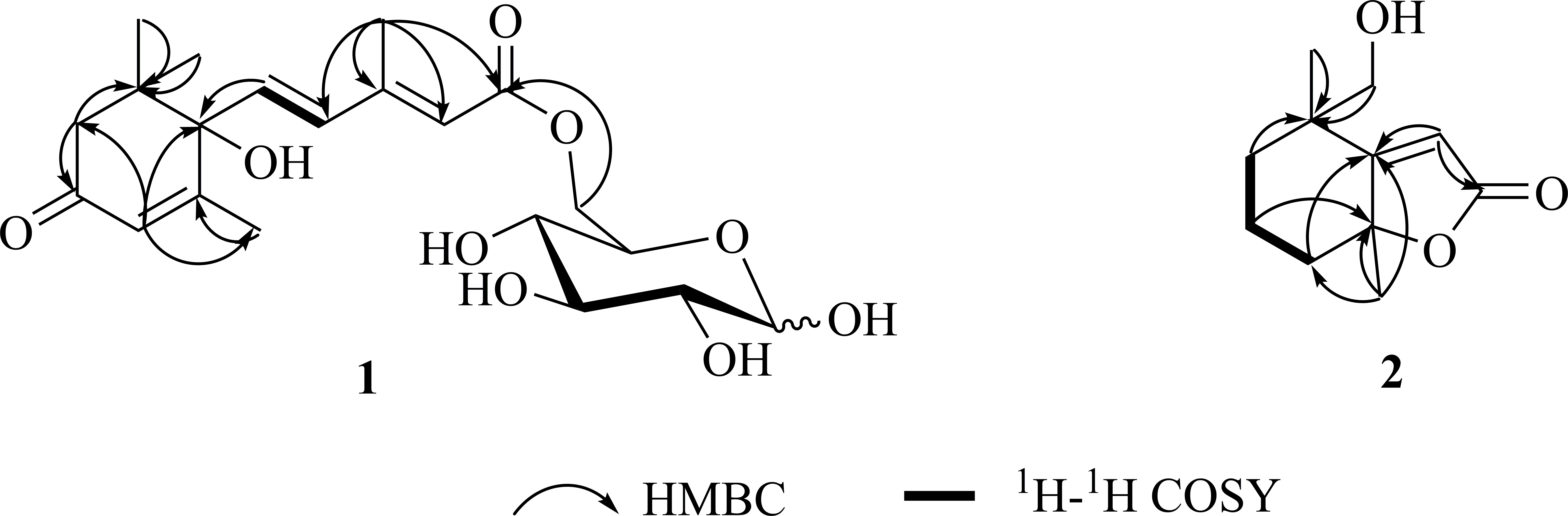
Key HMBC and 1H-1H COSY correlations of 1 and 2.
Compound 2, extracted as white powder, was determined to possess a molecular formula of C11H16O3 from the HR-ESI-MS [M + Na]+ ion at m/z 219.0992 (Calcd 219.0991) (Supplemental Figure S18). The UV spectrum of 2 exhibited absorptions at 213 and 265 nm (Supplemental Figure S20). In the 1H NMR spectrum of 2, the signals of one olefinic proton [δH 5.80 (1H, s, H-9)], one oxygenated methylene [δH3.57 (1H, d, J = 11.0 Hz, H-10α) and 3.55 (1H, d, J = 11.0 Hz, H-10β)], three methylenes [δH 2.24 (1H, dd, J = 1.6, 12.5 Hz, H-5α), 1.43 (1H, dd, J = 5.3, 12.5 Hz, H-5β), 1.80 (2H, m, H-4), 1.65 (1H, m, H-3α), and 1.35 (1H, m, H-3β)], and two methyl groups [δH 1.56 (3H, s, H-12) and 1.24 (3H, s, H-11)] were observed (Supplemental Figure S11). The 13C NMR and DEPT135 spectra indicated the presence of 11 carbons, including four quaternary [δC 182.0 (C-1), 174.7 (C-8), 89.5 (C-6), and 42.8 (C-2)], one methine group [δC 113.3(C-9)], four methylene groups [δC 70.9 (C-10), 40.9 (C-5), 36.8 (C-3), and 19.9 (C-4)] and two methyl groups [δC 20.0 (C-11) and 24.8 (C-12)] (Supplemental Figure S12 and S13). These spectroscopic features suggested that the structure of 2 was very similar to that of 3,9-dihydroxy dihydroactinidiolide.12 The obvious difference was that ahydroxyl group at C-4 of 3,9-dihydroxy dihydroactinidiolide was replaced by a proton in 2. The NOESY experiment showed the correlation between H-10 and H-12, confirming that H-10 and H-12 had a β-configuration (Figure 3 and Supplemental Figure S17). Therefore, compound 2 was characterized as chaenomelesterpenoid B (Figure 1).
Key NOESY correlations of 2.
The protective effects of the compounds were tested against corticosterone-induced damage in PC-12 cells using real-time cellular analysis (RTCA). Compounds 1 and 2 significantly improved cell viability and corticosterone-induced damage in PC-12 cells with EC50 values of 15.7 and 12.6 µM, respectively.
Experimental Part
Materials
NMR spectra (including 1D and 2D) were recorded on a Bruker Avance III 500 MHz spectrometer (500 MHz for 1H-NMR and 125 MHz for 13C-NMR), optical rotations on an APIV (Rudolph Research Analytical), and IR spectra on a Nicolet iS10 Microscope Spectrometer (Thermo Fisher Scientific). HR-ESI-MS were obtained on a Bruker maXis HD mass spectrometer and UV spectra on a Shimadzu UV-2401PC apparatus. Preparative HPLC was conducted using a Saipuruisi LC-50 instrument with an UV200 detector and YMC-Pack ODS-A column (250 × 20 mm, 5 µm and 250 × 10 mm, 5 µm). Column chromatography was performed using Diaion HP-20 (Mitsubishi Chemical Corporation), Toyopearl HW-40, MCI gel CHP-20 (TOSOH Corporation), Sephadex LH-20 (Amersham Pharmacia Biotech AB), LiChroprep RP-18 gel (Merck, Darmstadt), and silica gel (Marine Chemical Industry). For TLC, self-made silica gel G plates (Qingdao Marine Chemical Industry) were used. All of the chemical reagents were supplied by Beijing Chemical Plant and Tianjin No. 3 Reagent Plant. RTCA was measured with an xCELLLigence RTCA System (Acea Biosciences, Inc.).
Plantmaterials
The fruits of Chaenomeles sinensis (Thouin) Koehne were collected in November 2016 from Fangcheng County, Nanyang City, Henan Province, China. The plants were identified by Professor Chengming Dong of Henan University of Chinese Medicine. A voucher specimen (No. 20171101) has been deposited in the Department of Natural Medicinal Chemistry, School of Pharmacy, Henan University of Chinese Medicine, Zhengzhou, China.
Isolation
The fruits of C. sinensis (30.0 kg) were extracted thrice with 50% acetone-water using a tissue crushing extraction method. The filtrate was evaporated under vacuum to obtain the extract (2.67 kg), which was then precipitated using 80% ethanol (10 L × 5); the liquid supernatant was concentrated in a vacuum evaporator to yield the gross extract, which was resuspended in H2O (2.5 L).13 The extract was passed through a Diaion HP-20 macroporous resin column and eluted with 0%, 10%, 20%, 30%, 40%, 50%, 70%, and 95% EtOH-H2O successively to obtain eight fractions (A-H). Fr.C (39.3 g) was chromatographed on Toyopearl HW-40C eluting with water, and gradually decreasing the polarity with MeOH to yield 5 fractions (C1-C5). Fr.C3 was separated by Toyopearl HW-40C CC by eluting with MeOH-H2O (50:50) to yield 4 fractions (C3-1–C3-4). Fr. 3‐2 was subjected to ODS CC eluting with MeOH-H2O (0:100‐40:100) to generate 4 fractions (Fr. C3-2-1–Fr. C3-2-6). Fr. C3-2-5 was purified by semi-preparative HPLC (Saipuruisi LC-50) (MeCN-H2O, 18:82, v/v) to afford compound 1 (5.3 mg, tR = 41.33 minutes). Fr. C3-4 was purified by semi-preparative HPLC (MeCN-H2O, 10:90, v/v) to afford compound 2 (5.0 mg, tR = 130.85 minutes).
Chaenomelesterpenoid A (1). White powder; [α]20 D 214.838 (c 1.22, CH3OH); UV λmax (CH3OH)/nm (logε): 266 (2.12); IR (iTR) νmax/cm-1: 3385, 2960, 1657, 1237, 1161, 1030; ECD (CH3OH) λmax (Δε) nm: 233 (Δε –30.41) and 269 (Δε + 34.11); HR-ESI-MS m/z 449.1786 [M + Na]+ (calcd. for C21H30O9Na 449.1782). 1H-NMR (CD3OD, 500 MHz) and 13C-NMR (CD3OD, 125 MHz) see in Table 1.
Compound1 (1 mg) was treated with 2 mol/L aqueous HCl (2 ml) (sealed flask, heating in water bath, 80 °C, 3 hours). Then the acidic aqueous mixture was dried, H2O (2 ml) was added, and the mixture extracted with EtOAc (3 × 2 ml). The aqueous layer was subjected to chiral-phase HPLC analysis under the following conditions. The sugars of the hydrolysis were separated and detected using a CHIRALPAK AD-H column (250 × 4.6 mm) using n-hexane:EtOH:TFA (750:250:0.25) as the mobile phase (0.5 mL/min–1) and an evaporative light scattering detector (ELSD). Identification of D-glucose was carried out by comparison of its retention time with that of authentic samples (tR = 18.3 minutes; D-glucose).14
Biological Assay
PC-12 cells were cultured in RPMI 1640 medium containing 10% FBS, 50 units/mL penicillin, and 50 µg/mL streptomycin and penicillin in a humidified atmosphere at 37 °C in 5% CO2. The RTCA assay was performed for 60 hours using an xCELLLigence RTCA system. Background impedance signals were measured with 100 µL of cell culture medium per well. Exponentially growing PC-12 cells were digested with 0.25% trypsin to obtain a single-cell suspension in medium. The cells were seeded into 16-well E-plates with a target density of 2 × 10⁴ cells in 100 µL medium per well. After plating, impedance was routinely recorded at 15 minutes intervals. One day after seeding, the cells in 16-wall E-plates were treated with test compounds 1 and 2 at 5 different concentrations (0.1, 1, 10, 50, and 100 µM) and corticosterone (500 µM). All the incubations were performed with a final solvent concentration of 0.1% DMSO. After compound administration, impedance was measured every 5 minutes for the following time points, and afterward every 15 minutes until the end of the experiment.15,16
Conclusions
A new sesquiterpenoid, chaenomelesterpenoid A (1), and a new norisoprenoid, chaenomelesterpenoid B (2), were isolated from Chaenomeles fruits. The two compounds significantly improved cell viability and corticosterone-induced damage in PC-12 cells with EC50 values of 15.7 and 12.6 µM, respectively. Our study provides useful information on the chemistry of C. sinensis (Thouin) Koehne.
Supplemental Material
Supplementary Material 1 - Supplemental material for Two New Terpenoids From the Fruits of Chaenomeles sinensis (Thouin) Koehne
Supplemental material, Supplementary Material 1, for Two New Terpenoids From the Fruits of Chaenomeles sinensis (Thouin) Koehne by Meng Li, Zhi-guang Zhang, Jing-ya Shi, Ya-ge Li, Jing-ke Zhang, Yang Ying, Xiao-yan Deng, Xiao-ke Zheng and Wei-sheng Feng in Natural Product Communications
Footnotes
Declaration of Conflicting Interests
The author(s) declared no potential conflicts of interest with respect to the research, authorship, and/or publication of this article.
Funding
The author(s) disclosed receipt of the following financial support for the research, authorship, and/or publication of this article: The National Key Research and Development Project (2017YFC1702800), the Central Government Guide Local Science and Technology Development Funds (14104349), the Key Scientific Research Project of Institutions of Higher Learning in Henan (21B360003), and the Special Project of Scientific Research on Traditional Chinese Medicine in Henan (20-21ZY2151) supported this study.
ORCID ID
Meng Li
Supplemental Material
Supplemental material for this article is available online.
References
1.
ZhangR.ZhanS.LiSet al. Anti-hyperuricemic and nephroprotective effects of extracts from Chaenomeles sinensis (Thouin) Koehne in hyperuricemic mice. Food Funct. 2018;9(11):5778-5790.doi:10.1039/C8FO01480A30327809
2.
ZhangLH.XuHD.LiSF. Effects of micronization on properties of Chaenomeles sinensis (Thouin) Koehne fruit powder. Innovative Food Science & Emerging Technologies. 2009;10(4):633-637.doi:10.1016/j.ifset.2009.05.010
3.
ChaK-J.SongC-S.LeeJ-Set al. Chaenomeles sinensis Koehne extract suppresses the development of atopic dermatitis-like lesions by regulating cytokine and filaggrin expression in NC/Nga mice. Int J Med Sci. 2019;16(12):1604-1613.doi:10.7150/ijms.3785431839748
4.
SanchetiS.SanchetiS.SeoS-Y. Antidiabetic and antiacetylcholinesterase effects of ethyl acetate fraction of Chaenomeles sinensis (Thouin) Koehne fruits in streptozotocin-induced diabetic rats. Exp Toxicol Pathol. 2013;65(1-2):55-60.doi:10.1016/j.etp.2011.05.01021764274
5.
YinZH.ZhaoC.ZhangJJet al. Research progress on chemical constituents and pharmacological activities of Chaenomeles sinensis. Chin J Exp Tradit Med Form. 2017;23(9):234-242.doi:10.13422/j.cnki.syfjx.2017090221
6.
FengWS.ZhangZG.LiMet al. Chaenomelesalkaloid A, a new alkaloid from the fruits of Chaenomeles sinensis (Thouin) Koehne. Acta Pharm Sin. 2018;53(6):976-979.doi:10.16438/j.0513-4870.2018-0211
7.
BokernM.HeuerS.WrayVet al. Ferulic acid conjugates and betacyanins from cell cultures of Beta vulgaris. Phytochemistry. 1991;30(10):3261-3265.doi:10.1016/0031-9422(91)83189-R
8.
TianL.YJL.SWLet al. Chemical constituents of Syringaoblata leaves. Chin J Exp Tradit Med Form. 2013;19(01):144-147.doi:10.13422/j.cnki.syfjx.2013.01.051
9.
LiHB.YuY.WangZZet al. Research on chemical constituents from Re-Du-Ning injection (Ⅲ). Chin Tradit Herbal Drugs. 2016;47(10):1643-1649.doi:10.7501/j.issn.0253-2670.2016.10.002
10.
LiM.ZengM.ZhangZet al. Uridine derivatives from the seeds of Lepidium apetalum willd. and their estrogenic effects. Phytochemistry. 2018;155:45-52.doi:10.1016/j.phytochem.2018.07.01330075391
11.
ShiJY.ZhangZG.LiMet al. Phenolic constituents from the fruit of Chaenomeles sinensis. Chin Pharm J. 2020;55(20):1666-1672.doi:10.11669/cpj.2020.20.003
12.
BaiM.CaiY.WuS-Yet al. A new norisoprenoid from the leaves of Ficuspumila. Nat Prod Res. 2019;33(9):1292-1297.doi:10.1080/14786419.2018.147107729737877
13.
FengW.LvY.ZhengX.ZhangY.CaoY.PeiY. A new megastigmane from fresh roots of Rehmannia glutinosa. Acta Pharm Sin B. 2013;3(5):333-336.doi:10.1016/j.apsb.2013.07.001
14.
LopesJF.GasparEMSM. Simultaneous chromatographic separation of enantiomers, anomers and structural isomers of some biologically relevant monosaccharides. J Chromatogr A. 2008;1188(1):34-42.doi:10.1016/j.chroma.2007.12.01618177879
15.
LiG.WuK.LiYHet al. Evaluation of hypoxia/reoxygenation injury of H9c2 myocardial cells and intervention effect of notoginsenoside R1 based on RTCA technology. Chin Pharmacol Bull. 2019;35(3):436-440.doi:10.3969/j.issn.1001-1978.2019.03.027
16.
HeXY.ChenJL.XiangHet al. Comparative study of the corticosterone and glutamate induced PC12 cells depression model by 1H NMR metabolomics. Yao Xue Xue Bao. 2017;52(2):245-252.doi:10.16438/j.0513-4870.2016-054329979506
Supplementary Material
Please find the following supplemental material available below.
For Open Access articles published under a Creative Commons License, all supplemental material carries the same license as the article it is associated with.
For non-Open Access articles published, all supplemental material carries a non-exclusive license, and permission requests for re-use of supplemental material or any part of supplemental material shall be sent directly to the copyright owner as specified in the copyright notice associated with the article.