
Abstract
Caesalpinia sappan L. has been used as an herbal medicine to treat skin damage as a facial cleanser. In this study, 8 known compounds (
Caesalpinia sappan L. known as red wood, a member of the family Leguminosae, is widely distributed in China, India, Burma, and other countries. The dried heartwood of C. sappan has been used as traditional Chinese medicine for the treatment of various diseases including traumatic injury, fractures and muscle injury, blood stasis stagnation pain, dysmenorrhea, amenorrhea, postpartum blood stasis, and carbuncle swelling. 1 Pharmacological researches revealed that C. sappan had many bioactivities, such as inhibition of melanin production, anti-inflammation, antioxidant, antibacterial effects, and immune regulation.
Chemical constituents and pharmacological investigations of C. sappan heartwood had showed that the main bioactive components of C. sappan were phenolic compounds such as brazilin, protosappanin, and chalcone. 2 -7 Brazilin and 4-O-methylsappanol from the ethanol extracts of C. sappan exhibited effects on melanin synthesis in human malignant melanoma cell line (HMV-II). 4 It was found that brazilin possessed stronger anti-inflammatory effect than hematoxylin and berberine hydrochloride. 5,6 Brazilin exhibited hypoglycemic effect in diabetic animals through amelioration of glucose metabolisms in insulin-responsive tissues. 8 Another study also found that brazilin could reduce the bromotrichloromethane (BrCCl3)-induced toxicities on hepatocytes and depress BrCCl3-induced microsomal calcium sequestration. 9 In addition, the ethyl acetate, methanol, and water extracts of C. sappan heartwood exhibited strong antioxidant activity by the method of 1,1-diphenyl-2-picryl hydrazyl (DPPH) and nitric oxide. 10 It was also reported that the extract of C. sappan inhibited cancer cells in vitro and in vivo. 11 The aqueous extract of C. sappan could prolong the allograft survival time in murine skin allograft induced by immune tolerance. 12 It was also found that methanolic and 50% ethanolic extracts of C. sappan heartwood showed inhibitory effects on Propionibacterium acnes and lipase activity. 13
However, the effects of chemical constituents of C. sappan on tyrosinase activity and the inhibitory type have been rarely reported. At present, only Mitani et al, 4 Hridya et al, 14 Laksmiani et al, 15 and Chang et al 16 have reported it. Therefore, we investigated the chemical constituents of C. sappan and examined their effects on tyrosinase activity, types of inhibition, and mechanism in vitro and in vivo.
Eight compounds (

Chemical structures of 8 compounds isolated from Caesalpinia sappan.
Activation Rate of Tyrosinase of Different Fractions of Caesalpinia sappan.
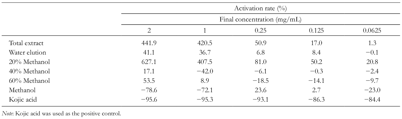
Note: Kojic acid was used as the positive control.
Activation Rate of Tyrosinase of Isolated Compounds From Caesalpinia sappan.

Note: Kojic acid was used as positive control.
In Table 1, the total extract, water elution fraction, and 20% methanol eluted fraction of C. sappan could remarkably activate tyrosinase. The effects of total extract and 20% methanol eluted fraction on tyrosinase activation rate were increased gradually with the increase of their concentrations. Overall, the order of their activation activities was 20% methanol eluted fraction > total extract > water elution fraction. The effects on tyrosinase activity of 40% and 60% methanol eluted fractions changed from inhibition to activation with the increase of their concentrations. The methanol-eluted fraction showed inhibitory activity at 0.0625, 1.000, and 2.000 mg/mL and activation activity at 0.125 and 0.25 mg/mL.
In Table 2, compounds
According to the results of tyrosinase activity in vitro, compounds
In Figure 2 and Table 3, Km
was increased and V
max was unchanged after compound
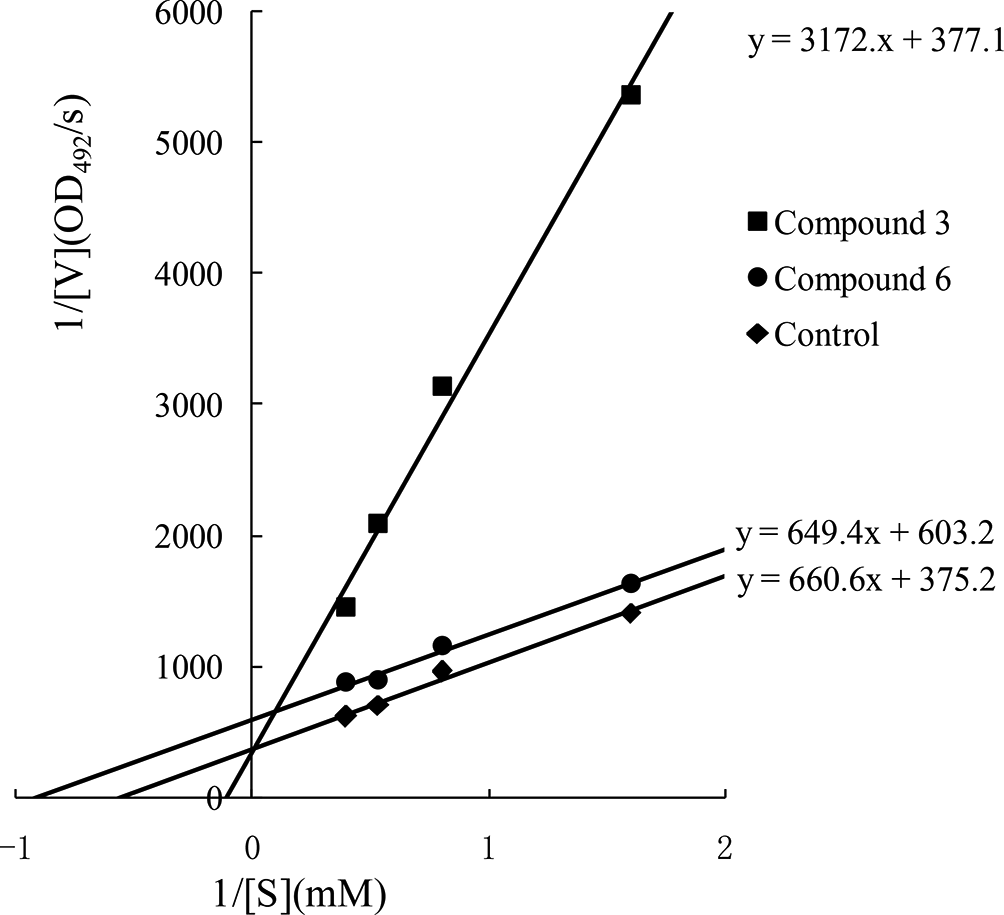
Inhibition mechanisms of 2 compounds on tyrosinase.
The Km , V max, and Ki of Compounds.


Contents of tyrosinase in rat serum samples after administration of different fractions of Caesalpinia sappan (n = 3). Compared with the control group: * P ≤ .05, ** P ≤ .01, and *** P ≤ .001. Compared with the positive control group: # P ≤ .05, ## P ≤ .01, and ### P ≤ .001.
The contents of tyrosinase in rat serum after administration of different fractions (total extraction, water elution, 20% methanol part, 40% methanol part, and 60% methanol part) and compounds

Contents of tyrosinase in rat serum samples after administration of compounds from Caesalpinia sappan (n = 3). Compared with the control group: * P ≤ .05, ** P ≤ .01, *** P ≤ .001. Compared with the positive control group: # P ≤ .05, ## P ≤ .01, ### P ≤ .001.
In Figure 3, the contents of tyrosinase in all of sample administration groups were lower than that in the control group except for the positive control group. Among them, there was no significant difference between the 40% and 60% methanol-eluted fractions compared with the control group. The total extract of C. sappan and 100% methanol-eluted fractions were significantly lower than that of the control group (P ≤ .05). Water elution and the 20% methanol-eluted fraction were significantly lower than control group (P ≤ .001). The results demonstrated that 70% ethanol extract of C. sappan could reduce the content of tyrosinase in rat serum.
In Figure 4, the contents of tyrosinase in all of administration group of serum were higher than that of the control group in addition except
Tyrosinase is one of the key enzymes involved in melanin synthesis. If its activity is inhibited, the production of melanin will be reduced. Compounds with these activities could be used in skin whitening and treated the face pigmentation sickness, such as freckle, chloasma, and senile plaque. While, the increase of tyrosinase activity can promote melanin synthesis to treat vitiligo and albinism. Most of the previous studies have reported that flavonoid compounds acted as potent tyrosinase inhibitors.
25,26
Studies have found that brazilin could inhibit melanin formation in human melanoma cell line (HMV-II) with the half-maximal effective concentration (EC50) of 3.0 ± 0.5 µM.
4
The results of this study were different from the reference, the discrepancy may be related to different administration modes of brazilin, experimental models, administration doses, etc. Iida K found that using
Enzyme kinetics experiments showed that the type of inhibition of compound
Compounds
Experimental
General
NMR spectra were measured on Bruker Ascend TM 400 superconducting nuclear magnetic resonance (NMR) spectrometer (Bruker Co., Germany). A CXTH LC-3000 (Beijing Innovation Technology Co., Ltd., Beijing, China) and Waters-2489 (Waters Co., Milford, MA, United States) preparative high-performance liquid chromatography (HPLC) system equipped with a YMC-C18 column (250 × 10 mm, 5 µm) were used for isolation. D101 macroporous resin was purchased from Tianjin City Sea Light Chemical Co., Ltd., Tianjin, China; GRP-9270 type water jacket incubator from Shanghai Senxin Experimental Equipment Co. Ltd., Shanghai, China; Multiskan GO microplate reader from Thermo Electron Inc, United States; AL104 type electronic balance and DELTA 320 type pH meter from Mettler-Toledo Inc, Columbus, OH, United States; and TGL-16gR high-speed desktop refrigerated centrifuge from Shanghai Anting Scientific Instrument Factory, Shanghai, China.
GF254 thin layer chromatography silica gel, silica gel H, 40 to 80 mesh and 200 to 300 mesh silica gel, was purchased from Qingdao Ocean Chemical Co., Ltd., Qingdao, Shandong, China; Sephadex LH-20 from Pharmacia Co., Sweden;
Plant Material
The heartwood of C. sappan L. was purchased from a medicine market in November 2014 and identified by Professor Changqin Li from Henan University. The specimen was deposited in Henan University.
Extraction and Isolation
Caesalpinia sappan heartwood (2.2 kg, dry weight) was extracted with 70% ethanol at 50°C for 3 times (2, 1, and 1 hour; 10, 6, and 6 L for each time, respectively) to obtain the total extract. The total extract was filtered and evaporated to dryness under reduced pressure with a rotary evaporator. The total extract was chromatographed on D101 macroporous resin by eluting with water, 20% methanol, 40% methanol, 60% methanol, and methanol, respectively. Then, 5 fractions were obtained, namely, water elution fraction, 20%, 40%, 60%, and 100% methanol eluted fractions with yields of 12.0, 26.3, 71.2, 45.7, and 48.3 g, respectively.
The 40% methanol eluted fraction (35.0 g) was subjected to flash column chromatography (CC) (40-80 mesh silica gel) eluting with petroleum ether-acetone (v/v = 50:1-1:1) to obtain 4 fractions (Fr.3.1-Fr.3.4). Fr.3.1 (0.796 g) was further subjected to CC over silica gel, eluted by gradient system of dichloromethane-methanol (50:1-30:1, v/v), to obtain Fr.3.1a. Subfraction Fr.3.1a was chromatographed over a column of silica gel, eluted with a gradient system of dichloromethane-methanol (70:1-50:1, v/v) to obtain Fr.3.1aa. Fr.3.1aa was purified by CC over Sephadex LH-20 using dichloromethane-methanol (1:1) as solvent to yield compound 2 (90.4 mg). Fr.3.2 (1.57 g) was further subjected to CC over silica gel, eluted with chloroform-acetone to afford Fr.3.2a (20:1) and then Fr.3.2a was subjected to CC over Sephadex LH-20, using dichloromethane-methanol (1:1) as eluent, to obtain compound
Fr.3.3 (1.85 g) was subjected to CC over silica gel, eluted with a gradient system of dichloromethane-acetone (8:1-4:1, v/v), to obtain compound
Fr.3.4 (9.85 g) was further separated on silica gel H medium pressure liquid chromatography, using petroleum ether-ethyl acetate (v/v = 10:1-1:1) as eluent, to obtain 2 fractions (Fr.3.4a-b). Fr.3.4a was subsequently subjected to pressure-reducing CC over silica gel, eluted with a gradient system of dichloromethane-ethyl acetate (40:1-4:1), to obtain compound
The 60% methanol-eluted fraction (30.0 g) was subjected to a silica gel CC (40-80 mesh) eluted with dichloromethane-methanol (100:0-3:2, v/v) to afford 3 fractions (Fr.4.1-Fr.4.3). Fr.4.1 was separated by recrystallization to yield Fr.4.1a (250 mg). Fr.4.1a was further purified by HPLC, eluted with methanol-water gradient [methanol-H2O, 60:40-68:32 (0-8 minutes) → 68:32-80:20 (8-20 minutes) → 80:20-100:0 (20-40 minutes), v/v] to obtain compound
Fr.4.3 was subjected to flash CC on 40 to 80 mesh silica gel and eluted with dichloromethane-acetone (v/v = 20:1-1:1) to afford Fr.4.3a, and subsequently separated by preparative HPLC, eluted with methanol-water gradient (methanol: 0-20 minutes, 50%-70%; 20-45 minutes, 70%-90%) as the mobile phase, 29.5 to 34.5 minutes as retention time to obtain compound
Tyrosinase Activity Assay in Vitro
Ninety-six-well microplate was used to assay tyrosinase activity according to the method reported. 30 The percentage of activation of the enzymatic activity was calculated as follows: tyrosinase activation activity was expressed as activation rate at a certain concentration. The formula for the activation rate of tyrosinase is as follows:

where A is the absorbance of sample with added test sample, substrate, and tyrosinase; B is the absorbance of sample with added test sample and substrate; C is the absorbance of sample with added DMSO, substrate, and tyrosinase; and D is the absorbance of sample with added DMSO and substrate.
According to the reaction system, phosphate buffer saline (PBS) (45 µL, pH 6.8), samples (5 µL) with the concentration of 2, 1, 0.25, 0.125, and 0.0625 mg/mL, and aqueous solution of tyrosinase (0.2 U/mL, 25 µL) were successively added in 96-well microplate, and incubated at 30°C for 10 minutes. Then, levodopa solution (0.5 mg/mL, 25 µL) was added to the mixture and oscillated at 30°C for 5 minutes, and the absorbance was measured at 492 nm. At the same time, the sample solution without adding the enzyme as sample blank, without adding the sample as blank control, and adding inhibitor kojic acid as positive control were carried out under the same conditions.
Tyrosinase Inhibitory Mode and Mechanism
According to Michaelis-Menten equation, a series concentrations (2.5-1.25/8 mmol/L) of dopa solution were prepared, the amount of tyrosinase was immobilized, pH 6.8, 25 µL dopa solution as substrate, cultivated for 10 minutes at the constant temperature of 30°C, and then tyrosinase (25 µL) was added with different concentrations of substrate, the initial reaction velocities were determined by 1/[S] as the abscissa and 1/V as the ordinate to produce a straight line, from which the Km and V max values were then calculated. 31,32
Tyrosinase Activity in Vivo
The SD female rats were randomly divided into 16 groups, with 3 rats in each group. These 16 groups were divided into 2 categories (I and II), one for 6 extracts of C. sappan groups (I) and the other one for 6 isolated compounds groups (II). Each category included blank group, positive control group, and control group. The rats in control group (I) were orally administered 1 time for 7 days, the single dose was calculated as 1.15 g/kg, according to the amount adult daily dose per kilogram of body weight. The blank group (I) and the positive control group (I) were given equal volume of distilled water and 0.0115 g/kg of vitamin C, respectively. The rats in control group II were intraperitoneally administered 1 time for 7 days, and the single injection dose was calculated as 5 mg/kg. The blank group (II) and the positive control group (II) were given an equal volume of 5% glucose injection and 1 mg/mL of vitamin C injection which was diluted by 5% glucose injection, respectively. The rats were fasted for 12 hours before the last drug administration and killed 1 hour after the last drug administration for collecting the blood sample from the abdominal aortas. The blood samples were rested for 2 to 3 hours at room temperature, and sequentially centrifuged 20 minutes at 3000 rpm. Then, the upper serum was taken to deposit at −40°C.
There were 4 parallel samples including the original serum of 3 SD rats in each group and the mixed serum with equal amount of serum. The contents of tyrosinase in rat serum were determined according to the manufacturer recommendations instruction manual. 33
Supplemental Material
Supplementary material - Supplemental material for Effective Compounds From Caesalpinia sappan L. on the Tyrosinase In Vitro and In Vivo
Supplemental material, Supplementary material, for Effective Compounds From Caesalpinia sappan L. on the Tyrosinase In Vitro and In Vivo by Yun Niu, Shengfeng Wang, Changqin Li, Jinmei Wang, Zhenhua Liu and Wenyi Kang in Natural Product Communications
Footnotes
Declaration of Conflicting Interests
The author(s) declared no potential conflicts of interest with respect to the research, authorship, and/or publication of this article.
Funding
The author(s) disclosed receipt of the following financial support for the research, authorship, and/or publication of this article: The project was supported by Henan University, National R&D Center for Edible Fungus Processing Technology.
References
Supplementary Material
Please find the following supplemental material available below.
For Open Access articles published under a Creative Commons License, all supplemental material carries the same license as the article it is associated with.
For non-Open Access articles published, all supplemental material carries a non-exclusive license, and permission requests for re-use of supplemental material or any part of supplemental material shall be sent directly to the copyright owner as specified in the copyright notice associated with the article.