Two new spirostane glycosides, (25R)-12β-hydroxyspirost-5-en-3β-yl O-β-D-glucopyranosyl-(1→4)-β-D-galactopyranoside (1) and (25S)-spirost-5-en-7-one-3β-yl O-β-D-glucopyranosyl-(1→2)-β-D-glucopyranosyl-(1→4)-β-D-galactopyranoside (2), and a known spirostane glycoside, funkioside C (3), were isolated from the roots of Polygonatum kingianum Collett & Hemsl. (Asparagaceae). Their structures were determined by extensive analysis of mass spectrometry high resolution electron spray ionization mass spectrum and nuclear magnetic resonance spectral data, as well as by comparison of the spectral data with those reported in the literature. Compound 2 showed inhibitory effects on nitric oxide production in the lipopolysaccharide stimulated RAW 264.7 cells with an IC50 value of 8.78 ± 0.05 µM compared to a value of 7.12 ± 0.08 µM for the positive control compound, NG-monomethyl-L-arginine.
The phytochemistry of plants belonging to the genus Polygonatum is mainly characterized by steroidal saponins (furostan and spirostan aglycone skeleton), such as kingianosides A-D, funkioside C, furostanol saponins, 22-hydroxylwattinoside C, (25R)-kingianoside G, (25R,S)-pratioside D1, (25R,S)-kingianoside A, kingianoside J, and kingianoside K, and some of which show many interesting activities such as antiviral, antitumor activity, anti-inflammatory, and anti-diabetes, variable effects on the immune system and anticoagulant activity.1-10 In our research program to screen medicinal plants with good activity for seedling through in vitro propagation, P kingianum plant has been chosen for the study. Continuing our studies on bioactive compounds from P kingianum plant,11 we report herein the isolation, the structure elucidation of 3 steroidal saponins (1-3), and their 3 anti-inflammatory activity evaluated by their inhibition of NO production in LPS stimulated RAW 264.7 cells. Compounds 1 and 2 have not been phytochemically investigated before.
Results and Discussion
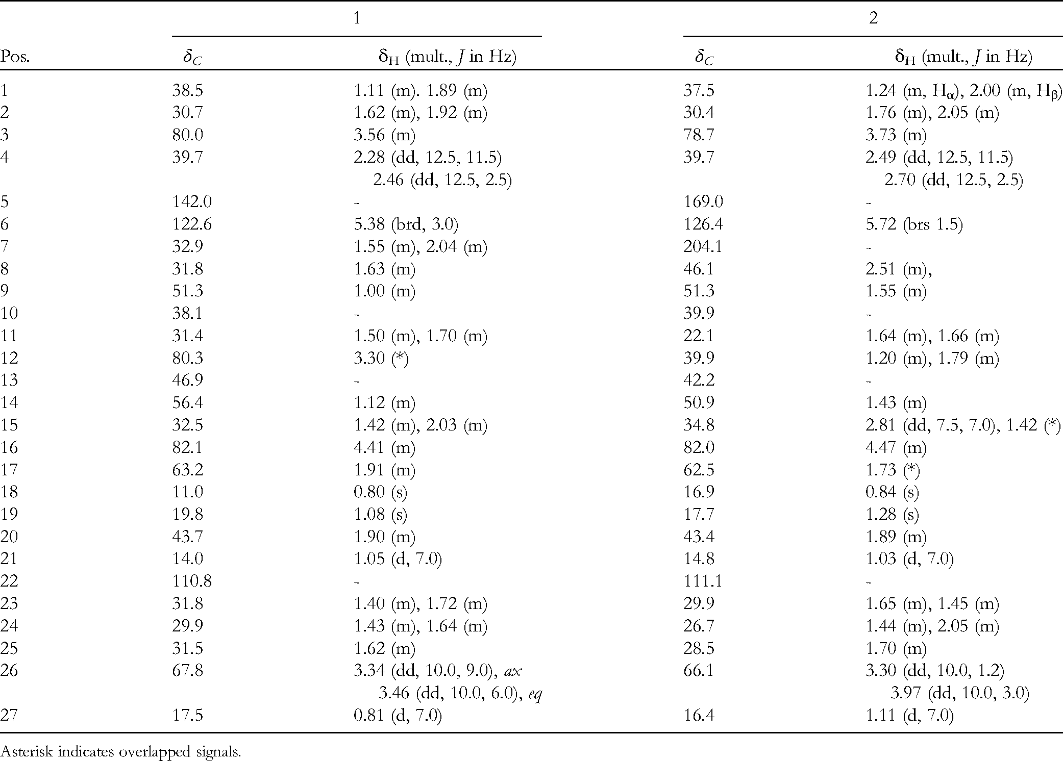
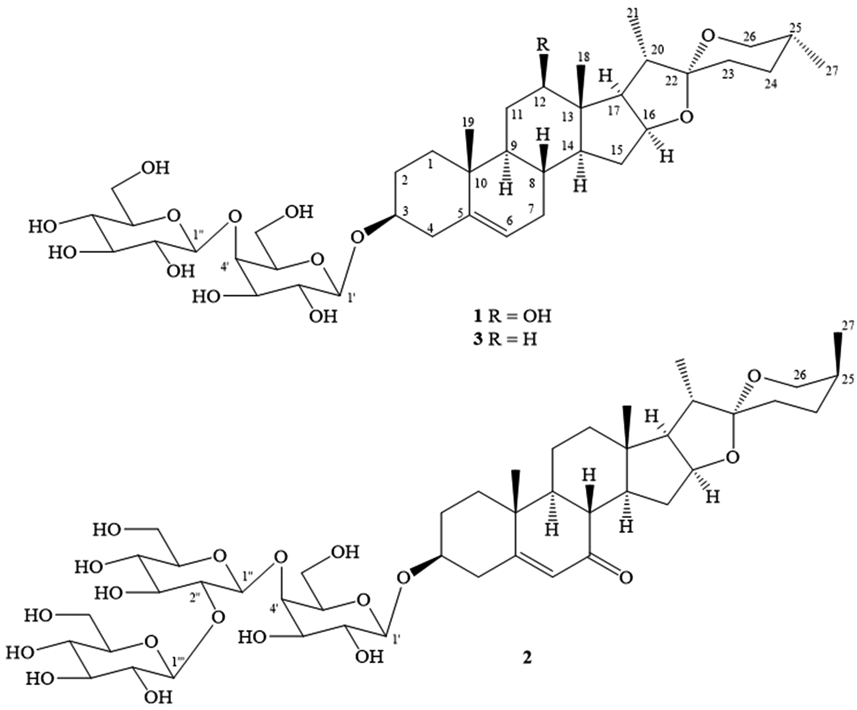
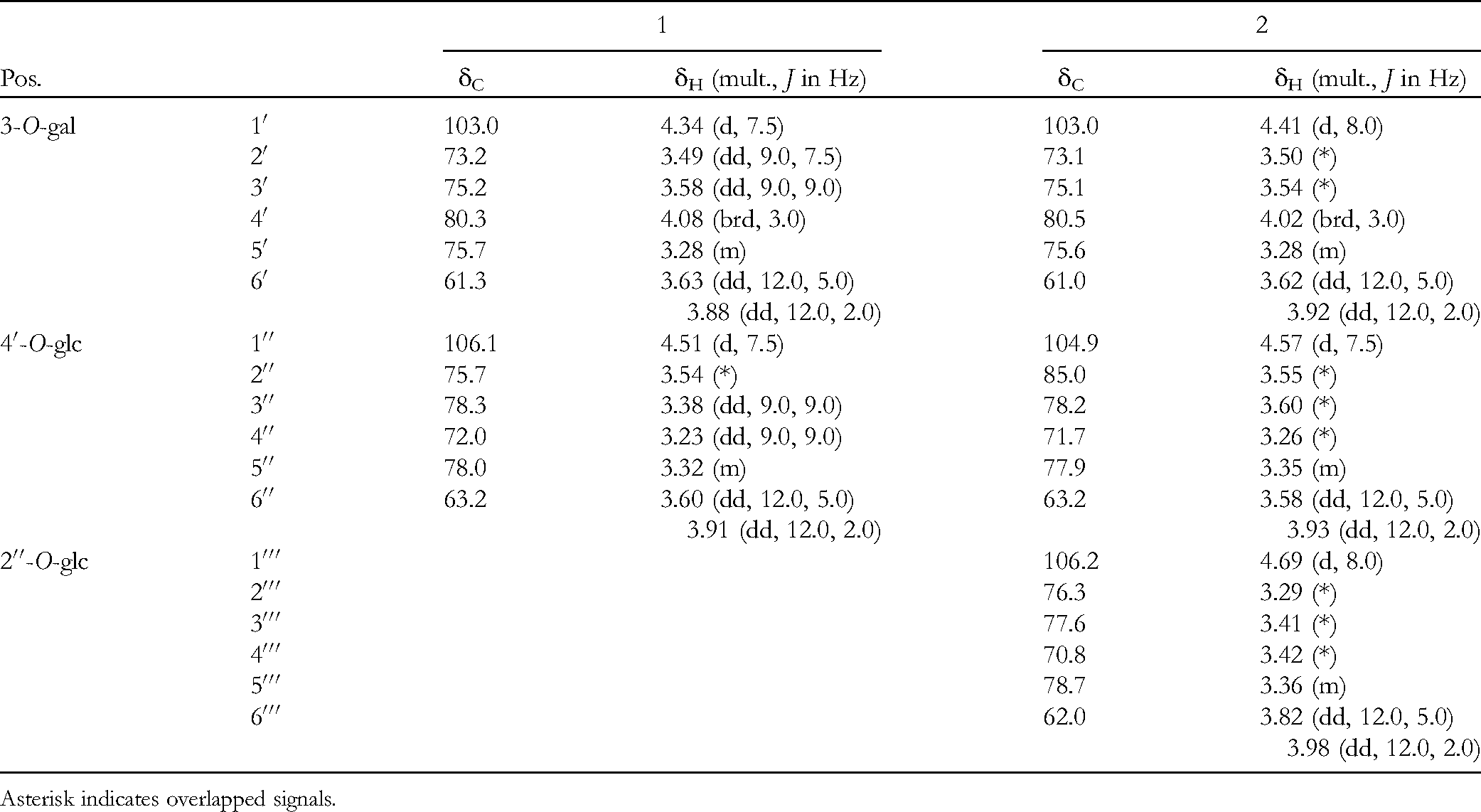
Compound 1 was obtained as a colorless amorphous powder and positive in the Liebermann-Burchard reaction. It showed a quasimolecular ion peak at m/z 777.4034 [M + Na]+ (calcd. for [C39H62O14Na]+, 777.4032, Δ = + 0.2 ppm) in the high-resolution electron spray ionization mass spectrum (HR-ESI-MS), indicating its molecular formula of C39H62O14 and 9° of unsaturation (see Supplemental Figure S1). The IR spectrum of 1 exhibited the presence of hydroxy (3371 cm−1) and C–O–C (1056 cm−1) groups. The nuclear magnetic resonance (NMR) spectra of 1 were similar to the corresponding spectra of 3 except for the additional signal of a methine carbinol group (δH 3.30/ δC 80.3) in the NMR spectra of 1, suggesting that 1 was a spirostan glycoside having 2 sugar moieties (see Supplemental Figures S2 and S3).4-7,9,11,12. The methine carbinol signal at δC 82.1/δH 4.41 (m) and the quaternary carbon signal at δC 110.8 were typical for C-16 and C-22, respectively, of the spirostan skeleton.9,11,12 In the heteronuclear multiple bond correlation (HMBC) spectrum, the H3 to 18 protons correlated to the oxygenated methine carbon at δC 80.3 confirming that the hydroxy group attached to C-12. Detailed analysis of the 1H NMR and 13C NMR spectra, combined with the heteronuclear single quantum coherence (HSQC) and HMBC spectra of 1 (see Supplemental Figures S4 and S5) indicated that the aglycone of this compound was similar to that of pratioside E1, isolated from Polygonatum prattii (see Table 1 and Figure S17).12 The carbon chemical shifts of C-25 (δC 31.5), C-26 (δC 67.8), C-27 (δC 17.5) were corresponded to that of ophiopogonin B [C-25 (δC 31.1), C-26 (δC 67.2), C-27 (δC 17.8)] suggested for 25R configuration of 1,13 which was further indicated from the large proton coupling constants of H-6 and Hax-5 (JHax-6/H−5 = 9.0 Hz, JHeq-6/H-5 = 6.0 Hz). In the nuclear Overhauser effect spectroscopy (NOESY) spectrum, H-12 (δH 3.30) had cross-peaks with Hα-9 (δH 1.10) and Hα-17 (δH 1.91) indicating Hα-12 and the hydroxyl group at C-12 was β-orientation (see Supplemental Figure S7). Furthermore, H3 to 19 (δH 1.08) had NOESY cross peak to Hβ-1 (δH 1.88) indicating Hα-1 was at δH 1.11, which had NOESY cross peak to H-3 (δH 3.56). This confirmed H-3 was α-orientation. In the 1H-1H COSY spectrum (see Supplemental Figure S6), the cross-peaks were observed between H-1′/H-2′/H-3′/ H-4′/ H-5′/ H-6′ and between H-1′′/H-2′′/H-3′′/H-4′′/H-5′′/H-6′′ (Figure 2). Moreover, the proton signal at δH 4.08 appeared as a broad doublet (J= 3.0 Hz) and their corresponding carbon was at δC 80.3 suggesting for a 4′-substituted galactose unit, which was further confirmed by the HMBC correlation from H-1′′ (δH 4.51) to C-4 gal (δC 80.3). The galactose unit linked to C-3 confirming by HMBC correlation from H-1′ (δH 4.34) to C-3 (δC 80.0) (Figure 2). Furthermore, the coupling constants (J= 7.5 Hz) observed for the anomeric protons in the 1H-NMR spectrum of 1 indicated that all the sugar linkages must be in the β-form. Acid hydrolysis of 1 obtained D-glucose and D-galactose, which were identified by TLC comparison with authentic samples and from the positive sign of the optical rotations.14 Thus, compound 1 was determined to be (25R)-12β-hydroxyspirost-5-en-3β-yl O-β-D-glucopyranosyl-(1→4)-β-D-galactopyranoside and named polygokingiaside C (Tables 1 and 2, Figure 1).
NMR Spectroscopic Data for the Aglycone Moieties of 1 and 2 in Deuterated Methanol.
Pos.
1
2
δC
δH (mult., J in Hz)
δC
δH (mult., J in Hz)
1
38.5
1.11 (m). 1.89 (m)
37.5
1.24 (m, Hα), 2.00 (m, Hβ)
2
30.7
1.62 (m), 1.92 (m)
30.4
1.76 (m), 2.05 (m)
3
80.0
3.56 (m)
78.7
3.73 (m)
4
39.7
2.28 (dd, 12.5, 11.5) 2.46 (dd, 12.5, 2.5)
39.7
2.49 (dd, 12.5, 11.5) 2.70 (dd, 12.5, 2.5)
5
142.0
-
169.0
-
6
122.6
5.38 (brd, 3.0)
126.4
5.72 (brs 1.5)
7
32.9
1.55 (m), 2.04 (m)
204.1
-
8
31.8
1.63 (m)
46.1
2.51 (m),
9
51.3
1.00 (m)
51.3
1.55 (m)
10
38.1
-
39.9
-
11
31.4
1.50 (m), 1.70 (m)
22.1
1.64 (m), 1.66 (m)
12
80.3
3.30 (*)
39.9
1.20 (m), 1.79 (m)
13
46.9
-
42.2
-
14
56.4
1.12 (m)
50.9
1.43 (m)
15
32.5
1.42 (m), 2.03 (m)
34.8
2.81 (dd, 7.5, 7.0), 1.42 (*)
16
82.1
4.41 (m)
82.0
4.47 (m)
17
63.2
1.91 (m)
62.5
1.73 (*)
18
11.0
0.80 (s)
16.9
0.84 (s)
19
19.8
1.08 (s)
17.7
1.28 (s)
20
43.7
1.90 (m)
43.4
1.89 (m)
21
14.0
1.05 (d, 7.0)
14.8
1.03 (d, 7.0)
22
110.8
-
111.1
-
23
31.8
1.40 (m), 1.72 (m)
29.9
1.65 (m), 1.45 (m)
24
29.9
1.43 (m), 1.64 (m)
26.7
1.44 (m), 2.05 (m)
25
31.5
1.62 (m)
28.5
1.70 (m)
26
67.8
3.34 (dd, 10.0, 9.0), ax 3.46 (dd, 10.0, 6.0), eq
66.1
3.30 (dd, 10.0, 1.2) 3.97 (dd, 10.0, 3.0)
27
17.5
0.81 (d, 7.0)
16.4
1.11 (d, 7.0)
Asterisk indicates overlapped signals.
Compound 2 was obtained as a colorless amorphous powder, positive in the Liebermann-Burchard reaction, and its molecular formula was deduced to be C45H74O19 based on the HR-ESI-MS results (see Supplemental Figure S10). The IR spectrum of 2 exhibited the presence of hydroxyl (3381 cm−1), carbonyl (1667 cm−1), and C–O–C (1070 cm−1) groups. The NMR spectra of 2 (see Supplemental Figures S11 and S12) were similar to those of 1 except for the disappearance of the 12-OH group signals and additional typical signals of one hexose unit [δC/δH: 106.2/4.69, 76.3/3.29, 77.6/3.41, 70.8/3.41, 78.7/3.36, 62.0/(3.98 and 3.82)] and 1 carbonyl group (δC 204.1). The signals at δC 169.0, 126.4, and 204.1 were assigned for C-5, C-6, and C-7, respectively, by comparing with the corresponding values of 7-oxodioscin15 and further confirmed by the HMBC correlations from H3 to 19 (δH 1.28) to C-5 (δC 169.0), from H-6 (δH 5.72) to C-4 (δC 39.7)/C-8 (δC 46.1), and from H-8 (δH 2.51) to C-7 (δC 204.1). The methine carbinol signals at δC 82.0/δH 4.47 (m) and the quaternary carbon at δC 111.1 were typical for C-16 and C-22 of the spirostan skeleton.9,11,12 The (25S)-configuration was suggested by comparing the carbon chemical shifts of C-24, C-25, C-26 of 2 with those of OJV-IV (25S) and OJV-III (25R),13 and further confirmed by the small JH-5/H-6 values (J = 3.0 Hz, 1.2 Hz). Moreover, the aglycone of 2 was determined to be spirostan-3β-hydroxy-5-en-7-one, similar to the aglycone of kingianoside K isolated from P kingianum,4 confirmed by analyzing the 1H-1H COSY and HMBC spectra as shown in Supplemental Figure S17 (see Supplemental Figures S14 and S15). Furthermore, H3 to 19 (δH 1.28) had NOESY cross peak to Hβ-1 (δH 2.00) indicating that Hα-1 was at δH 1.24, which had NOESY cross-peaks to H-3 (δH 3.73). This confirmed Hα-3 (see Supplemental Figure S14). The additional sugar was suggested to link to C-2′′ indicated from HSQC cross-peaks of H-1 (δH 4.57)/C-1 (δC 104.9) and H-2 (δH 3.55)/C-2 (δC 85.0) (see Supplemental Figure S11), the 1H-1H COSY cross peak of H-1′′ (δH 4.57) and H-2′′ (δH 3.55), and from the HMBC correlation from H-1′ (δH 4.69) to C-2′′ (δC 85.0). In addition, HMBC correlations from H-1′′ to C-4′ and from H-1′ to C-3 determined the second sugar attached to C-4′ of the galactose, and the galactose linked to C-3 of the aglycon. The large J values of the anomeric proton (J= 7.5-8.0 Hz) indicated β-glycoside linkages for the sugar moieties. Finally, acid hydrolysis of 2 obtained D-glucose and D-galactose, which were identified by TLC comparison with authentic samples and from the positive sign of the optical rotations.14 Thus, compound 2 was determined to be (25S)-spirost-5-en-7-one-3β-yl O-β-D-glucopyranosyl-(1→2)-β-D-glucopyranosyl-(1→4)-β-D-galactopyranoside and named polygokingiaside D (Tables 1 and 2, Figure 1).
Chemical structure of compounds 1 to 3.
NMR Spectroscopic Data for the Sugar Moieties of 1 and 2 in Deuterated Methanol.
Pos.
1
2
δC
δH (mult., J in Hz)
δC
δH (mult., J in Hz)
3-O-gal
1′
103.0
4.34 (d, 7.5)
103.0
4.41 (d, 8.0)
2′
73.2
3.49 (dd, 9.0, 7.5)
73.1
3.50 (*)
3′
75.2
3.58 (dd, 9.0, 9.0)
75.1
3.54 (*)
4′
80.3
4.08 (brd, 3.0)
80.5
4.02 (brd, 3.0)
5′
75.7
3.28 (m)
75.6
3.28 (m)
6′
61.3
3.63 (dd, 12.0, 5.0) 3.88 (dd, 12.0, 2.0)
61.0
3.62 (dd, 12.0, 5.0) 3.92 (dd, 12.0, 2.0)
4′-O-glc
1′′
106.1
4.51 (d, 7.5)
104.9
4.57 (d, 7.5)
2′′
75.7
3.54 (*)
85.0
3.55 (*)
3′′
78.3
3.38 (dd, 9.0, 9.0)
78.2
3.60 (*)
4′′
72.0
3.23 (dd, 9.0, 9.0)
71.7
3.26 (*)
5′′
78.0
3.32 (m)
77.9
3.35 (m)
6′′
63.2
3.60 (dd, 12.0, 5.0) 3.91 (dd, 12.0, 2.0)
63.2
3.58 (dd, 12.0, 5.0) 3.93 (dd, 12.0, 2.0)
2′′-O-glc
1′′′
106.2
4.69 (d, 8.0)
2′′′
76.3
3.29 (*)
3′′′
77.6
3.41 (*)
4′′′
70.8
3.42 (*)
5′′′
78.7
3.36 (m)
6′′′
62.0
3.82 (dd, 12.0, 5.0) 3.98 (dd, 12.0, 2.0)
Asterisk indicates overlapped signals.
Compound 3 was identified as funkioside C.7 The NMR spectral data of 3 were consistent with those previously reported in the literature (see Supplemental Figures S15 and S16).
The anti-inflammatory activity of compounds 1 to 3 was evaluated by their ability to inhibit NO production in LPS stimulated RAW 264.7 cells. Each compound was assessed at a concentration of 20 µM. No cytotoxic effect on the RAW 264.7 cells was found after treating the cells with compounds 1 to 3 (20 µM). L-NMMA (NG-monomethyl-L-arginine) was used as a positive control. Compound 2 exhibited the highest inhibitory, 68.3%, comparing to the positive control, L-NMMA: 83.1% (Supplemental Table S1). Therefore, compound 2 was further evaluated for its IC50 value. Compound 2 showed the inhibitory effects on NO production with an IC50 value of 8.78 ± 0.05 µM compared to a value of 7.12 ± 0.08 µM for the positive control compound, L-NMMA.
Material and Methods
General Experimental Procedures
The general experimental procedures are the same as described in our previous work.11 Refer to Supplemental Material.
Plant Material
The plant materials are the same as described in our previous work.11
Extraction and Isolation
Continue studying the water layer as described in our previous work,11 the fraction IPK2C was chromatographed on a silica gel column eluting with CH2Cl2/MeOH/H2O (3/1/0.1,v/v/v) to give 5 subfractions (IPK2C1-IPK2C5). The IPK2C3 was further chromatographed by HPLC using a J'sphere ODS H-80, 250 mm × 20 mm column, MeCN in H2O (30%, v/v), and a flow rate of 3 mL/min to yield compounds 1 (17 mg) and 3 (14.5 mg). The IPK2C4 was further chromatographed by HPLC using a J'sphere ODS H-80, 250 mm × 20 mm column, MeCN in H2O (35%, v/v), and a flow rate of 3 mL/min to yield compound 2 (14.0 mg).
Polygokingiaside C (1)
Colorless amorphous powder, : −43.5° (c 0.05, MeOH); IR (KBr) νmax: 3371 (broad), 2928, 2873, 1455, 1056 cm−1. HR-ESI-MS m/z 777.4032 [M + Na]+ (calcd. for [C39H62O14Na]+, 777.4032, Δ = +0.2 ppm); 1H NMR (CD3OD, 500 MHz) and 13C NMR (CD3OD, 125 MHz) data are given in Tables 1 and 2.
Polygokingiaside D (2)
Colorless amorphous powder, : −52.0° (c 0.05, MeOH); IR (KBr) νmax: 3381 (broad), 2930, 1667, 1452, 1070 cm−1. HR-ESI-MS m/z 949.4192 [M + 35Cl]− (calcd. for [C45H70O1935Cl]−, 949.4200, Δ = −0.8 ppm, m/z 951.4194 [M + 37Cl]− [calcd. for [C45H70O1937Cl]−, 951.4170, Δ = +2.5 ppm]. 1H NMR [CD3OD, 500 MHz] and 13C NMR [CD3OD, 125 MHz] data are given in Tables 1 and 2.
Acid Hydrolysis of Compounds 1 and 2
Compounds 1 and 2 (each, 9.0 mg) were dissolved in 0.5 mL of 6 M HCl and heated at 60°C for 1.5 h. After cooling, the mixtures were extracted with EtOAc. The acid aqueous layer was neutralized with 1 M NaOH and freeze-dried. Two sugars were identified as glucose and galactose by comparison with authentic samples by TLC using MeCOEt–isoPrOH–Me2CO–H2O (20:10:7:6). After preparative TLC of the sugar mixture (for each 1 and 2, 3.0 mg) using this solvent, each isolated sugar was filtered to eliminate SiOH residue. The optical rotation of each purified sugar was measured to afford glucose and galactose as +19.5 (c 0.15, H2O) and+45.7 (c 0.15, H2O), respectively. By comparing the optical rotations with D-glucose :+18.0 (c 0.1, H2O) and D-galactose : +45.0 (c 0.08, H2O), the glucose and galactose in compounds 1 and 2 were determined to have D-configurations.14
Phytochemical study on the methanol extracts of the roots of P kingianum Collett & Hemsl. (Asparagaceae) led to the isolations of 2 new spirostane glycosides, (25R)-12β-hydroxyspirost-5-en-3β-yl O-β-D-glucopyranosyl-(1→4)-β-D-galactopyranoside (1) and (25S)-spirost-5-en-7-one-3β-yl O-β-D-glucopyranosyl-(1→2)-β-D-glucopyranosyl-(1→4)-β-D-galactopyranoside (2), and a known spirostane glycoside, funkioside C (3). Their structures were determined by extensive analysis of HR-ESI-MS and NMR spectral data, as well as by comparison of the spectral data with those reported in the literature. The anti-inflammatory activity of the isolated compounds was evaluated by their inhibition of NO production in LPS stimulated RAW 264.7 cells. Interestingly, compound 2 showed inhibitory effects on NO production with an IC50 value of 8.78 ± 0.05 µM compared to a value of 7.12 ± 0.08 µM for the positive control compound, L-NMMA. These results suggested the selection of the medicinal plant varieties for further breeding in future.
Supplemental Material
sj-docx-1-npx-10.1177_1934578X211041258 - Supplemental material for New Nitric Oxide Inhibitory Spirostane Glycosides from Polygonatum kingianum
Collett & Hemsl
Supplemental material, sj-docx-1-npx-10.1177_1934578X211041258 for New Nitric Oxide Inhibitory Spirostane Glycosides from Polygonatum kingianum
Collett & Hemsl by Tran Thi Thu Ha and Phan Van Kiem in Natural Product Communications
Footnotes
Acknowledgments
The authors express their sincere thanks to the Ministry of Science and Technology of Vietnam for supporting research funding through the topic: “Research on seed selection and developing on industrial scale breeding technology for some high-value non-timber forest products toward socio-economic development in the Northern mountainous region of Vietnam” (grant no.: DTDL-CN-12/18). The authors thank Prof Dang Kim Vui at The Institute of Forestry Research and Development, Thai Nguyen University of Agriculture and Forestry for the identification of the plant sample.
Declaration of Conflicting Interests
The authors declared no potential conflicts of interest with respect to the research, authorship, and/or publication of this article.
Funding
The authors disclosed receipt of the following financial support for the research, authorship, and/or publication of this article: This work was supported by the Ministry of Science and Technology of Vietnam (grant no. DTDL-CN-12/18).
ORCID iD
Phan Van Kiem
Supplemental Material
Supplemental material for this article is available online.
References
1.
ZhaoPZhaoCLiX, et al.The genus Polygonatum: a review of ethnopharmacology, phytochemistry and pharmacology. J Ethnopharmacol. 2018;214:274-291. doi:10.1016/j.jep.2017.12.006
2.
BichDChungDChuongB, et al. The medicinal plants and animals in Vietnam. vol 1. Sci. Technol. Publ. House; 2004: 960-963.
3.
XiaoweiCWangSCaoH, et al. A review: the bioactivities and pharmacological applications of Polygonatum sibiricum polysaccharides. Molecules. 2018;23(5):1170. doi:10.3390/molecules23051170
4.
YuHSMaBPSongXB, et al.Two new steroidal saponins from the processed Polygonatum kingianum. Helv Chim Acta. 2010;93(6):1086-1092. doi:10.1002/hlca.200900308
5.
ZhangJMaBPKangLP, et al.Furostanol saponins from the fresh rhizomes of Polygonatum kingianum. Chem Pharm Bull. 2006;54(7):931-935.
6.
YuHSZhangJKangLP, et al.Three new saponins from the fresh rhizomes of Polygonatum kingianum. Chem Pharm Bull. 2009;57(1):1-4.
7.
LiXCYangCRIchikawaMMatsuuraHKasaiRYamasakiK. Steroid saponins from Polygonatum kingianum. Phytochemistry. 1992;31(10):3559-3563.
8.
LuJMWangYFYanHLLinPGuWYuJ. Antidiabetic effect of total saponins from Polygonatum kingianum in streptozotocin-induced diabetic rats. J Ethnopharmacol. 2016;179:291-300.
9.
ZhaoXLiJ. Chemical constituents of the genus Polygonatum and their role in medicinal treatment. Nat Prod Commun. 2015;10(4):683-688. doi:10.1177/1934578X1501000439
10.
YuHSMaBPKangLP, et al.Saponins from the processed rhizomes of Polygonatum kingianum. Chem Pharm Bull. 2009;57(9):1011-1014.
11.
HaTTTHuyTQChinhPTVan KiemP. Polygokingiasides A and B: two new spirostane glycosides from the roots of Polygonatum kingianum with nitric oxide inhibitory activity. Nat Prod Commun2021;16(1):1-6. doi:10.1177/1934578X20985634
12.
LiXCYangCRMatsuuraHKasaiRYamasakiK. Steroid glycosides from Polygonatum prattii. Phytochemistry. 1993;33(2):465-470.
13.
AsanoTMurayamaTHiraiYShojiJ. Comparative studies on the constituents of Ophiopogonis tuber and its congeners. VIII. Studies on the glycosides of the subterranean part of Ophiopogon japonicus Ker-Gawler cv. Nanus. Chem Pharm Bull. 1993;41(3):566-570.
14.
Voutquenne-NazabadiokoLGevrenovaRBorieN, et al. Triterpenoid saponins from the roots of Gypsophila trichotoma Wender. Phytochemistry. 2013;90:114-127. doi:10.1016/j.phytochem.2013.03.001
15.
AliZSmillie TJIAK. 7-Oxodioscin, a new spirostan steroid glycoside from the rhizomes of Dioscorea nipponica. Nat Prod Commun. 2013;8(3):319-321.
Supplementary Material
Please find the following supplemental material available below.
For Open Access articles published under a Creative Commons License, all supplemental material carries the same license as the article it is associated with.
For non-Open Access articles published, all supplemental material carries a non-exclusive license, and permission requests for re-use of supplemental material or any part of supplemental material shall be sent directly to the copyright owner as specified in the copyright notice associated with the article.