Two new spirostane glycosides, named polygokingiaside A (1) and polygokingiaside B (2), and 6 known spirostane glycosides, (25R)-26-O-(β-d-glucopyranosyl)-furost-5-en-3β,22α,26-triol 3-O-β-d-glucopyranosyl-(1→4)-β-d-galactopyranoside (3), (25S)-26-O-(β-d-glucopyranosyl)-furost-5-en-3β,22α,26-triol 3-O-β-d-glucopyranosyl-(1→2)-β-d-glucopyranosyl-(1→4)-β-d-galactopyranoside (4), (25R)-26-O-(β-d-glucopyranosyl)-furost-5-en-3β,22α,26-triol 3-O-β-d-glucopyranosyl-(1→2)-β-d-glucopyranosyl-(1→4)-β-d-galactopyranoside (5), (23S,25R)-spirostan-5-en-3β,23-dihydroxy-12-one-3-O-β-d-glucopyranosyl-(1→2)-β-d-glucopyranosyl-(1→4)-β-d-galactopyranoside (6), (24S,25R)-spirostan-5-en-3β,24-dihydroxy-12-one-3-O-β-d-glucopyranosyl-(1→2)-β-d-glucopyranosyl-(1→4)-β-d-galactopyranoside (7), and (25R)-spirostan-5-en-3β-hydroxy-12-one-3-O-β-d-glucopyranosyl-(1→4)-β-d-galactopyranoside (8), were isolated from the roots of Polygonatum kingianum Collett & Hemsl. (Asparagaceae). Their structures were determined by extensive analysis of high-resolution electron spray ionization mass spectrometry and nuclear magnetic resonance spectral data, as well as by comparison of the spectral data with those reported in the literature. Anti-inflammatory activity of compounds 1-8 was evaluated by their inhibition of nitric oxide (NO) production in lipopolysaccharide (LPS)-stimulated RAW 264.7 cells. At a concentration of 20 µM, compounds 1-8 exhibited a modest inhibitory effect on NO production in LPS-stimulated RAW246.7 cells with inhibitory values ranging from 9.5 ± 0.8% to 33.8 ± 2.1%.
The genus Polygonatum comprises 74 accepted species belonging to the Asparagaceae family. Polygonatum kingianum Collett & Hemsl has 13 synonyms: P. agglutinatum Hua, P. cavaleriei H. Lév., P. darrisii H. Lév., P. ericoideum H. Lév., P. esquirolii H. Lév., P. huanum H. Lév., P. kingianum var. cavaleriei (H. Lév.) C. Jeffrey & McEwan, P. kingianum var. ericoideum (H. Lév.) C. Jeffrey & McEwan, P. kingianum var. grandifolium D. M. Liu & W. Z. Zeng, P. kingianum var. uncinatum (Diels) C. Jeffrey & McEwan, P. uncinatum Diels, P. vietnamicum L. I. Abramova, and Galium tuberosum Lour. The species is a precious medicinal plant that is distributed in south-central China, southeast China, Myanmar, Thailand, and in the north of Vietnam.1,2 The roots of P. kingianum are well known as a traditional medicinal herb and functional food in China and Vietnam for the treatment of lung diseases, back pain, and body weakness.1-3 As reported earlier, the roots of P. kingianum contain mainly steroidal saponins.3-7 Especially, saponins from P. kingianum may be used as adjuvant therapy to control blood glucose and insulin resistance in type 2 diabetic individuals.8 With the aim of finding natural bioactive constituents from Vietnamese medicinal plants, we describe herein the isolation and chemical structure elucidation of 8 spirostane glycosides, including 2 that are new, from the methanol extract of the roots of P. kingianum. The anti-inflammatory activity of these compounds was evaluated by their inhibition of nitric oxide (NO) production in lipopolysaccharide (LPS)-stimulated RAW 264.7 cells.
Results and Discussion
Compound 1 was obtained as a colorless amorphous powder. Its molecular formula was deduced to be C45H72O19 based on the cluster of quasi-molecular ion peaks in the high-resolution electron spray ionization mass spectrometry (HR-ESI-MS), including those at m/z 951.4325 [M + 35 Cl]− (Calcd. for [C45H72O1935Cl]−, 951.4356), m/z 952.4373 [M + 35+37 Cl]− (Calcd. for [C45H72O1935+37 Cl], 952.4390), m/z 953.4361 [M + 37 Cl]− (Calcd. for [C45H72O1937Cl]−, 953.4327), and m/z 917.4749 [M + H]+ (Calcd. for [C45H73O19]+, 917.4741) (Supplemental Figure S2). The 1H-nuclear magnetic resonance spectrum of 1 exhibited 3 methyl singlets at δH 0.84, 1.07, and 1.25 (each, 3H), 1 methyl doublet at 1.02 (3H), and 1 olefinic broad singlet at δH 5.40 corresponding to a steroidal aglycon having a double bond at C-5/C-6.44-7,9 Besides, 3 sugar units were identified by 3 anomeric protons at δH 4.32, 4.37, and 4.53 (each, 1H, doublet, J = 7.5 Hz) (Supplemental Figure S3). The 13C-NMR spectrum of 1 (Supplemental Figure S4) indicated the presence of 45 carbon signals, which were then further classified from the heteronuclear single-quantum correlation spectroscopy (HSQC) (Supplemental Figure S5), including 27 carbons of the steroidal aglycon and 18 carbons of 3 sugar molecules. The 13C NMR spectrum of compound 1 indicated signals for C-3, C-5, C-6, C-22, C-25, and C-26 (δC 80.0, 142.0, 122.5, 121.8, 85.2, and 77.6, respectively) confirming that the aglycone of 1 was in good accordance with that of nuatigenin [(22S,25S)-22,25-epoxyfurost-5-en-3β,26-diol]10 having a sugar moiety linked to C-3, as with chonglouoside SL-11, chonglouoside SL-12, chonglouoside SL-13, chonglouoside SL-14, chonglouoside SL-15, and chonglouoside SL-16.9 Detailed analysis of the 1H, 13C NMR, HSQC, heteronuclear multiple bond correlation (HMBC), and 1H-1H correlation spectroscopy (COSY) of 1 (Supplemental Figures S3-S7) and comparison of the sugar NMR data of 1 with the corresponding data of kinggianoside A (8),7 (25R)-26-O-(β-d-glucopyranosyl)-furost-5-en-3β,22α,26-triol 3-O-β-d-glucopyranosyl-(1→4)-β-d-galactopyranoside (3),7 chonglouoside SL-10, and chonglouoside SL-119 suggested that 1 glucosyl unit was attached to C-26 and the other sugar moiety, including 1 glucose and 1 galactose, was linked to C-3, which were further confirmed by the HMBC spectrum (Supplemental Figure S6). In the 1H-1H COSY spectrum, cross-peaks were observed from H-2″ (δH 3.51) to H-1″ (δH 4.37)/H-3″ (δH 3.58), from H-4″ (δH 4.07) to H-3″/H-5″ (δH 3.53), and from H-5″ to H-6″ (δH 3.62/3.88). This evidence, together with the small coupling constant (J = 3.0 Hz) of H-4″, confirmed that H-4″ was in an equatorial orientation and the sugar was galactose. In the HMBC spectrum, the anomeric proton H-1′ (δH 4.32) correlated with C-26 (δC 77.6), while proton H-1″′ (δH 4.53) correlated with gal C-4 (δC 79.2), and gal H-1″ (δH 4.37) correlated with C-3 (δC 80.0) confirming that the sugar linkages of compound 1 are as shown in Figure 1. Furthermore, the coupling constant (J = 7.5 Hz) observed for the anomeric protons in the 1H NMR spectrum of 1 indicated that the sugar linkages must be in the β-form. In the nuclear Overhauser effect spectroscopy (NOESY) (Supplemental Figure S8), cross-peaks from H-21 (1.02) to H-17 (1.79), H-18 (0.84) to H-23 (1.31/1.62), and from H-17 to H-16 (4.48) reconfirmed the aglycon structure of 1 as nuatigenin [(22S,25S)−22,25-epoxyfurost-5-en-3β,26-diol]. Finally, the presence of d-glucose and d-galactose in the acid hydrolysis products of compound 1 was confirmed by gas chromatographic (GC) analysis of their trimethylsilyl derivatives, as previously described.11 Consequently, compound 1 was determined to be 26-O-β-d-glucopyranosyl nuatigenin 3-O-β-d-glucopyranosyl-(1→4)-β-d-galactopyranoside and named as polygokingiaside A.
Chemical structure of compounds 1‐8 isolated from the roots of Polygonatum kingianum.
Compound 2 was obtained as a colorless amorphous powder, and its molecular formula was deduced to be C45H74O19 based on the cluster of quasi-molecular ion peaks in the HR-ESI-MS at m/z 941.4717 [M + Na]+ (Calcd. for [C45H74O19Na]+, 941.4717) (Supplemental Figure S8). The NMR spectra of 2 (Supplemental Figures S10-S16) were similar to those of compounds 1, 3,7 and 412 suggesting that compound 2 was a spirostane glycoside. The sugar moieties of 2 were also found to be similar to 1 and 3, identified from anomeric protons and carbons at δH 4.27 (d, J = 7.5 Hz)/δC 104.7, 4.37 (d, J = 7.5 Hz)/δC 103.0, and 4.53 (d, J = 7.5 Hz)/δC 106.1. One glucose was attached to C-26 and a β-d-glucopyranosyl-(1→4)-β-d-galactopyranoside moiety was linked to C-3, which were confirmed by 1H-1H COSY and HMBC spectra, as shown in Supplemental Figure S1. Comparing the NMR data of 2 with that of 3 showed that they were very similar except for the proton signals of C-26 at δH3.80 (1H, Ha-26) and δH 3.36 (1H, Hb-26) (Δ = 0.44) in compound 2, instead of proton signals at δH 3.75 (1H, Ha-26) and 3.41 (1H, Hb-26) (Δ = 0.34) in 3. This evidence indicated that the configuration of the methyl group at C-25 of 2 is S and that of 3 is R.5,13,14 In addition, NOE cross-peaks from H-18 (0.85) to H-23 (1.67/1.70) were observed, reconfirming the 22α-OH group (Supplemental Figure S16). Finally, the presence of d-glucose and d-galactose in the acid hydrolysis products of compound 2 were confirmed by GC analysis of their trimethylsilyl derivatives, as previously described.11 Consequently, compound 2 was determined to be (25S)-26-O-(β-d-glucopyranosyl)-furost-5-en-3β,22α,26-triol 3-O-β-d-glucopyranosyl-(1→4)-β-d-galactopyranoside, and named as polygokingiaside B.
Compounds 3-8 were identified as (25R)-26-O-(β-d-glucopyranosyl)-furost-5-en-3β,22α,26-triol 3-O-β-d-glucopyranosyl-(1→4)-β-d-galactopyranoside (3),7 (25S)−26-O-(β-d-glucopyranosyl)-furost-5-en-3β,22α,26-triol 3-O-β-d-glucopyranosyl-(1→2)-β-d-glucopyranosyl-(1→4)-β-d-galactopyranoside or dehydrotomatoside (4),12 (25R)-26-O-(β-d-glucopyranosyl)-furost-5-en-3β,22α,26-triol 3-O-β-d-glucopyranosyl-(1→2)-β-d-glucopyranosyl-(1→4)-β-d-galactopyranoside (5),7 (23S,25R)-spirostan-5-en-3β,23-dihydroxy-12-one-3-O-β-d-glucopyranosyl-(1→2)-β-d-glucopyranosyl-(1→4)-β-d-galactopyranoside (6),6 (24S,25R)-spirostan-5-en-3β,24-dihydroxy-12-one-3-O-β-d-glucopyranosyl-(1→2)-β-d-glucopyranosyl-(1→4)-β-d-galactopyranoside (7),15 and (25R)-spirostan-5-en-3β-hydroxy-12-one-3-O-β-d-glucopyranosyl-(1→4)-β-d-galactopyranoside (8).7 The NMR spectral data of compounds 2-8 were consistent with those previously reported in the literature (Supplemental Figures S17-S38). Anti-inflammatory activity of compounds 1-8 was evaluated by their ability to inhibit NO production in LPS-stimulated RAW 264.7 cells. Each compound was assessed at a concentration of 20 µM. Compounds 1-8 moderately inhibited NO production with inhibitory rates from 9.5 ± 0.8% to 33.8 ± 2.1%. No cytotoxic effect on the RAW 264.7 cells was found after treating the cells with compounds 1-8 (20 µM). l-NG-monomethyl-l-arginine was used as a positive control. Its NO inhibitory value was 83.1 ± 2.3% at a concentration of 20 µM (Table 1).
Effect of Compounds 1-8 (20 µM) on Nitric Oxide Production in LPS-Stimulated RAW 264.7 cells.
Optical rotation was measured on a Jasco P-2000 polarimeter, NMR spectra on a Bruker 500 MHz spectrometer, and HR-ESI-MS on an Agilent 6530 Accurate Mass Q-TOF LC/MS. The QTOF instrument was set at 2 GHz extended dynamic range resolution mode, with a negative ESI capillary voltage of 3500 V, fragmentor voltage of 175 V, MS scan ranging from m/z 100-1700, and an MS acquisition rate of 1.0 spectra/second. Flash column chromatography was performed using either silica gel or reversed-phase (RP-18) resins as adsorbent. The ratio between the amount of silica gel and fraction was 20/1 (w/w). A fraction collector was set by volume per tube (15 mL/tube). Fractionation was monitored by thin-layer chromatography (TLC) to combine test tubes showing similar TLC pattern. TLC was carried out on either precoated silica gel 60 F254 and/or RP-18 F254S plates. Compounds were visualized by ultraviolet irradiation (254 and 365 nm) and by spraying with a sulfuric acid solution (5%) followed by heating with a heat gun. High-performance liquid chromatography (HPLC) was carried out using an AGILENT 1100 HPLC system.
Plant Material
The roots of P. kingianum Collett & Hemsl. (Asparagaceae) were collected at Ho Thau Commune, Hoang Su Phi District, Ha Giang Province, Vietnam, in September 2020, and identified by Professor Dang Kim Vui at the Institute of Forestry Research and Development, Thai Nguyen University of Agriculture and Forestry, Thai Nguyen City, Vietnam. A voucher specimen (HTD.09.2020) is kept at the Institute of Forestry Research and Development.
Extraction and Isolation
Dried, powdered P. kingianum roots (3.4 kg) were ultrasonically extracted with hot methanol (MeOH), 3 times. The extracts were filtered through filter paper, and the filtrate was evaporated to dryness to yield 550 g of a dark solid extract. This extract was suspended in water (H2O) and successively partitioned with dichloromethane (CH2Cl2) and ethyl acetate giving the CH2Cl2 extract (IPK1A, 22 g), ethyl acetate extract (IPK1B, 1.4 g), and H2O layer (IPK1C). The IPK1A fraction was chromatographed on a silica gel column, eluting with CH2Cl2/MeOH (100:0→ 0:1, v/v), to give 7 subfractions (IPK5A-IPK5G). The IPK5F (3.5 g) and IPK5G (1.5 g) fractions were combined and chromatographed on a silica gel column eluting with CH2Cl2/MeOH/H2O (6/1/0.1, v/v/v) to give 8 smaller fractions (IPK9A-IPK9H). IPK9E (300 mg) was chromatographed on an RP-18 column eluting with acetone/H2O (2/1, v/v) to give 2 fractions, IPK10A and IPK10B. Compound 8 (7.0 mg, tR = 35.52′) was obtained from IPK10A by HPLC using a J’sphere ODS H-80, 250 mm × 2 0 mm column, acetonitrile (ACN) in H2O (50%, v/v), and a flow rate of 3 mL/min. Continuously, the H2O layer was chromatographed on a Diaion HP-20 column eluting with H2O to remove sugar, then with increasing concentration of MeOH in H2O (25%, 50%, 75%, and 100%) to obtain 4 fractions, IPK2A (12.0 g), IPK2B (6.3 g), IPK2C (5.3 g), and IPK2D (1.6 g). Fraction IPK2D was chromatographed on a silica gel column eluting with CH2Cl2/MeOH/H2O (4/1/0.1,v/v/v) to give 6 subfractions IPK3A-IPK3F. IPK3B was further chromatographed by HPLC using a J’sphere ODS H-80, 250 mm × 20 mm column, ACN in H2O (35%, v/v), and a flow rate of 3 mL/min to yield compound 6 (12.0 mg, tR=30.82′). The IPK3C fraction was chromatographed on HPLC using the same conditions to yield 7 (4.6 mg, tR = 29.10′)). IPK3D was chromatographed on a Sephadex column eluting with MeOH/H2O (1/1, v/v) to give 2 smaller fractions, IPK4A-IPK4B. Compound 1 (7.0 mg, tR = 52.62′) was obtained from IPK4A chromatographed on an RP-18 column eluting with acetone/H2O (1/1.2, v/v). IPK4B was further chromatographed by HPLC using a J’sphere ODS H-80, 250 mm × 20 mm column, ACN in H2O (27 %) to yield 2 (11.4 mg, tR = 62.14′) and 3 (20.0 mg, tR = 65.79′). Finally, the IPK3F fraction was chromatographed on HPLC using the same conditions to yield 4 (8.0 mg, tR = 47.03′) and 5 (7.0 mg, tR = 48.99′).
Polygokingiaside A (1)
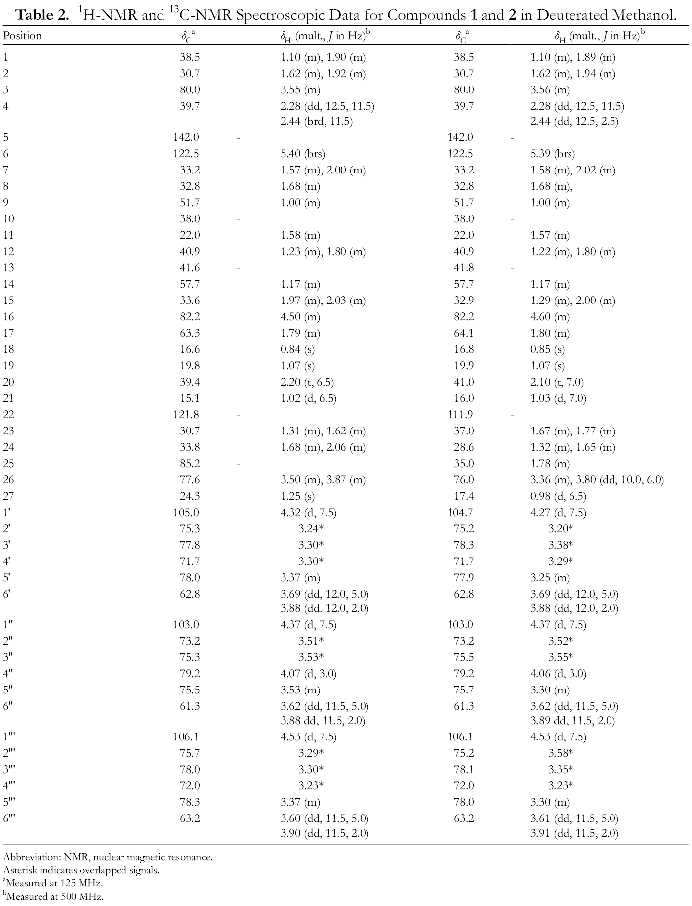
Colorless amorphous powder, − 45.0° (c 0.1, MeOH); HR-ESI-MS m/z 951.4325 [M + 35Cl]− (Calcd. for [C45H72O1935Cl]−, 951.4356), m/z 952.4373 [M + 35+37Cl]− (Calcd. for [C45H72O1935+37Cl], 952.4390), m/z 953.4361 [M + 37Cl]− (Calcd. for [C45H72O1937Cl]−, 953.4327), and m/z 917.4749 [M + H]+ (Calcd. for [C45H73O19]+, 917.4741); 1H-NMR (CD3OD, 500 MHz) and 13C-NMR (CD3OD, 125 MHz) data are given in Table 2.
1H-NMR and 13C-NMR Spectroscopic Data for Compounds 1 and 2 in Deuterated Methanol.
Polygonatum kingianum Collett & Hemsl. (Asparagaceae) is a valuable medicinal plant, used in traditional remedies to treat some diseases. From the methanol extract of the roots of this plant, 2 new spirostane glycosides, named polygokingiaside A (1) and polygokingiaside B (2), and the 6 known spirostane glycosides (25R)-26-O-(β-d-glucopyranosyl)-furost-5-en-3β,22α,26-triol 3-O-β-d-glucopyranosyl-(1→4)-β-d-galactopyranoside (3), (25S)-26-O-(β-d-glucopyranosyl)-furost-5-en-3β,22α,26-triol 3-O-β-d-glucopyranosyl-(1→2)-β-d-glucopyranosyl-(1→4)-β-d-galactopyranoside (4), (25R)-26-O-(β-d-glucopyranosyl)-furost-5-en-3β,22α,26-triol 3-O-β-d-glucopyranosyl-(1→2)-β-d-glucopyranosyl-(1→4)-β-d-galactopyranoside (5), (23S,25R)-spirostan-5-en-3β,23-dihydroxy-12-one-3-O-β-d-glucopyranosyl-(1→2)-β-d-glucopyranosyl-(1→4)-β-d-galactopyranoside (6), (24S,25R)-spirostan-5-en-3β,24-dihydroxy-12-one-3-O-β-d-glucopyranosyl-(1→2)-β-d-glucopyranosyl-(1→4)-β-d-galactopyranoside (7), and (25R)-spirostan-5-en-3β-hydroxy-12-one-3-O-β-d-glucopyranosyl-(1→4)-β-d-galactopyranoside (8) were isolated. Their structures were determined by extensive analysis of HR-ESI-MS and NMR spectral data, as well as by comparison of the spectral data with those reported in the literature. Anti-inflammatory activity of compounds 1-8 was evaluated by their inhibition of NO production in LPS-stimulated RAW 264.7 cells. At a concentration of 20 µM, compounds 1-8 moderately inhibited NO production with inhibitory rates from 9.5 ± 0.8% to 33.8 ± 2.1%.
Supplemental Material
Figure S1 - Supplemental material for Polygokingiasides A and B: Two New Spirostane Glycosides From the Roots of Polygonatum kingianum With Nitric Oxide Inhibitory Activity
Supplemental material, Figure S1, for Polygokingiasides A and B: Two New Spirostane Glycosides From the Roots of Polygonatum kingianum With Nitric Oxide Inhibitory Activity by Tran Thi Thu Ha, Trinh Quang Huy, Pham The Chinh and Phan Van Kiem in Natural Product Communications
Footnotes
Acknowledgments
The authors would like to thank Professor Dang Kim Vui at The Institute of Forestry Research and Development, Thai Nguyen University of Agriculture and Forestry, for the identification of the plant sample.
Declaration of Conflicting Interests
The author(s) declared no potential conflicts of interest with respect to the research, authorship, and/or publication of this article.
Funding
The author(s) received no financial support for the research, authorship, and/or publication of this article.
ORCID iD
Phan Van Kiem
Supplemental Material
Supplemental material for this article is available online.
References
1.
ZhaoP.ZhaoC.LiXet al. The genus Polygonatum: A review of ethnopharmacology, phytochemistry and pharmacology. J Ethnopharmacol. 2018;214:274-291.doi:10.1016/j.jep.2017.12.00629246502
2.
BichDH.ChungDQ.ChuongBXet al. The Medicinal Plants and Animals in Vietnam. Scientific and Technical Publishing House; 2004:960-963.
3.
CuiX.WangS.CaoHet al. A review: the bioactivities and pharmacological applications of Polygonatum sibiricum polysaccharides. Molecules. 2018;23(5):1170.doi:10.3390/molecules2305117029757991
4.
YuH-S.MaB-P.SongX-Bet al. Two new steroidal saponins from the processed Polygonatum kingianum. Helv Chim Acta. 2010;93(6):1086-1092.doi:10.1002/hlca.200900308
5.
ZhangJ.MaB-P.KangL-Pet al. Furostanol saponins from the fresh rhizomes of Polygonatum kingianum. Chem Pharm Bull. 2006;54(7):931-935.doi:10.1248/cpb.54.93116819206
6.
YuH-S.ZhangJ.KangL-Pet al. Three new saponins from the fresh rhizomes of Polygonatum kingianum. Chem Pharm Bull. 2009;57(1):1-4.doi:10.1248/cpb.57.119122309
7.
LiXC.YangCR.IchikawaM.MatsuuraH.KasaiR.YamasakiK. Steroid saponins from Polygonatum kingianum. Phytochemistry. 1992;31(10):3559-3563.doi:10.1016/0031-9422(92)83727-g1368862
8.
LuMJ.WangYF.YanHL.LinP.GuW.YuJ. Antidiabetic effect of total saponins from Polygonatum kingianum in streptozotocin-induced daibetic rats. J Ethnopharmacol. 2016;179:291-300.doi:10.1016/j.jep.2015.12.05726743227
9.
QinX-J.YuM-Y.NiWet al. Steroidal saponins from stems and leaves of Paris polyphylla var. yunnanensis. Phytochemistry. 2016;121:20-29.doi:10.1016/j.phytochem.2015.10.00826546502
10.
MimakiY.SashidaY. Steroidal saponins from the bulbs of Lilium brownii. Phytochemistry. 1990;29(7):2267-2271.doi:10.1016/0031-9422(90)83050-B
11.
ThuVK.Van ThangN.NhiemNXet al. Oleanane-type saponins from Glochidion glomerulatum and their cytotoxic activities. Phytochemistry. 2015;116:213-220.doi:10.1016/j.phytochem.2015.05.00126007323
12.
YamanakaT.VinckenJ-P.de WaardP.SandersM.TakadaN.GruppenH. Isolation, characterization, and surfactant properties of the major triterpenoid glycosides from unripe tomato fruits. J Agric Food Chem. 2008;56(23):11432-11440.doi:10.1021/jf802351c18998702
13.
AgrawalPK. Dependence of 1H NMR chemical shifts of geminal protons of glycosyloxy methylene (H2-26) on the orientation of the 27-methyl group of furostane-type steroidal saponins. Magn Reson Chem. 2004;42(11):990-993.doi:10.1002/mrc.147415386548
14.
AgrawalPK. Assigning stereodiversity of the 27-Me group of furostane-type steroidal saponins via NMR chemical shifts. Steroids. 2005;70(10):715-724.doi:10.1016/j.steroids.2005.04.00115923015
15.
YuH-S.MaB-P.KangL-Pet al. Saponins from the processed rhizomes of Polygonatum kingianum. Chem Pharm Bull. 2009;57(9):1011-1014.doi:10.1248/cpb.57.101119721268
Supplementary Material
Please find the following supplemental material available below.
For Open Access articles published under a Creative Commons License, all supplemental material carries the same license as the article it is associated with.
For non-Open Access articles published, all supplemental material carries a non-exclusive license, and permission requests for re-use of supplemental material or any part of supplemental material shall be sent directly to the copyright owner as specified in the copyright notice associated with the article.