
Abstract
Demand for sunscreen products has been rising in recent years due to increasing cases of skin damage caused byultraviolet radiation (UVR). Recent studies have proved that some materials in commercial sunscreen products are harmful to the marine ecosystem. Therefore, the development of a photoprotective and environmentally friendly screening agent has been a leading direction for the cosmetic industry. Calophyllum inophyllum is a seaside plant found in the Pacific Rim, regarded as a potential source of biodiesel feedstock given the high oil content of its seeds. Due to the harsh environment of its natural habitat, C. inophyllum has developed UV-absorbing secondary metabolites, which are natural sources for screening agents. In this study, samples of seed extracts of C. inophyllum were subjected to in vitro UV, Fourier-transform infrared (FTIR) spectroscopy, and cytotoxicity analysis; all the samples extracted by solvents of various polarities showed different chemical compositions by FTIR spectroscopy and levels of cytotoxicity. The n-hexane seed extract showed the highest UVA and UVB absorption efficiencies. In the in vitro cytotoxicity test on human skin fibroblast cells, seed oil of C. inophyllum demonstrated low cytotoxicity. Results have shown that seed extracts of C. inophyllum can be an ideal material for a natural high-efficiency screening agent.
Sunlight is a crucial energy source for the whole biota and human metabolism, but excessive exposure to sunlight could cause a deleterious impact on health, and each year, there are about 2-3 million cases of skin cancer reported. 1 Besides, there are also other dermatological injuries induced by sunlight exposure, such as erythema, pigmentation, reddening, and swelling of human skin. 2
Among the entire spectrum of solar radiation, the invisible ultraviolet (UV) spectrum, or the so-called UV radiation (UVR), is responsible for damage to human skin. The spectrum is commonly subdivided into 3 subspectra according to ranges of wavelength, which are UVA (wavelength 320-400 nm), UVB (wavelength 290-320 nm), and UVC (wavelength 200-290 nm). A major part of the UVC is absorbed by the atmospheric ozone layer, while UVA and UVB reach the terrestrial surface and penetrate human skin. 3 At the molecular level, UVR causes deoxyribonucleic acid (DNA) damage. UVB induces the synthesis of pyrimidine dimers and photoproducts, while exposure to UVA triggers reactive oxygen species (ROS) generation and DNA breakage. 4 Following an increasing awareness of the need for sun protection, the demand for sunscreen is rising, and the global sun care market is predicted to reach a value of US$24.9 billion by the end of 2024. 5 Meanwhile, the high quantity use of sun protection products also brings negative health impacts on people and deleterious bioecological impacts on other organisms; research has demonstrated that the common chemical UV filters in commercial sunscreen products, such as oxybenzone and caprylic triglyceride, could negatively influence human and animal endocrine systems. 6 Another study by Ruszkiewicz et al 7 reported that after zebrafishes had been exposed to benzophenone (oxybenzone components; UVB-UVA filters) and cinnamate (octinoxate components; UVB filter), their estrogen secretion, progestogen secretion, and androgen receptors were altered. Recently, significant bioaccumulation of the benzophenone derivatives has been discovered in the placenta of pregnant women, as well as in human urine and semen. 8 -10
Regarding the environmental aspect, related studies found that some chemical UV-filter compounds also contribute to coral bleaching. The study of Danovaro and Corinaldesi 11 indicated that the addition of sunscreen products to seawater will increase significantly the viral abundance in all microcosms compared with the control group, while the organisms in coral reef ecosystems are susceptible and sensitive to environmental change. He et al 12 showed that exposure of the coral species Seriatopora caliendrum to certain benzophenone UV filters (2,4-dihydroxybenzophenone and dioxybenzone) at concentrations ranging from 0.1 to 1000 g/L caused significant settlement failure, bleaching, and mortality of the coral larvae. Due to scientific evidence and environmental concern, authorities of Hawaii and Palau passed a bill to restrict the sale of sunscreen with formulas that contain oxybenzone and octinoxate. 13 As a result, researchers and the cosmetic industry are dedicated to figuring out possible alternative UV filters from analyzing the chemical composition of plant extracts.
Calophyllum inophyllum L., or “Alexandrian Laurel”, family Clusiaceae, has multiorigins and is found along the coasts of East Africa, India, South East Asia, Australia, and the South Pacific. 14 In the tropical and subtropical world, it is a well diffused and domesticated species. In Taiwan, arbors of this versatile species are widely planted in cities as roadside trees that provide bio corridors for animals and maintain the functions of the urban ecosystem. The timber harvested from C. inophyllum is characterized by its high density and resistance to the salinity of seawater and pests, and hence it is often utilized as an important material for shipbuilding and furniture manufacturing. Traditionally, fresh-pressed C. inophyllum seed oil is applied to cure burned skin, remove scars, and disinfect inflamed skin injuries; commonly it is used as a crucial material of cosmetics and hygienic products. 15
At a global level, C. inophyllum is estimated to produce seed oil in the total amount of 4680 kg/ha/year, which equals 11.7 kg/tree/year of oil production. 16 The high yield of C. inophyllum oil is only lower than that of palm trees, which are currently the main source of biofuel. Its long production cycle for at least 50 years also made it well recognized as a possible feedstock for biodiesel production, together with castor (Ricinus communis L.), sunflower (Helianthus annuus L.), and cotton (Gossypium arboretum). 16 Recently, C. inophyllum oil has been considered as an ideal source of biofuel for its properties such as biodegradability, nontoxic, high flash point, high calorific value, and less competitive with arable land. The fatty acid composition of C. inophyllum oil is generally dominated by monounsaturated fatty acids (oleic acid C18:1, 39.1 ± 1.4%), polyunsaturated fatty acids (linoleic acid, C18:2, 31.1 ± 1.4%), and saturated fatty acid (stearic acid, C18:0, 14.3 ± 0.8%, and palmitic acid, C16:0, 13.7 ± 0.8%). 14
Due to the high content of unsaturated fatty acids, C. inophyllum oil tends to oxidize rapidly, with rising acid value and viscosity; this unstable attribute shows its downside as a biofuel. Researchers are still seeking novel methods for the pretreatment of C. inophyllum crude oil to overcome its defects such as deposit formation and diesel injector coking due to its high viscosity and low volatility. 16 While being a potential feedstock of biofuel with a quality yet to be improved, other innovative commercial ways of utilizing C. inophyllum oil are being developed to accommodate the constantly fluctuating world energy market. Up to date studies of bioactivities of C. inophyllum seed extracts revealed a few extraordinary results: various compounds and derivatives in its extracts are proving to be HIV inhibitory, 17,18 antitumor, 19,20 antimicrobial, 21 anti-inflammatory, 22,23 and antioxidant. 24,25 As a widely used seaside plant species for wound healing in Polynesian culture, it is believed to have photoprotective functions; however, there are few studies to prove this.
For assessing C. inophyllum seed extract (CISE) as a potential ingredient for the cosmetic industry, our survey focused on the UVA and UVB absorption effectivity of CISEs by different methods. By adopting solvent extraction methods, pigments were first extracted from C. inophyllum crude oil and were homogenized in organic solvents and separated into different fractions by their polarities. Extracted fractions were evaluated using their spectroscopic properties, UV-protection performances, and in vitro cytotoxicity evaluations at different concentrations. Further inspection of the properties of different fractions of CISEs was conducted to know its potential as a novel renewable natural resource for screening agents.
Results and Discussion
Physicochemical Properties of CISEs
The yield of CISE was around 52.8% (dried weight basis), which was consistent with other research which showed that the yield of seed extract fell between 40% and 70%. 14 CISE was extracted with ethanol and separated into pigment (CIP) and refined oil (CIRO) fractions; the yield of CIP was around 19%-22%.
The fatty acid profile of CISE, determined by gas chromatography (GC)-flame ionization detector (FID), is shown in Table 1. The content of oleic acid was the highest, followed by linoleic acid. C18 fatty acids accounted for 83.0% of the total fatty acid content, and the percentage of oleic acid together with linoleic acid was 62.6%, accounting for 75.4% of C18 fatty acids. Thus, CISE is rich in unsaturated fatty acids. This observation was broadly consistent with other studies. 26
Fatty Acid Profiles of Calophyllum inophyllum in Eastern Taiwan.

Abbreviations: CIRO, C. inophyllum refined oil; CISE, C. inophyllum seed extract; OO, olive oil.
In addition, low concentrations of squalene and docosanoic acid were detected in the CISE (2% and 2.9%, respectively), and eicosanoic acid (20:0) was discovered in both CISE and CIRO (1.1% and 1.6%, respectively), which have not been observed in previous studies of C. inophyllum seeds. Hence, the chemical compositions of CISEs could vary with environmental conditions and habitats.
Compared with the control group, the proportion of oleic acid in the monounsaturated fatty acids of olive oil was 80.8%, and in CISE from 39.4% to 46.1%. The proportion of saturated fatty acids of olive oil was less than half that of CISE, showing the difference in fatty acid profiles. On the other hand, the CIRO sample was mainly composed of palmitic acid, stearic acid, oleic acid, and linoleic acid, with a relative percentage of 11.9%, 26.7%, 46.1%, and 11.4%. C18 acids accounted for 84.3% of the total fatty acid content, and oleic and linoleic acids combined were 57.6%, which is about 68.3% of the C18 acid and is also different from the fatty acid profile of CISE.
Extracted with n-hexane, ethyl acetate, and methanol, CIP was separated into pigment samples C. inophyllum n-hexane (CIH), C. inophyllum ethyl acetate (CIEA), and C. inophyllum methanol (CIM); the observed yields were 71.3%, 22.0%, and 5.3% (w/v), respectively.
UV-Vis of CISEs
Ultraviolet-visible (UV-Vis) spectroscopy is an analytical method for discovering the chemical composition of experimental samples by observing their characteristic absorptions in the UV and visible spectra. Owing to the almost complete UVC absorption by the atmospheric ozone layer, the main studies of UVR absorption usually focus on the absorption of UVA (wavelength 320-400 nm) and UVB spectra (wavelength 290-320 nm), which often cause dermatological injuries such as erythema and pigmentation of the skin. 2
Compared with the yellowish olive oil (OO), the CISEs are grass green. The major compounds discovered in the seed extracts are fatty acids and thus are hydrophobic and soluble in organic solvents such as n-hexane, ethyl acetate, and alcohols. Compared with the golden color of refined OO, the colors of the different extracted samples of C. inophyllum are distinguishable with the naked eyes; the seed oil extract (CISE) is light green, the pigment (CIP) dark green, the refined seed oil extract (CIRO) light yellow, the n-hexane extract fraction (CIH) light green, and the ethyl acetate (CIEA), and methanol fractions (CIM) dark green. The colors observed for the C. inophyllum extracts showed the high reproducibility of the C. inophyllum seeds’ visual colorimetric assay compared with previous studies. 14
For UV-Vis spectroscopy, the concentration of all the samples was calibrated to 20 mg/L using n-hexane as the solvent; only refined OO and refined seed oil extract (CIRO) remained undiluted; all samples were scanned by UVA and UVB spectroscopy to determine their UVR absorption characteristics (Figure 1). Among the samples, OO and CIRO demonstrated a lower absorption ratio of UVA and UVB comparing with the other samples, which indicates that neither are effective UV filters; CISE demonstrated a high percentage of UVR absorption within the targeted spectral range, where the absorption peak of UVA occurred at a wavelength of 305 nm, which coincided with the result of the previous study of Said et al. 27 CIP also showed significant UVR absorption in the target spectral range, where the absorption peak of UVA occurred at 300 nm; the absorption intensity was about 4.5 times higher than that of CISE. At the same concentration, the UVR absorption ratio of 4-methylbenzylidene camphor is lower than that of CIP but higher than that of CISE.

Ultraviolet spectra of Calophyllum inophyllum seed extract and its different polarity fractions from 250 to 400 nm. (A) C. inophyllum pigment; (B) C. inophyllum n-hexane; (C) C. inophyllum ethyl acetate; (D) C. inophyllum methanol; (E) CISE; (F) C. inophyllum refined oil; (G) olive oil; and (H) 4-methylbenzylidene camphor.
Summarizing the UV-Vis spectral study of all the CISE samples, CIP had an elevated UV shield capacity due to its increased concentration of pigments obtained during the separation process, whereas CIRO lost its UVR absorbing capability after the refining process. It can thus be deduced that the pigments are the main substances which contribute to the UV protection of CISE. The pigments extracted by the different solvents of various polarities: n-hexane (nonpolar; CIH), ethyl acetate (semipolar; CIEA), and methanol (polar; CIM) were also subjected to UV-Vis spectroscopic measurement. The absorption peaks of these 3 fractions varied from those of CISE and CIP, which occurred inside the UVA spectrum, demonstrating more effective UVA absorption capability than that of CISE and CIP. Among these 3 samples, CIEA had its absorption peak at 310 nm, with an absorbance ratio 4 times higher than that of CISE; the absorbance ratio of CIEA in the spectral band 360-400 nm was higher than that of any of the other samples, which means CIEA probably contains more phenolic components with UVR absorption characteristics. 28 -30
ATR-FTIR Spectroscopy of CISEs
Attenuated Total Reflectance Fourier Transform Infrared (ATR-FTIR) spectroscopy is a widely adopted analysis method since pretreatment of test samples is not required, and the procedure is highly efficient and nondestructive to samples. ATR-FTIR spectroscopy has been universally used in recent studies of liquid and material surfaces for an understanding of characteristic functional groups in samples and is widely considered a convenient and fast method to obtain important information. FTIR spectra of all the samples are displayed in Figure 2; the scanning spectrum is the fingerprint region between 600 and 1800 cm−1. Significant characters of samples were observed. OO and CIRO had vibration peaks at 720-750 cm−1 (C-H bending) and 1740-1747 cm−1 (C = O stretching), which are common features of fatty acid groups.

Attenuated Total Reflectance Fourier Transform Infrared spectra of Calophyllum inophyllum seed extract (CISE) and its different polarity fractions. (A) C. inophyllum pigment; (B) C. inophyllum n-hexane; (C) C. inophyllum ethyl acetate; (D) C. inophyllum methanol; (E) CISE ; (F) C. inophyllum refined oil; (G) olive oil; and (H) 4-methylbenzylidene camphor.
CISE demonstrated its characteristic absorptions at 1742 cm−1 (C = O stretching), 1612 cm−1 (C = C stretching of aromatic rings), 1451 cm−1 (C-H bending of alkane methyl group), 1376 cm−1 (O-H bending of phenol), 1158 cm−1 (C-O stretching), 966 cm−1 (C = C bending of alkene disubstituted (trans), 892 cm−1 (C = C bending of alkene) and 720 cm−1 (benzene derivatives), 31 which indicate that CISE has abundant carbon and hydrogen content. Comparing the ATR-FTIR spectroscopy results of CISE and CIRO, a significant difference occurred at 1612 cm−1 (C-C stretching of aromatic ring); only CISE showed characteristic absorption. CIP and the other 3 extract fractions also showed characteristic absorption at 1612 cm−1. From this result, it was deduced that CIRO is mainly composed of fatty acids, whereas CISE was found to contain a certain amount of benzene functional groups or derivatives; this corresponds with previous results of high UV-absorption of the aromatic group and its derivatives . 32,33
As a result, the different fractions of CISEs showed distinct IR absorption spectra, which means that the fractions extracted by solvents of various polarities have very different chemical properties. It was also proved that the solvents adopted in the C. inophyllum seed extraction process could be factors that influence the UVR absorption capacity.
UV Shielding Performance (SPF/PA)
SPF (Sun Protection Factor) and PA (Protection Grade of UVA) values are broadly adopted international systems of evaluating photoprotective efficiencies of sunscreen samples; SPF and PA values represent the UVA and UVB protection capability of the samples, respectively; both systems show the radiation energies required to induce pigmentation or erythema, defined by the ratio of required energy when sunscreen is adopted compared with required energy with bare skin. Therefore, SPF and PA values can demonstrate the UV-protection capability of sunscreen samples. 34,35 Currently, SPF and UV evaluations made via in vivo experiments with skin from human volunteers are the most common approach in many countries. 36 However, problems such as time requirements, high expenses, low reproducibility, and ethical issues are also critical aspects of in vivo experimenting. To avoid these inconveniences, in vitro experimental design has become an alternative in recent years. In this study, the UV-absorption performances were evaluated of refined OO, CISE, CIRO, CIP, and 4-methylbenzylidene camphor. Figure 3 shows the UV transmission spectrum of every sample.

Ultraviolet (UV) transmittance spectrum of (A) olive oil; (B) C. inophyllum refined oil; (C) CISE; (D) C. inophyllum pigment; and (E) 4-methylbenzylidene camphor from C. inophyllum seed extract in UVA and UVB area.
Among them, the refined OO sample demonstrated no obvious absorption of ultraviolet radiation at 290-400 nm; the seed oil sample of C. inophyllum also demonstrated low absorption (almost 0) at 290-380 nm, but considerable absorption between 350 and 400 nm, and at under wavelengths higher than 400 nm, the sample’s transmittance ratio was augmented as well.
When SPF equals 1, the photoprotective efficiency of sunscreen for UVB is almost equal to that of bare skin; when the transmittance ratio of UVA equals 99.6%, the PA value will be lower than 2, which means the tested sunscreen is almost without UVA protective function. On the other hand, when the UVR absorption of the sample is high and SPF equals 50+, the protection of the sunscreen for UVB is 50 times more effective than bare skin, which is capable of protecting the skin from about 98% of UVB (Australian Radiation Protection and Nuclear Safety Agency).
When the UVA transmittance ratio is about 10.1%, the PA value will be around 8-16. Then the expression PA+++ is adopted to indicate its basic photoprotective function; when the UVA adsorption is high and the transmittance ratio equals 0.2%
According to a study by Said et al, 27 CISE possess photoprotective function, with an estimated SPF value of about 18-22. 27 This value for the seed extracts surveyed in this study was higher than that in previous studies, and as there have been only a few studies about PA values of CISEs, this survey could be a reference for further studies. The same analysis was applied to CISE with solvents of various polarities. Their SPF and PF values are shown in Table 2. Before dilution, the n-hexane extract demonstrated an SPF of 50+ and PA++++, and the transmittance ratio for UVA was 0.6%, which shows highly effective UV protective potential. Samples extracted with ethyl acetate and methanol also showed SPF 50+ and PA++++, and the UVA transmittance ratios of them were 1.2% and 5.0%, respectively.
SPF and PA Values of CISE and With Different Added Amounts of Different Polarity Fractions.
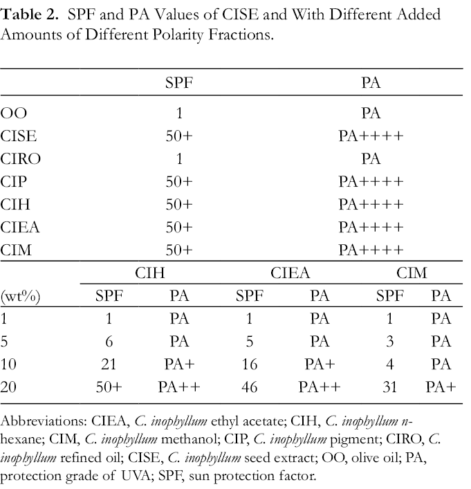
Abbreviations: CIEA, C. inophyllum ethyl acetate; CIH, C. inophyllum n-hexane; CIM, C. inophyllum methanol; CIP, C. inophyllum pigment; CIRO, C. inophyllum refined oil; CISE, C. inophyllum seed extract; OO, olive oil; PA, protection grade of UVA; SPF, sun protection factor.
Addition of Pigments in Different Concentrations
In the pigments-adding study, the different seed extract samples of C. inophyllum were added to refined OO at different concentrations (1%, 5%, 10%, 20%, w/v) and then subjected to SPF and PF evaluation to know their UV-protective capability. 4-Methylbenzylidene camphor at the same concentrations was also subjected to SPF and PF assay as a reference.
The results showed that the concentrations of extracts added to the refined OO were positively correlated with both the SPF and PA values observed but without an exact linear correlation. The UV-absorbing character of oleic acid and linoleic acid has been proved by research, 37 and these 2 compounds share high relative proportions in the fatty acid profiles of OO and CIRO samples. However, deduced from this study, the anti-UV function of CISE is mainly attributed to its pigments according to a cross-comparison of the fatty acid profiles and UV-shielding performances of CIH, CIEA, and CIM.
The CISE sample showed a lower relative concentration of oleic acid and linoleic acid comparing with OO but showed higher SPF and PA values than OO. After the separation of CISE into CIRO and CIP, the CIP sample demonstrated considerably elevated SPF and PA values, while CIRO showed a less photoprotective function than CISE. Also, SPF and PA values of pigment samples of CIH, CIEA, and CIM at different concentrations shared a positive correlation.
Furthermore, it was proved that extracts prepared with the 3 solvents of different polarities contained various substances, which led to the different UVA and UVB absorption ratios observed in SPF/PA study. Compared with 4-methylbenzylidene camphor at the same levels of concentrations, CIH and CIEA showed higher SPF/PA values, which proved that C. inophyllum extracts separated by n-hexane and ethyl acetate cover a wide range of UVA and UVB absorption.
Cytotoxicity of C. inophyllum Extracts
Human fibroblast cells exposed to different substances using WST-1 demonstrated a decrease in cell viability with increasing dose. Table 3 shows the no observable adverse effect concentration, half-maximal inhibitory concentration (IC50), and total lethal concentration (TLC) biotoxicity parameters of human skin fibroblasts exposed to CISE, CIRO, and CIP for 4 and 24 hours, respectively. 38 A slight decrease in cell viability was observed after 4 hours of exposure, and a more obvious cytotoxic effect after 24 hours.
Toxicity Values of CISE, CIRO and CIP.

Abbreviations: CIP, C. inophyllum pigment; CIRO, C. inophyllum refined oil; CISE, C. inophyllum seed extract; IC50, half-maximal inhibitory concentration; n/a, not available; NOEAC, no observable adverse effect concentration; TLC, total lethal concentration.
After the 4-hour exposure to CISE and CIRO solution at concentrations from 2500 to 10 000 ppm, regression drawn by viability results and concentrations observed was evaluated with a test of significance. There was no significant correlation between the concentrations of samples and cell viability (P > 0.1). As a result, IC50 values for both samples are shown as n/a. After 4-hours of exposure, the IC50 of CIP was 460.7 ± 12.2 ppm.
In the 24 hours of exposure survey, the IC50 observed was 997.9 ± 12.8 ppm for CISE, 2762.1 ± 75.1 ppm for CIRO, and 198.9 ± 39.3 ppm for CIP. A comparison of cytotoxicities was then set between CISE, CIRO, the in vitro cytotoxicity assay of ZnO studied by Dechsakulthorn et al, 38 and 4-methylbenzylidene camphor; the last 2 are often used as UV filters in commercial sunscreen. After 4 hours of exposure to 4-methylbenzylidene camphor, the observed IC50 was 32.7 ± 0.5 ppm and 32.4 ± 0.6 ppm after 24 hours. In the cytotoxicity assays of 4-methylbenzylidene camphor studied by Broniowska et al, 39 the IC50 values of neuroblastoma (SH-SY5Y) cells at 24 hours of exposure were between 10−7 and 10−8 M. Also, CISE and CIRO showed a lower cytotoxic effect to the nano-size ZnO in the cytotoxicity assays. In the NOAEC assay, after 4 and 24 hours, cells showed apoptosis at a higher concentration of CIP than that of ZnO. As well, IC50 assay results showed that CIP, CISE, and CIRO are less toxic than ZnO to human skin fibroblasts after either 4 or 24 hours of exposure. IC50 values of CIH, CIEA, and CIM isolated from CIP were observed at 214.2 ± 22.5/89±1.5, 161.5 ± 25.0/32.7±2.0, and 188.5 ± 8.1/49.5±0.3 (ppm), respectively, after 4 and 24 hours of exposure.
This assay provides rapid, low-cost, and reliable results. 40 CISE, CIRO, and CIP outperformed ZnO and 4-methylbenzylidene camphor in the preliminary assessment of biotoxicity after 4 and 24 hours of exposure. The relatively low cytotoxicity, but high photoprotective performances of CISE and CIP, showed that C. inophyllum extracts have high potential to become a cosmetic material as an effective UV filter.
Experimental
Materials and Chemicals
Calophyllum inophyllum fruits were collected from the southern part of Taitung County, Taiwan, in late 2019. Around 100 kg of mature fallen fruits with brown and wrinkled appearance were randomly collected from the ground. Fruits were subjected to qualitative selection to remove the moldy, incompletely developed, and damaged ones. The selected fruits were subjected to mechanical crushing to remove the flesh and shell to obtain seeds, which were eventually preserved at −20 °C before the following procedures. Methanol, ethanol, ethyl acetate, n-hexane, and olive oil were obtained from ECHO CHEMICAL (Taiwan) and kept at room temperature. Silica gel was purchased from Merck (Germany). The reagents for cytotoxicity assessment included Minimum Essential Medium (MEM; Simply, cat. no. CC116-0500), qualified fetal bovine serum (Gibco, cat. no. 26140079), sodium pyruvate-100X (Simply, cat. no. CC518-0100), nonessential amino acids-100X (Simply, cat. no. CC517-0100), Antibiotic-antimycotic-100X (Simply, cat. no. CC501-0100) and WST-1 Cell Proliferation Reagent (Roche, cat. no. 05015944001). The UV filter, 4-methylbenzylidene camphor, was obtained from DSM (Netherlands).
Solvent Extraction of C. inophyllum Seeds
The sample preparation protocol was developed by following protocols of former research and previous personal experience. 16 Calophyllum inophyllum fruits were crushed and then oven-dried under 105 °C till the moisture content was down to 15%. The dried seeds were smashed and homogenized with n-hexane for 8 hours to ensure complete extraction. Solid and liquid phases were separated by filtration, and the pellet was again subjected to n-hexane extraction following the previous procedure. Both n-hexane solutions were combined and the solvent removed in a vacuum concentrator (BÜCHI, R-215, Switzerland) to obtain the seed oil (CISE). The solvent-extracted seed oil was mixed with ethanol to extract the pigments, forming one layer of pigment (CIP) and another of refined oil (CIRO). The pigments extract was removed and purified in a vacuum concentrator (BÜCHI, R-215, Switzerland) till a purified seed cake was obtained, while the refined oil was kept for ongoing analysis. The seed cake was placed into a 1 kg silica gel column and separated into soluble and insoluble fractions with n-hexane. The insoluble part was further extracted with ethyl acetate and methanol. Finally, the various solvents were removed in a vacuum concentrator obtaining paste-like samples ready for analysis.
Characterization of CISE
An UV-Vis spectrometer (JascoV-750) was used to analyze the characteristic absorption of the sample. The sample was measured at 250‐400 nm using a 1 mL cuvette with n-hexane and ethanol as blank reference values, respectively. The sample was also analyzed by ATR-FTIR spectroscopy (FTIR-2000, Varian). The resolution of the spectrometer was 4 cm−1, the number of scans 32, the spectral range 4000-600 cm−1, and the crystal material of the ATR optical attachment ZnSe. A gas chromatograph (Hewlett Packard 5890 Series II GC) was used to analyze the fatty acid composition and content of fatty acids in the samples using a DB-5 capillary column (30 m × 0.25 mm×0.25 μm, Agilent Technologies). The inlet temperature was 120 °C, column oven temperature 250 °C, nitrogen was the carrier gas at a flow rate of 1 mL/min, the detector temperature 260 °C, and with an FID.
Spectroscopic Evaluation of UV-Protection Performance
The determination (measurement) of SPF and PA were conducted with a UV-Vis spectrophotometer with an integrated sphere, and calculation software provided by Jasco V750 (Japan). The sample was placed in a quartz cell containing 2 µL/cm2 analytes, and the analytic spectrum was prepared from 290 to 400 nm with a spectral resolution of 1 nm. An empty quartz cell without analytes was also tested as a control. As for the UV Shield Factor calculation, the same quartz cuvette used for the analytes was employed and surveyed at wavelengths in the range 290-400 nm, which includes the UVA and UVB spectra. The SPF equivalent value was calculated by the following equation:

where E(λ) is the radiation intensity of sunlight (AS/NZA 4399:1996), R(λ) is the CIE reference erythema action spectrum (CIE J.6: 17‐22, 1987), and T(λ) is the diffuse transmittance spectrum (%). If the value is greater than 50, the SPF is defined as 50+. The PA equivalent value is calculated from the following equation:

where E(λ) is the radiation intensity of sunlight (AS/NZA 4399:1996), M(λ) is the minimum sustained immediate blackening action spectrum (1.00), and T(λ) is the diffuse transmittance spectrum (%). The PA values are classified into 5 categories according to their PA equivalent value and are labeled corresponding to their grade: “PA” represents PA equivalent value of less than 2; “PA+” represents PA equivalent value between 2 and 4; “PA++” represents PA equivalent value between 4 and 8; “PA+++” represents PA equivalent value between 8 and 16, and “PA++++” represents PA equivalent value of more than 16.
Cytotoxicity Assessment
The WS1 cell strain (human skin normal fibroblast cells, BCRC 60300) was maintained in MEM at a concentration of 5 × 10⁴ cells/mL. WS1 suspension (100 µL) was seeded in triplicate in 96-well tissue culture plates and incubated at 37 °C under 5% CO2 in a humidified incubator for 24 hours.
After incubation, the upper-medium layer was removed. Following this, the C. inophyllum extract samples, diluted with dimethyl sulfoxide (DMSO) to achieve homogeneous concentration, were added to the cells (100 µL each). After 4 and 24 hours, WST-1 reagent (Roche) solution was added to the cell culture (100 µL cell culture medium plus 10 µL WST-1) for an indication of cell viability. The plate was read by a microplate reader (Tecan Sunrise, Austria), and the absorbance ratio was measured at a wavelength of 450 nm and a reference wavelength of 690 nm after 4 hours of incubation. Abs(sample) and Abs (DMSO) are the absorbances of sample extract and DMSO, which served as the blank or 0 ppm solution. Viability was calculated as a ratio of Abs(sample) to Abs(DMSO) and the average value was calculated from three independent experiments (means ± SD). The correlation between cell viability and concentrations of samples was evaluated by analysis of variance, and the F test by adopting SPSS software 12.0 (SPSS Inc, Chicago). The significance level used was P < 0.05.
Footnotes
Acknowledgments
The authors would like to thank Fu-Ching Ma for valuable suggestions and Zheng-Hui Lai of Taimalee Research Center, Taiwan Forestry Research Institute for helping in the collection of the plant materials. The authors also thank Chun-Ying Liao, Po-Feng Lin and Huei-Wun Fong for sample preparation and their technical assistance.
Declaration of Conflicting Interests
The author(s) declared no potential conflicts of interest with respect to the research, authorship, and/or publication of this article.
Funding
The author(s) disclosed receipt of the following financial support for the research, authorship, and/or publication of this article: We appreciate the financial support under grant no. 109AS-10.5.1-FI-G2 from Taiwan Forestry Research Institute, the Council of Agriculture, Taiwan.