
Abstract
Orthosiphon aristatus (Blume) Miq. is traditionally used for wound healing in South East Asia and scientifically proven for its antidiabetic potential. Wounds due to diabetes, especially diabetic foot ulcer (DFU), always involve a complicated healing process. The present work aims to review the information on the rationale of the phytochemicals from O. aristatus in promoting DFU healing. The findings showed that the DFU healing potential of O. aristatus was characterized by a reduction in the blood glucose level, mainly attributed to the significant concentration of constituents such as caffeic acid, rosmarinic acid, and sinensetin in the plant extract. These phytochemicals possibly induce insulin secretion and sensitivity, improve the lipid profile, and stimulate glucose uptake. Furthermore, the healing effect may also be contributed to the antioxidant, anti-inflammatory, and antihyperglycemic properties of the plant. The roles of phytochemicals have been systematically postulated in the 4 phases of the healing process. Moreover, no adverse toxic sign or abnormality has been reported upon oral administration of the plant extract. This suggests that O. aristatus extract could be a potential diabetic wound healing phytomedicine for further preclinical and clinical studies.
Introduction
About 10% of 250 000 plant species have been scientifically studied and found to have potential uses in healthcare. 1,2 Some of the plant-derived compounds such as vinblastine, vincristine, taxol, and digoxin have been synthesized because of the high demand and limited supply from natural resources. 3,4 Recent development of traditional medicines has led to the extensive use of natural products and their derivatives which contribute to more than half of the total medicines consumed worldwide. 5
One of the widely used medicinal plants in Asia is Orthosiphon aristatus (Blume) Miq. This herb, belonging to the family Lamiaceae, is a traditional medicinal plant originating from tropical East Asian countries such as Thailand, Indonesia, Vietnam, and Malaysia. 6 The leaves of the herb are popularly consumed as tea to promote overall well-being. A decoction of the herb is traditionally prepared as a remedy for arteriosclerosis, kidney stones, diabetes, and nephritis. 7 The herb is also extensively used as an ethnomedicine to treat rheumatism, hypertension, tonsillitis, epilepsy, menstrual disorder, gonorrhea, syphilis, renal calculus, gallstone, lithiasis, edema, eruptive fever, influenza, hepatitis, and jaundice. 8 -12
Many pharmacological studies have demonstrated that this herb exhibits antimicrobial, antioxidant, hepatoprotective, antigenotoxic, antiplasmodial, cytotoxic, cardioactive, antidiabetic, and anti-inflammatory activities. 13 The pharmacological properties are most probably attributable to the presence of various groups of phenolic acids, 14 -19 terpenoids (diterpenes and triterpenes), 7,20 -22 flavonoids, 14,15 and benzochromenes. 23 Phenolic compounds, such as caffeic acid, rosmarinic acid, sinensetin, and eupatorin, have been frequently reported to be chemically bioactive constituents of O. aristatus. 7,24 -27 These compounds are likely responsible for the wound contraction and enhancement of the rate of epithelialization. 28 -33 Moreover, O. aristatus is popularly known as an alternative antidiabetic medicine, specifically for type II diabetes. 34 Many in vitro and in vivo studies have also been conducted to evaluate the antidiabetic activity and toxicity of O. aristatus. 35 -39 Hence, this current review aims to rationalize the potential of O. aristatus as a phytomedicine for the healing of diabetic food ulcer (DFU), mainly focusing on the role of bioactive phytochemicals, their mechanisms of action, and toxicity studies on diabetic-induced animal models.
An intensive literature survey was conducted using the following databases: PubMed, Google Scholar, Scopus, and Science Direct. The keywords used for the search were “Orthosiphon aristatus,” its synonym “Orthosiphon stamineus,” “phytochemical,” “caffeic acid,” “rosmarinic acid,” “sinensetin,” “antimicrobial,” “antidiabetic” and “diabetic foot ulcer healing.” No limit was set for the time frame of published articles in order to retrieve all relevant papers, and the last search was performed on February 21, 2020.
Wound Formation and Healing
Skin is the body’s largest organ and plays a crucial role as a protective barrier against environmental insult. 40 Wounds are a type of injury that results in either the opening or breaking in the epidermis layer of skin. 41,42 An acute wound is a tissue injury for which the reparative process follows an orderly process to sustain restoration and functional integrity. Usually, an acute wound can be healed within 3 weeks, starting from the initial insult. 40,43 Possibly, an acute wound can progress into a chronic wound when the healing process is delayed by up to 12 weeks. This will result in the interruption of all skin layers and a delay in closing the wound gap. A chronic wound does not follow the normal stages of healing and requires a prolonged time to heal or it will recur frequently. 44 -46 Chronic wounds usually happen due to the complication of some diseases such as diabetes, spinal cord injuries, and Pick’s disease. 40,47
Wound healing is a natural body reaction in response to an injury involving biochemical and physiological phenomena that behave in a synergistic manner. Wound healing occurs in 4 stages. The 4 overlapping district phases of healthy wound healing can be categorized as homeostasis, inflammation, proliferation, and remodeling. 48,49 The first phase is homeostasis, which controls excessive blood loss from the damaged vessels. During the homeostasis stage, the body will release chemical mediators and intercellular messengers (growth factors) to begin the wound cleaning and healing process.
The second phase of the wound healing process is inflammation and debridement. After stopping blood loss (coagulating), the body will immediately send plasma proteins, blood cells, and antibodies to the wound area as a defense mechanism at the inflammatory phase. This causes swelling, pain, fever, and redness around the wound site, and the symptoms could last for up to 2-4 days. 46,48,50
The proliferation stage starts when the inflammation subsides. Dermal fibroblasts migrate to the wound site and start granulating until the wound is healed. 46,48,50 The scab sloughs off when the epidermis has been restored to standard thickness during the remodeling or maturation phase. Therefore, the third phase is re-epithelialization, which includes proliferation, migration, and differentiation of squamous epithelial cells in the epidermis. The final stage of wound healing involves collagen deposition and remodeling within the dermis layer of skin. 51 Ultimately, collagen fibers become more organized, fibroblasts decrease in number, and blood vessels are restored to normal. 46,48,50 Understanding the process of wound formation and healing would be of importance in the search for natural alternatives for addressing this complication.
Complications of Diabetic Foot Ulcer
Diabetic patients are facing a complicated wound healing cascade. The healing process is delayed and disrupted from following the normal wound-healing process. 44,52 The healing process of diabetic wounds is relatively slow. Previous studies explained that a high glucose concentration was the main factor inhibiting the wound-healing process and was always associated with a prolonged inflammatory phase. 46 This is because the cell walls become rigid, which causes difficulty for blood flow through small vessels at the wound surface. This also obstructs the permeability and flow of red blood cells. Such a condition will deteriorate oxygen release and nutrient deficit at the wound site. 53 When this happens, blood glucose will be elevated, and chemotaxis and phagocytosis are being agonized to control the infection in the wound area. Consequently, this delays macrophage introduction and diminishes leukocyte migration, as well as prolonging the inflammatory stage in the wound healing cascade. 53
DFU is characterized by the presence of a full-thickness, prolonged wound below the ankle of people with diabetes, or a lesion of the foot penetrating through the dermis layer. 54 DFU does not follow the orderly process of wound healing. It is always associated with poor glycemic (blood glucose) control. A study showed that 49% of participants who had foot ulcers had a glycated hemoglobin (glycemic measure) level above 8.4%. 55 Thus, chronic hyperglycemia appears to be one of the most important factors in the development and delayed healing of DFUs. 56 -59
Hyperglycemia results in the activation of the polyol pathway, nonenzymatic glycosylation, and formation of advanced glycation end products (AGEs), diacylglycerol-(DAG) protein kinase C pathway, and overactivity of the hexosamine pathway. 60,61 All 4 mechanistic pathways will lead to mitochondrial overproduction of reactive oxygen species (ROS). 62 ROS are known to promote cellular dysfunction, thus leading to damage of deoxyribonucleic acid synthesis, lipid and amino acid oxidation, and enzyme inactivation involved in metabolic function. Moreover, hyperglycemia also leads to the activation of an inflammatory response via the activation of nuclear factor kappa-light-chain-enhancer of activated B cells (NF-κB). 63 -65 All these factors delay the healing of foot ulcers. Orthosiphon aristatus and related plant species in the same family have been scientifically proven to lower blood glucose level 66,67 and also possess antioxidant 32,68 and anti-inflammatory 69 -71 activities. Therefore, O. aristatus and its phytochemicals can be a potential lead in healing DFU.
Low Glycemic Index Helps Diabetic Foot Ulcer
It is known that the glycemic measure is frequently poor in people with diabetes, and, therefore, normoglycemia is important for managing foot ulcer, in the belief that it will enhance healing. 72 The adverse effects on cellular immunity and infection can be reduced by controlling the blood glucose level. 73
Several observational studies found a positive correlation of glycemic control and wound healing. 74 -76 Type 2 diabetic patients with proper glycemic control could have a 35% reduction of amputation risk in the lower extremity of the body. 77 Intensive glycemic control also leads to a reduction in the progression and development of microvascular (small vessel) complications, including diabetic peripheral neuropathy. 78
The wound morphology and proliferation of fibroblasts is abnormal for people with DFU. There is also a glucose-dependent reduction of keratinocyte proliferation and differentiation. 79,80 Both insulin and insulin-like growth factor-1 were observed to have a beneficial effect on wound healing in experimental animals. 81,82 Therefore, a low glycemic index is strongly recommended for wound healing. This is because hyperglycemia, insulin resistance, dyslipidemia, and oxidative stress play a dominant function in the pathogenesis of DFU. 73,83,84
Herbs With Diabetic Foot Ulcer Healing Potential
The wound healing potential of a plant extract may be due to the presence of bioactive phytochemicals which have been reported to improve its repair mechanism. Many medicinal plant species synthesize equivalent or closely related compounds with similar biological properties and share the same biological targets and pathways. For example, acemannan from Aloe vera, hydroxysafflor yellow A from Carthamus tinctorius, polysaccharides from Ganoderma lucidum and Sanguisorba officinalis, phthalide lactones and alkaloids from Ligusticum striatum, saponins from Panax ginseng, shikonin and arnebin-1 from Lithospermum erythrorhizon, salvianolic acid from Salvia miltiorrhiza, and alkaloid and stilbenoid from Stemona tuberosa have been well characterized and demonstrated to exhibit the properties of wound healing. Salvianolic acid was also detected in O. aristatus in the study of Nuengchamnong et al. 17 The compounds mostly target mitogenic pathways (eg, phosphokinase B, phosphatidylinositol-3-kinase, SMAD, and cyclins), proinflammatory NF-κB pathways (eg, caspases, interleukins, tumor necrosis factor-α, and tumor growth factor-β1), angiogenesis pathways (eg, vascular endothelial growth factor), extracellular matrix synthesis (eg, matrix metalloproteinases), and differentiation pathways (eg, α-smooth muscle actin), which are the key routes in the mammalian wound healing cascade. 5
As suggested by the Chinese Pharmacopeia, 85 the combination of Cornus officinalis (dried ripe sarcocarp of Fructus Corni), Schisandra chinensis (dried ripe fruit of Fructus Schisandrae Chinensis), Poria cocos (dried sclerotium of Poria), Alisma orientalis (dried tuber of Rhizoma alismatis), and Dioscorea opposita (dried rhizome of Rhizoma dioscoreae) are traditionally used for diabetic treatment, either as a single herb or a cluster of herbs in a traditional formula (Liuwei Dihuang Wan) for diabetes treatment. 86 They are also commonly used in traditional Chinese medicine for the treatment of antidiabetic foot ulcer with proven clinical efficacy. The effectiveness was associated with the normalization of glycemic control in diabetes. 87 A simple herbal formula consisting of Astragalus spp. (Astragali Radix) and Rehmannia glutinosa (Rehmanniae Radix) could expedite the healing of DFUs by inducing gene expression implicated in fibroblast regeneration, angiogenesis, and anti-inflammation, thus promoting vascularization and granulation, as well as modulating the inflammatory response. 5
Involvement of Phytochemicals in the Healing Mechanism
The majority of phytochemicals found in O. aristatus are phenolic acids, flavonoids, and terpenes, as presented in Table 1. The major constituents are rosmarinic acid, eupatorin, and sinensetin. 16,24 Flavonoids (sinensetin) and terpenoids (limonene, borneol, linalool, camphor, and eugenol) are known to promote wound healing due to their antimicrobial properties. 88 -91 These compounds have remarkable antioxidant and antiulcer activities. 92 The contribution of phytochemicals in O. aristatus in wound healing can be explained in Figure 1. The figure postulates and explains the role of each class of phytochemicals in detail. In the homeostasis phase, triterpenes (oleanolic acid and ursolic acid) help in wound healing by producing and activating inflammatory mediators and growth factors, and thus enhancing wound contraction and the rate of epithelialization. 93 -95 Since the inflammatory phase is overlapping with the proliferative phase, the presence of excessive ROS may delay the wound-healing process. Plant-derived antioxidants, such as triterpenes, flavonoids, and phenolics, can be potent radical scavengers, and thereby preventing the damage due to free radicals during the wound-healing process. 33,96 Triterpenes also function to modulate the production of ROS in the wound microenvironment and induce cell migration, cell proliferation, and collagen deposition, thus accelerating the process of tissue repair. 97 Phenolics (chicoric acid, lithospermic acid, and rosmarinic acid) and flavonoids (eupatorin) have been shown to significantly reduce tissue lipid peroxidation level. 98 -100 This will enhance the viability of collagen fibrils by increasing the strength of collagen fibers, preventing cell damage, and accelerating DNA synthesis. 51 During the remodeling phase, caffeic acid was found to help an increase in collagen synthesis in fibroblast cells and controlling melanin production by inhibiting tyrosinase activity. 101 A group of researchers from Indonesia prepared an O. aristatus-based functional drink and reported that it could restrain the increase in blood glucose and inhibit the damaging rate of pancreatic beta cells in diabetic mice. 102 One of the reported bioactive compounds in the functional drink was sinensetin, besides other plant constituents from the polyherbal formulation.
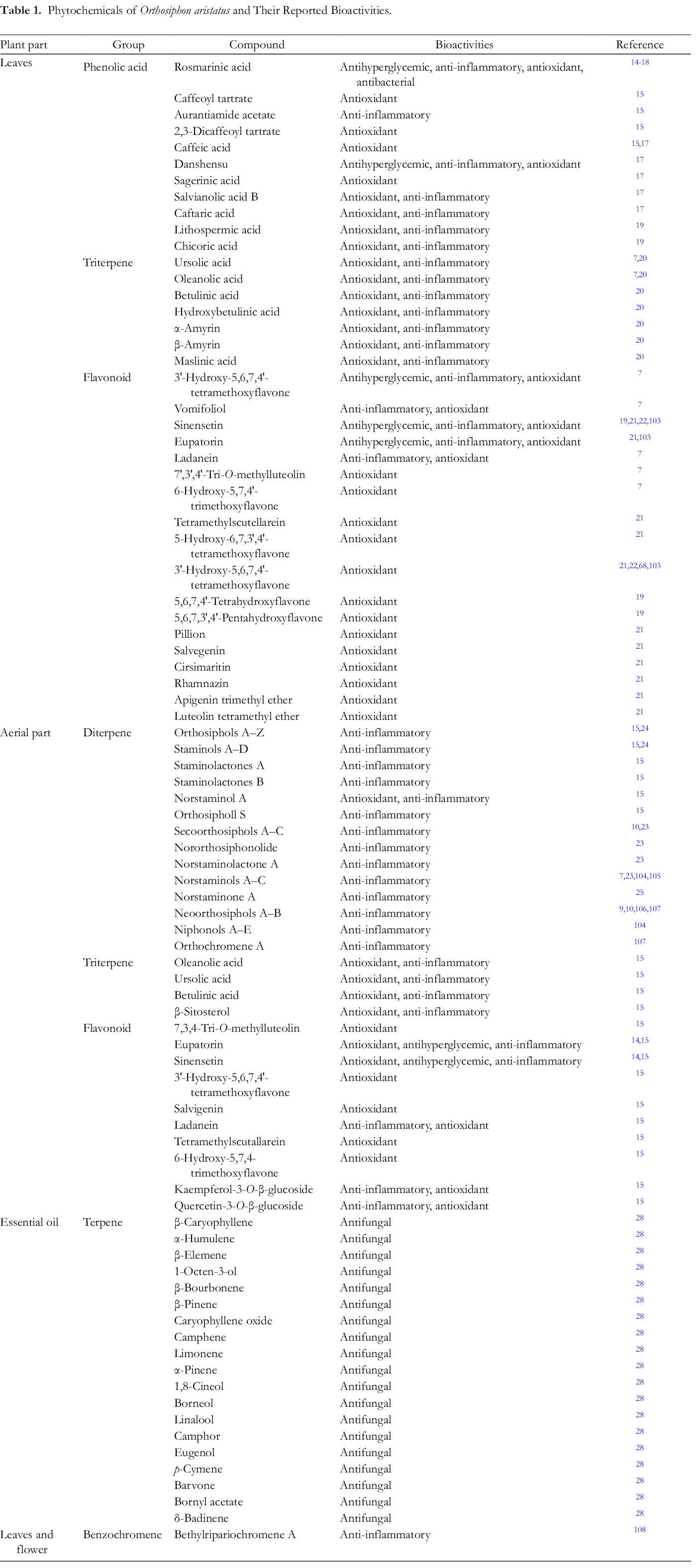
Phytochemicals of Orthosiphon aristatus and Their Reported Bioactivities.
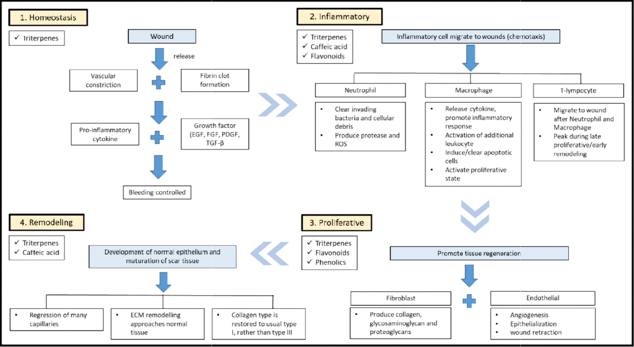
The contribution of phytochemicals in Orthosiphon aristatus extract in wound healing. In the homeostasis phase, triterpenes in O. aristatus are believed to enhance wound contraction and the rate of epithelialization. The inflammatory phase is overlapping with the proliferative phase, and thus, triterpenes, flavonoids, and phenolics can be potent radical scavengers to enhance the wound-healing process by inducing cell migration, cell proliferation, and collagen deposition, enhancing the viability of collagen fibrils to increase the strength of collagen fibers and accelerate the process of tissue repair. In the remodeling phase, caffeic acid helps to increase collagen synthesis in fibroblast cells and control melanin production by inhibiting tyrosinase activity.
Antihyperglycemic Property of Plant Extract
Orthosiphon aristatus is popularly known as an antidiabetic alternative medicine for type II diabetes. Many in vitro and in vivo studies have been conducted to evaluate the antidiabetic activity and toxicity of O. aristatus, mostly using aqueous or aqueous ethanol extract of the plant leaves. The 50% ethanolic extract of the herb was found to exert in vitro antidiabetic activity by inhibiting α-glucosidase (half-maximal inhibitory concentration [IC50] 4.63 mg/mL) and α-amylase (IC50 36.70 mg/mL). 34,66 Earlier, a 14-day oral treatment was carried out using an aqueous extract of the herb. Investigation was conducted on plasma glucose and lipid profile in normal and streptozotocin-induced diabetic Wistar rats. The results showed that the administration of O. aristatus aqueous extract at 1000 mg/kg exerted hypoglycemic and antihyperglycemic effects. The reduction of plasma glucose levels in both euglycemic and hyperglycemic animals was also observed when the plant extract was administrated orally at 200-1000 mg/kg. 13,109 A similar finding was also obtained from the study conducted by Sriplang et al 110 who used an aqueous extract of O. aristatus to alleviate hyperglycemia and improve the lipid profile in diabetic rats. The extract at 1 g/kg was effective in decreasing the plasma glucose concentration, and the response was close to the result of glibenclamide (5 mg/kg). By the end of the study, plasma triglyceride concentration was lower in the extract-treated diabetic rats than untreated rats. 110 Furthermore, plasma high-density lipopolysaccharide-cholesterol concentration was significantly increased in diabetic rats treated with the extract. Moreover, the plant extract (100 µg/mL) could stimulate glucose-induced insulin secretion. Therefore, the use of O. aristatus extract is beneficial to diabetic patients, especially for those who have a defect in insulinotropic response. 110 -112
Orthosiphon aristatus aqueous extract was given orally to streptozotocin-induced Sprague Dawley rats. The experiments were conducted to evaluate the potential of the extract for managing maternal hyperglycemia and to understand the mechanism of actions in lowering blood glucose levels. The extract was effective in lowering the blood glucose level in both nonpregnant and pregnant rats, partly via the stimulation of insulin release which could probably be triggered by several peptide interactions such as ghrelin and glucagon-like peptide 1. Moreover, no sign of toxicity and mortality was recorded on nonpregnant and pregnant rats throughout the study. This indicated that O. aristatus did not induce systematic toxicity. 37 The researchers also suggested that this herb is likely to be a potential antidiabetic agent to treat glucose intolerance during pregnancy. The antidiabetic activity of O. aristatus extract was most probably due to the presence of rosmarinic acid and eupatorin. Chemical screening of the extract showed it to have phenolic and flavonoid contents of 13.24 ± 0.33 mg/g and 1.73 ± 0.14 mg/g, respectively. 110 The researchers suggested that sinensetin could be the compound responsible for the glucose reduction with an IC50 value (50% inhibition) of 0.66 g/mL for α-glucosidase and 1.13 mg/mL for α-amylase. They also stated that sinensetin outperformed the reference drug, acarbose. 66 However, the blood glucose reduction might not be caused by sinensetin only because the water extract has an almost undetectable amount of sinensetin. 14,24
Based on bioactivity-guided fractionation, the chloroform fraction of the plant exhibited a blood glucose-lowering effect in fasting, treated rats, when compared with controls, after glucose loading at 150 mg/kg. 35 Terpenoids and flavonoids, including sinensetin, were identified in the crude extract and the chloroform fraction. Hence, these compounds either acted separately or synergistically. Sinensetin and other compounds in the chloroform fraction were also found to be responsible for the antihyperglycemic effect. 35 Another team of researchers reported that the rosmarinic rich fraction of the ethanolic extract could achieve up to 100% inhibition of α-amylase and α-glucosidase; this was about 5 times more active than the antidiabetic drug, acarbose. 113
Antioxidant Property of Plant Extract
Rosmarinic acid, which is an ester of caffeic acid with 3,4-dihydroxyphenylacetic acid (danshensu), is the characteristic plant constituent of O. aristatus. 114 Its content was found to be in the range of 5.1%-29.9% of the total dry leaf weight. 15 Rosmarinic acid is well known for its potential to improve insulin sensitivity, lower plasma lipid level, 115 and its antidiabetic effect. 113,116 The compound is also popular for its anti-inflammatory, antioxidative, antiviral, and antibacterial activities, which had been proven in many in vitro and in vivo studies. 117 In line with the review of Shahidi and Chandrasekara, 118 compounds with a hydroxycinnamyl group are potent antioxidants. Both caffeic acid and rosmarinic acid have a hydrocinnamyl molecular structure with 2 and 4 hydroxyl groups, respectively. This explains the higher antioxidant activity of rosmarinic acid than caffeic acid. 119
The pharmacological properties of the compounds are important to understand the wound-healing mechanism. Detailed studies demonstrated the inhibitory effects of rosmarinic acid on 5-lipoxygenase and 12-lipoxygenase and gene expression of cyclooxygenase-2 (COX-2). 120 -122 The IC50 value of the rosmarinic acid-rich extract on 5-lipoxygenase was 0.69 µg/mL and 3.25 µg/mL for p38α; the compound had only moderate inhibitory effects on TNFα release. 117 Moreover, rosmarinic acid (5-20 mmol/L) showed an anti-inflammatory effect by reducing 12-O-tetradecanoylphorbol-13-acetate-induced COX-2 promoter activity and protein levels. 120
Orthosiphon stamineus has also been proven to have excellent antioxidant properties. The methanol extract of the herb showed a variation in total phenolics ranging from 6.7 to 10.1 mg caffeic acid/g dry weight, in line with the antioxidant activities, which ranged from 55.5% to 84.2%. The antioxidative potency of the methanol extract was comparable to that of quercetin and the synthetic antioxidant, butylated hydroxylanisole (BHA). 68 Research conducted by Akowuah et al 32 also found that different solvent systems with varied polarities resulted in different radical scavenging activities based on the in vitro 1,1-diphenyl-2-picrylhydrazyl (DPPH) assay. The acetone extract showed the highest activity. The free radical-scavenging activities of the extracts were also comparable to those of quercetin and BHA.
The antioxidative potency of the methanol/water extract of O. stamineus has been demonstrated in terms of DPPH radical scavenging, Fe3+ induced lipid peroxidation inhibition, and Trolox equivalent antioxidant capacity in in vitro models. 123 An aqueous extract of O. stamineus exhibited significant free radical scavenging activity with an IC50 of 9.6 µg/mL, whereas the IC50 of a 50% ethanol extract was 21.4 µg/mL. These results showed that O. stamineus possessed high antioxidant activity and could be considered as an immunomodulatory agent. 30
Ultrasound-assisted extraction was used to extract antioxidant compounds from O. stamineus. The antioxidant activities of the extract were evaluated using 2,2′-azinobis-(3-ethylbenzothiazoline-6-sulfonic acid) (ABTS) radical scavenging and DPPH radical scavenging assays. The results were found to be 1961.3 and 2,423.3 µmol Trolox Equivalent Antioxidant Capacity (TEAC)/100 g dry weight (DW), respectively. Rosmarinic acid, kaempferol-rutinoside, and sinensetin were identified by high-performance liquid chromatography-mass spectrometry in the study. 124 This shows again that the high antioxidative capacity of the plant extract could be due to the presence of these phenolic compounds.
Polymethoxy and polyhydroxy flavonoids were found to possess remarkable health-promoting benefits. 125 -127 It is believed that most of the bioactivities are attributed to the chemical structure of the compounds, especially the polyhydroxyl groups as radical scavengers. 128 Polymethoxylated flavonoids are also readily absorbed in the intestine and have been shown to have a wide tissue distribution and metabolic stability. 129
Polyhydroxy flavonoids were found to have better antioxidative activity than their monohydroxy derivatives. This is because polyhydroxy substituted flavonoids can easily donate electrons to scavenge free radicals and have remarkable inhibitory actions on lipid peroxidation and significantly to be more potent antioxidants, being about 2.5-4 times higher than the reference compound Trolox. 127
The polymethoxylated and/or polyhydroxylated flavonoids in O. aristatus are sinensetin, eupatorin, and 3′‐hydroxy-5,6,7,4′-tetra-methoxyflavone (TMF). These compounds were found to be the dominant compounds in the chloroform fractions 69 and were chosen as markers to standardize various leaf extracts. 16,24,68,103,130 Eupatorin and sinensetin were found to have in vivo anti-inflammatory properties. The expression of inflammatory genes such as inducible nitric oxide synthase (iNOS), COX-2, and TNF-α, as well as the production of inflammatory mediators like nitric oxide (NO) and prostaglandin E2 (PGE2), were suppressed in vitro, probably by inhibiting the enzyme activity and the activity of a transcription factor STAT1α. 131
Moreover, an ethanolic extract of O. stamineus containing 1.02% TMF, 3.76% sinensetin, and 3.03% eupatorin possessed an inhibitory activity toward α-glucosidase. 132 In addition, the 50% ethanolic extract of O. stamineus, and isolated sinensetin, were shown to have equal preference for both α-glucosidase and α-amylase. The 50% inhibitory activities were 4.63 and 0.66 mg/mL, respectively for α-glucosidase, and 36.7 mg/mL and 1.13 mg/mL, respectively for α-amylase. 66 Therefore, the presence of polymethoxylated and/or polyhydroxylated flavonoids is very important for the potential of the plant extract as a DFU remedy.
Anti-inflammatory Property of Plant Extract
Previous in vitro and in vivo studies revealed that O. aristatus possesses remarkable anti-inflammatory activity. 131 Its ethanolic extract inhibited lipopolysaccharide (LPS)-stimulated NO, PGE2, and intracellular ROS production in RAW 264.7 cells. Moreover, the plant extract was able to inhibit protein and messenger ribonucleic acid expression of iNOS and COX-2 in LPS-stimulated RAW 264.7 cells. Ursolic acid, detected in the extract by high-performance liquid chromatography, was shown to suppress LPS-induced NO and PGE2 production by inhibiting ROS generation, along with reducing the expression of iNOS and COX-2 in RAW 264.7 cells. Therefore, the ethanol extract of O. aristatus is likely to have promising effects on the metabolic pathway of inflammatory-mediated diseases. 133
The NO inhibitory activity was probably due to the presence of 47 diterpenes isolated from O. stamineus. 134 All the diterpenes displayed significant concentration-dependent inhibition of NO production in macrophage-like J774.1 cells. The activities of the compounds varied depending upon the chemical structure. Although NO is an important signaling molecule, its excessive production triggers tissue damage and the release of proinflammatory cytokines such as tumor necrosis factor, interferon, and interleukin-1. 71
The findings reported by Yam et al 70 justified the traditional use of O. aristatus in treating pain and inflammation. The 50% methanol extract of O. stamineus was found to possess anti-inflammatory and analgesic activities. Oral administration at doses of 500 and 1000 mg/kg significantly reduced hind paw edema in rats at 3 and 5 hours after carrageenan administration and produced significant analgesic activity (P < 0.05) in both the acetic acid-induced writhing test and the formalin-induced licking test.
No Adverse Effects of Plant Extract
Till now, no sign of toxicity has been reported in the literature for O. aristatus extract. Previously, 4 test groups of female Sprague-Dawley rats were treated up to 14 days with a methanolic extract of the plant at concentrations from 0.5 to 5 g/kg body weight. 135 No lethality nor adverse toxic signs were noticed during the experimental period. Abdullah et al 136 also reported similar results, with no death record. The animals that were administered with 5000 mg/kg body weight of the plant extract did not show signs of toxicity during the experimental period. The median lethal dose (LD50) was estimated to be more than 5000 mg/kg body weight, with no relative change in the general behavior, body weight, food and water intake, relative organ weight per 100 g body weight, and hematological and clinical biochemistry analyses. 136 The methanolic extract of O. aristatus was also reported to produce no sign of toxicity. 36 With oral administration at doses of 1250, 2500, and 5000 mg/kg/day for 28 days, no abnormality of internal organs was observed between the treatment and control groups. The oral lethal dose was more than 5000 mg/kg and the no-observed-adverse-effect level (NOAEL) of the methanol extract for both male and female rats was considered to be 5000 mg/kg/day. 36,45 The standardized aqueous extract with a concentration up to 2000 mg/kg/day did not alter pregnancy body weight gain, food and water consumption, and any other sign of maternal toxicity. 137 Therefore, O. aristatus extract was considered to have a NOAEL of up to 5000 mg/kg/day.
Conclusion
DFUs continue to be a significant burden for people with diabetes, caregivers, and the health care system. Traditional Chinese medicines have been used for diabetes treatment and antidiabetic foot ulcer with proven clinical efficacy. The effectiveness was associated with the normalization of glycemic control in diabetes. Thus, proper management of blood glucose levels is the key factor to heal DFUs. Many phytochemicals in O. aristatus have been postulated to promote DFU healing, mainly due to their capability of lowering blood glucose. The presence of compounds such as phenolic acids, flavonoids, and triterpenes are postulated to be involved in the 4 phases of wound healing, due to their remarkable antioxidant, anti-inflammatory, and antihyperglycemic properties. No adverse toxic effect of the plant extract was observed and the LD50 was estimated to be more than 5000 mg/kg body weight. Therefore, O. aristatus extract could be a potential DFU phytomedicine for further pre-clinical and clinical studies.
Footnotes
Declaration of Conflicting Interests
The author(s) declared no potential conflicts of interest with respect to the research, authorship, and/or publication of this article.
Funding
The author(s) disclosed receipt of the following financial support for the research, authorship, and/or publication of this article: The authors thank the Ministry of Higher Education, Malaysia, and Universiti Teknologi Malaysia for giving the research grant of HICoE-4J263 and TDR-07G21.