
Abstract
According to the WHO, cancer is the second leading cause of death globally and the third most common cancer is colorectal. A significant etiological factor for carcinogenesis might be oxidative stress. Chemoprevention by consuming natural antioxidants has great perspectives in the struggle to control cancer because it is available and affordable for the wide population. Studies by diverse research groups discovered that grapes, as well as grape-based products, are exceptional sources of the polyphenolic compound resveratrol, which has powerful antioxidant properties. Despite the great number of publications on the anticancer effectiveness of resveratrol, they were all aimed at studying its action once the condition was established. This experiment was the first to study the dynamics of the anticancer activity of resveratrol in the development of chemically induced colorectal cancer. Administrating resveratrol along with 1,2-dimethylhydrazine (DMH) during 30 weeks led to the inhibition of oxidative stress manifestations, in particular, lipid peroxidation. Our research showed that the level of thiobarbituric acid reactive substances in blood serum was 85.1%, 214.6%, and 276.9% lower on the third, fifth, and seventh months of the experiment in the group of rats that obtained resveratrol, compared with the animals affected only by DMH. In the fifth month of the experiment, we noticed that the GPx activity in blood serum was 1.54 times higher than the DMH-control level. During the next 8 weeks, this indicator decreased. The activity of glutathione reductase increased by 2 times in the seventh month, compared with the DMH-control. Histologically resveratrol decelerated the development of the tumor. After 30 weeks of experiment, rats that were receiving only DMH had developed colon adenocarcinoma in situ. In contrast to them, morphological changes in the colon tissue of the animals that obtained resveratrol + DMH could be characterized as signs of mucous colitis.
Colorectal cancer (CRC) is one of the most prevalent types of cancer, being the third most common type. According to GLOBOCAN appraisals, in 2020, the number of new cases of CRC will expand to over 1.8 million, and the number of deaths will reach 10 502 507. 1,2 Colorectal cancer is an after effect of manifold genetic occurrences. 3 One of the key mechanisms responsible for DNA damage is oxidative stress, 4 which is outlined as a disproportion between the production of reactive oxygen species (ROS) and reactive nitrogen species (RNS) and the efficiency of enzymatic and nonenzymatic antioxidant protection. 5
The conception that oxidative stress might be a significant etiological factor for carcinogenesis is receiving increasing support. 6 Several routes trigger CRC pathogenesis, but the core 3 directions are the chromosomal instability (CIN) pathway, the microsatellite instability (MSI) pathway, and the serrated pathway. 7 Of these, the CIN pathway is the major one. It is distinguished by failings in chromosomal segregation, telomere stability, and the DNA damage response. Yet, MSI commences from the loss of DNA mismatch repair and is acquired in about 15% of all CRCs. 8 Some behavioral and environmental risk factors are related to the inception and progression of CRC. The latter are solar UV radiation, automobile exhaust pollutants, vocational exposure to carcinogens and mutagens, bacterial/viral infection, and genetic predisposition. 9 A cross talk between these known risk factors can result in oxidative stress, with an accompanying overproduction of ROS. 10
More risk factors of excessive ROS production include smoking, alcohol, stress, toxins, and inflammatory processes caused by metabolic diseases, as well as lifestyle factors, diet, and dysbiosis, which are also related to the development of CRC. 6,11,12 The rise in ROS level may lead to redox disproportion and cause tumor formation and progression by activation of redox-responsive signaling cascades directed to cell growth promotion. 13
Reactive oxygen species, such as hydrogen peroxide, hydroxyl radical, superoxide anion, and peroxynitrite, arise as by-products of normal energy metabolism from the incomplete reduction of oxygen. 14 They are continuously produced in aerobic organisms from both endogenous, such as mitochondria, cytochrome P450 metabolism, peroxisomes, and inflammatory cell activation, as well as exogenous sources. 15 These species may react with biomolecules, such as lipids, carbohydrates, proteins, and nucleic acids, which interfere with cell function. Therefore, damage may proceed in the chain of nucleotides causing DNA strand breaks, oxidation of purine and pyrimidine bases, genetic instability, and alterations in DNA methylation resulting in chromosomal instability and aneuploidy. This resulting oxidative damage is the first step involved in mutagenesis, carcinogenesis, and aging. 7,16
In numerous systemic diseases, such as obesity, insulin resistance, diabetes, chronic kidney disease, and dementia, as well as in some types of cancer, such as gastric, ovarian, and melanoma, the usefulness of oxidative stress biomarkers has been established. 17 -22 However, still, little is known about the diagnostic benefit of oxidative stress parameters in patients with CRC. 1
Because of evolving trends in the incidence of cancer of various organ sites, doctors are in need of extra methods to control malignant tumors in humans. Intervention or prevention of cancer by consuming dietary constituents, a strategy defined as chemoprevention, has great prospects in our struggle to control cancer because it is simple and economically viable. Several epidemiological studies have shown that populations with diets rich in fruits and vegetables generally have lower cancer incidence. Therefore, the efforts of researchers worldwide have focused on identifying, characterizing, and providing a scientific basis for the anticancer efficacy of various phytonutrients. Cancer induction, growth, and progression are a sequence of events and various studies have demonstrated that numerous dietary agents hold up these stages. Fruits and vegetables contain several nutritive and non-nutritive phytochemicals with potential cancer chemopreventive activity. Studies by diverse research groups discovered that grapes, as well as grape-based products, are exceptional sources of various anticancer substances, so their regular intake could be profitable for the general population. 23 One such substance is resveratrol.
Although its putative cardioprotective effects were first noted in 1982, only in 1997 was resveratrol isolated in a screen for cyclooxygenase inhibitors and demonstrated to have effective chemotherapeutic and chemopreventive proficiencies, including anti-inflammatory, antioxidant, and anticancer activities. 24,25 Attention to this compound was renewed in recent years after reports that it activates sirtuin deacetylases and prolongs the lifecycles of lower organisms.
In vitro, resveratrol has shown the ability to induce cellular apoptosis 26 and inhibit proliferation 27,28 of CRC cell lines. It acts mainly through the inhibition of cell proliferation, induction of apoptotic mechanisms, and downregulation of K-ras. 14 Despite skepticism concerning its bioavailability, a large number of in vivo experiments show that resveratrol has protective effects in rodent stress and disease models. 24 The anticancer activities of resveratrol are interceded through modulation of several cell-signaling molecules regulating cell cycle progression, inflammation, proliferation, apoptosis, invasion, metastasis, and angiogenesis of tumor cells. 22,29,30 It has been revealed that resveratrol can overcome 1 or more mechanisms of chemoresistance, sensitizing previously resistant cells. 25
Several excellent reviews on animal models of colon carcinogenesis, including chemically induced carcinogenesis, 31 have recently been published. However, in order to detect and interpret correctly all the alterations that occur in the colon mucosa by introducing natural or pharmacological compounds in an animal model, we need to understand both the morphological and molecular development of the CRC process in this model. 32
Numerous studies have proved that, unfortunately, none of the tumor markers is specific. Almost every healthy person under some circumstances can detect a slight increase in these indicators. A significant increase in the concentration of tumor markers is observed in the later stages of the disease when the malignant oncological formation is sufficiently developed.
Thereby, a universal marker that can detect bowel cancer at an early stage of the process to this day has still not been found. Procedures to determine tumor markers such as ELISA or PCR are quite expensive. That is why in the middle- and low-income countries, any of the existing affordable methods to alert the doctor and the patient about the possible neoplastic injury on the early stage of its development are highly appreciated.
Despite the large number of publications on resveratrol anticancer effectiveness, they are all aimed at studying its action once the condition has been established, but it is important to study resveratrol activity in the dynamics of the carcinogenesis. Due to its significant antioxidative activity, resveratrol might have a significant effect on suppressing tumor progression, especially colon adenocarcinoma.
Taking into consideration all the above-mentioned points, our research aim was to answer 2 most important questions:
Can determination of the oxidative stress indicators be helpful in the early diagnostics of the carcinogenesis?
Can consumption of resveratrol from different sources (like food, dietary supplements, or drugs) have a positive effect, not only on the oxidative system, but also on the structure of the organ, injured by the neoplastic process, eg, colon?
Materials and Methods
Animals
The research was carried out on 140 white outbred male rats with body weight 190 ± 5 g. The animals were kept in standard vivarium conditions. Body weights and survival were monitored throughout. Rats had free access to drinking water and basal diet ad libitum. Animal experiments conducted in this study conformed to internationally accepted standards and were approved by the Bioethical Committee of Ternopil National Medical University. All experiments were performed in the animal facility according to institutional guidelines in compliance with the requirements of the “European Convention for the protection of vertebrate animals used for experimental and other scientific purposes. 33 ”
Colorectal Cancer Model
Among all models of chemically induced colon cancer in animals, 1,2-dimethylhydrazine (DMH) seems to be the most commonly used. The DMH model is a well-established, well-appreciated, and widely used model of experimental colon carcinogenesis. It has many morphological as well as molecular similarities to human sporadic CRC. 32
The rats were randomly allocated into 3 groups: 1—control (20 animals), 2—DMH alone (60 animals), and 3—60 animals obtained DMH with resveratrol (Resverasin, Nutrimed Ltd., Ukraine; standardized plant extracts EUSA, France). 1,2-Dimethylhydrazine-induced colon adenocarcinoma was modeled by introducing dimethylhydrazine hydrochloride (Sigma-Aldrich Chemie, Japan, series D161802) dissolved in isotonic sodium chloride solution. The chemical carcinogen was injected subcutaneously into the interscapular region at a dose of 7.2 mg/kg body weight (based on active substance) once a week for 30 weeks. Control group animals were treated with subcutaneous injections of 0.1 mL of physiological saline with the above frequency. 34,35 Every 30 days, 24 hours after the each last scheduled DMH administration, equal numbers of rats from each experimental group were deeply anesthetized with Thiopental (50 mg/kg, intraperitoneally, Arterium, NUA/3916/01/02) and sacrificed by cervical displacement and exsanguination. At the end of the experimental period, colon adenocarcinoma in situ was histologically identified in all DMH-treated rats.
Lipid Peroxidation Determination
Oxidizing agents can transform lipid structure, creating lipid peroxides that result in the formation of malondialdehyde (MDA), which can be measured as thiobarbituric acid reactive substances (TBARS). The concentration of MDA was determined colorimetrically at 535 nm using the thiobarbituric acid method. 36
Reduced Glutathione (GSH) Determination
The concentration of GSH was measured colorimetrically based on the reaction of GSH from the blood sample with 5,5′-dithiobis-2-nitrobenzoic acid at 412 nm. 37
Glutathione Peroxidase (GPx) Activity Determination
The activity of GPx was analyzed colorimetrically at 340 nm based on the reduction of organic peroxides in the presence of nicotinamide adenine dinucleotide phosphate (NADPH) and sodium azide. One unit of GPx activity was determined as the amount of GPx catalyzing the oxidation of 1 μmol of NADPH per 1 minute. 38
Glutathione Reductase Activity Determination
The activity of glutathione reductase (GR) was defined colorimetrically at 340 nm by measuring the decrease in NADPH absorbance. One unit of GR activity was identified as the quantity of GR that catalyzes the oxidation of 1 μmol of NADPH per 1 minute. 39
Resveratrol human equivalent dose (HED) and Administration
The daily HED of resveratrol was calculated using a representative surface area to weight ratios (km) normalization method, 40 which was 20 mg/kg of body weight. An administrated dose of resveratrol was well tolerated and showed no toxic effects.
Histopathology
Colonic tissues were harvested from animals and fixed overnight in 10% neutral buffered formalin. The tissue processing procedure was performed in a histoprocessor LOGOSone (Milestone). For histological analysis, all colon and liver paraffin sections (5 µm thickness) were stained with Hematoxylin and Eosin (Biognost) and evaluated with a light microscope (Nikon Eclipse Ci). The liver was examined by macroscopy and microscopy to determine either the presence or absence of metastasis (absent).
Statistical Analyses
All data are presented as the mean ± standard deviation. The significance of the difference between groups was evaluated with Prism 5.0 software. P < .05 were considered to be statistically significant.
Results and Discussion
Growth and Survival
The average body weight of the control group (Group 1) rats was 190.1 ± 2.8 g; DMH-only treated (Group 2)—(192.9 ± 2.7) g; and DMH + resveratrol (Group 3)—(193.2 ± 2.9) g. During the 30 weeks of experiment, untreated control animals gained approximately 42 g. In the other 2 groups, the weight responses were as follows: Group 2, –54 g and Group 3, +40 g.
At the end of the experiment, the survival of the untreated control animals (Group 1) and of the DMH + resveratrol treated rats (Group 3) was 100%. In the DMH-only treated group, the survival rate markedly differed before and after the fifth month of the experiment. Until week 22 of the DMH administration, the survival of this group was 100%, and after the next 8 weeks it reduced to 66.7%.
The development of a tumor is always accompanied by changes in the oxidation-reduction equilibrium, along with ROS. This leads to the activation of lipid peroxidation (LPO) and violations of the antioxidative system. 41 It is scientifically proved that free radical reaction activities in humans change significantly during tumor development. Activation of LPO in the case of such pathology may be caused by the development of a stress reaction. Stress is a representation of all the body’s adaptive responses to all kinds of stimuli. Long-term stress, especially with the development of pathological processes, causes an increase in the level of ROS. As a result, free radical oxidation processes are activated. All these reactions inhibit the abilities of the cells’ protective systems, which can lead to the development of either necrosis or apoptosis.
In our case, such a stimulus was the injection of DMH, which induced the formation of numerous radical metabolites—active oxidants of biological substrates. They have a significant cytotoxic effect and initiate the processes of LPO and protein peroxidation.
Thiobarbituric acid reactive substance is one of the intermediate metabolites of LPO. It can form polymer molecules with proteins and phospholipids, which lead to an increase in cell membrane permeability and a decrease in the activity of membrane-bound enzymes and phospholipid metabolism rate.
In the condition of DMH-induced carcinogenesis, a credible (P ≤ .05) increase in the level of TBARS in blood serum was observed during the entire experiment. The level of TBARS in blood serum of the affected animals increased by 4.7 times and by 3.8 times compared with the control group animals after 3 months of the experiment; after 5 months, these numbers increased by 5.3 and 4.7 times, respectively. After 7 months of investigation, this indicator was 6.0 times higher than the level in the serum of the control group animals (Table 1).
The Levels of Thiobarbituric Acid Reactive Substances and Reduced Glutathione, and Activities of Glutathione Peroxidase and Glutathione Reductase in Blood Serum of Rats During the Development of Colon Carcinogenesis (M ± m, N = 6).

GR, glutathione reductase; TBARS, thiobarbituric acid reactive substances.
aSignificant changes between the indexes of control and 1,2-dimethylhydrazine-affected animal groups (P ≤ .05).
The functional base for the antioxidant protection system is the glutathione system, which consists of glutathione (GSH) and enzymes, which catalyze the reaction of its reverse conversion (oxidation or reduction)—glutathione peroxidase (GPx) and GR. Reduced glutathione is the main sulfur-containing antioxidant in the animal’s body. It protects the sulfhydryl groups of globin, erythrocyte membranes, and ferrous iron from oxidizing agents. It is a central component of antioxidant protection systems of almost all cells and organs. Its antioxidant effect is related to the transfer of sulfhydryl groups. Reduced glutathione is not only a substrate for chemical reactions but also is needed for a permanent reduction of selenolate groups, located in the enzyme catalytic center, which become oxidized during the glutathione peroxidase reaction. 42
One of the mechanisms that determines the resistance of the body to the negative effects of both exogenous and endogenous toxins is the involvement of glutathione and its related systems into biotransformation and detoxification processes.
It was scientifically proved that changes in GSH concentration, as well as GPx and GR activities, can be used as a marker of negative influence.
We investigated the level of reduced glutathione in rats’ blood serum in the dynamics of the development of induced CRC (Table 1).
In the conditions of induced carcinogenesis, we observed a significant (P ≤ .05) increase in the studied indicator for the first 3 months of the experiment. The level of reduced glutathione began to decrease in the fourth month and its lowest level was observed at the end of the experiment (1.5 times lower than that of the control group).
Long-term activation of glutathione peroxidase is possible only with an appropriately high level of intracellular reduced glutathione (this is demonstrated in our research) (Table 1).
In our opinion, the next decrease in enzyme activity is caused by various failures in the body’s defense mechanisms and a decrease in the functional capacity of the liver to synthesize enzymes de novo in the case of the pathological process. A decrease in the glutathione peroxidase activity compared with the control group of animals was caused by a gradual exhaustion of the reduced glutathione pool during the antiradical activity, as well as by increased sensitivity to О2−, which can inhibit GPx.
Also, the activity of GR was investigated. This enzyme maintains a high intracellular concentration of reduced glutathione due to reduction of the oxidized disulfide form of glutathione involving NADPH. After 2 months long injection of DMH, the activity of GR increased in rats’ serum. This indicates the active involvement of this enzyme in the process of reducing the oxidized form of glutathione. This indicator was by 1.24 times higher than that in the control group in the second month of the study. The activity of GR decreased significantly (P ≤ .05) from the third month of the experiment, and by the end of the research, was 2.23 times lower than normal (Table 1).
After 30 weeks of DMH injection, severe dysplasia of the colon tunica mucosa epithelium was histologically revealed, which was proved by many criteria including nucleic hyperchromism, destruction of epitheliocyte rows, cell atypism, which was manifested by the wide dimension of the nucleal forms and volume, and violation of the nucleocytoplasmic ratio toward the nucleus. The highly differentiated epithelium of the crypts and goblet cells lost its usual outline. The degree of differentiation of the gland linings was different. Goblet cells with signs of atypism and polymorphism, and anaplastic cells were identified. The glands were close to each other. The stroma was poorly developed and contained thin-walled vessels and single lymphocytes. The submucosal stroma was well developed, with slight lymphocytic infiltrate. The smooth muscle myocytes were tightly positioned. Adventitia was full-blooded (Figure 1).
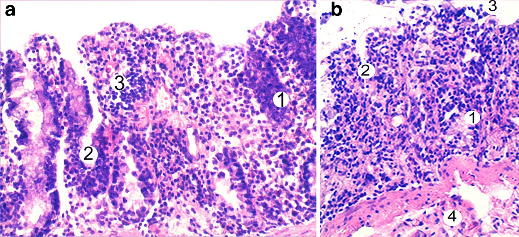
Colon tissue histological changes after 30 weeks of 1,2-dimethylhydrazine administration. (a) Atypia cells of mucosa (1), crypt (2), and lymphocyte infiltration (3). (b) Epithelial dysplasia of the mucous membrane (1), crypt (2), erosion of the mucous epithelium (3), and submucosa (4). Hematoxylin & Eosin, ×200.
Resveratrol (3,4,5-trihydroxystilbene) is a phytoalexin produced by plants, such as Vitis vinifera, in response to different pathogens. The structure of resveratrol includes 2 phenolic rings connected by double bonds, which have cis-(Z) and trans-(T) orientation. The antioxidant effect of resveratrol is the basis of its various pharmacological effects, 43 which are predetermined by its molecular structure. 44 The antioxidant activity of resveratrol is 5 times better than that of beta-carotene, 50 times better than vitamin E, and 20 times better than vitamin C.
Resveratrol has a powerful inhibitory effect on the formation of superoxide anion and hydrogen peroxide in the body. Also, resveratrol can neutralize hydroxyl radicals. 45 The compound suppresses certain processes in cells, which lead to the development and progression of oncological conditions. Resveratrol has shown the ability to inhibit the proliferation of most human cancer cell lines, including breast cancer, prostate, stomach, colorectal, pancreas, and thyroid gland cancer. 14,46,47
We used resveratrol to decrease the processes of LPO, which in the future should help to reduce destructive damage in the mucous membrane of the colon, thereby preventing the development of a neoplastic process in it.
Our research showed that the level of TBARS in blood serum was 85.1%, 214.6%, and 276.9% lower on the third, fifth, and seventh months of the experiment during the usage of resveratrol, compared with the same indicator of animals without administration of this substance (Figure 2). The efficiency of resveratrol treatment on the level of GSH manifested in the third month of development of the pathological condition is shown in Figure 3.

Changes in thiobarbituric acid reactive substances concentration under the conditions of 1,2-dimethylhydrazine and resveratrol + 1,2-dimethylhydrazine administration in the dynamics of the experiment (n = 10 in each group of analysis except the 1,2-dimethylhydrazine-only treated animals on the sixth and seventh months, where n = 7).

Changes in GSH concentration under conditions of 1,2-dimethylhydrazine and resveratrol + 1,2-dimethylhydrazine administration in the dynamics of the experiment (n = 10 in each group of analysis except the 1,2-dimethylhydrazine-only treated animals on the sixth and seventh months, where n = 7).
In the initial stages, the antioxidant caused a decrease in the activity of glutathione peroxidase, because the antioxidant had taken over the function of GPx (Figure 4).
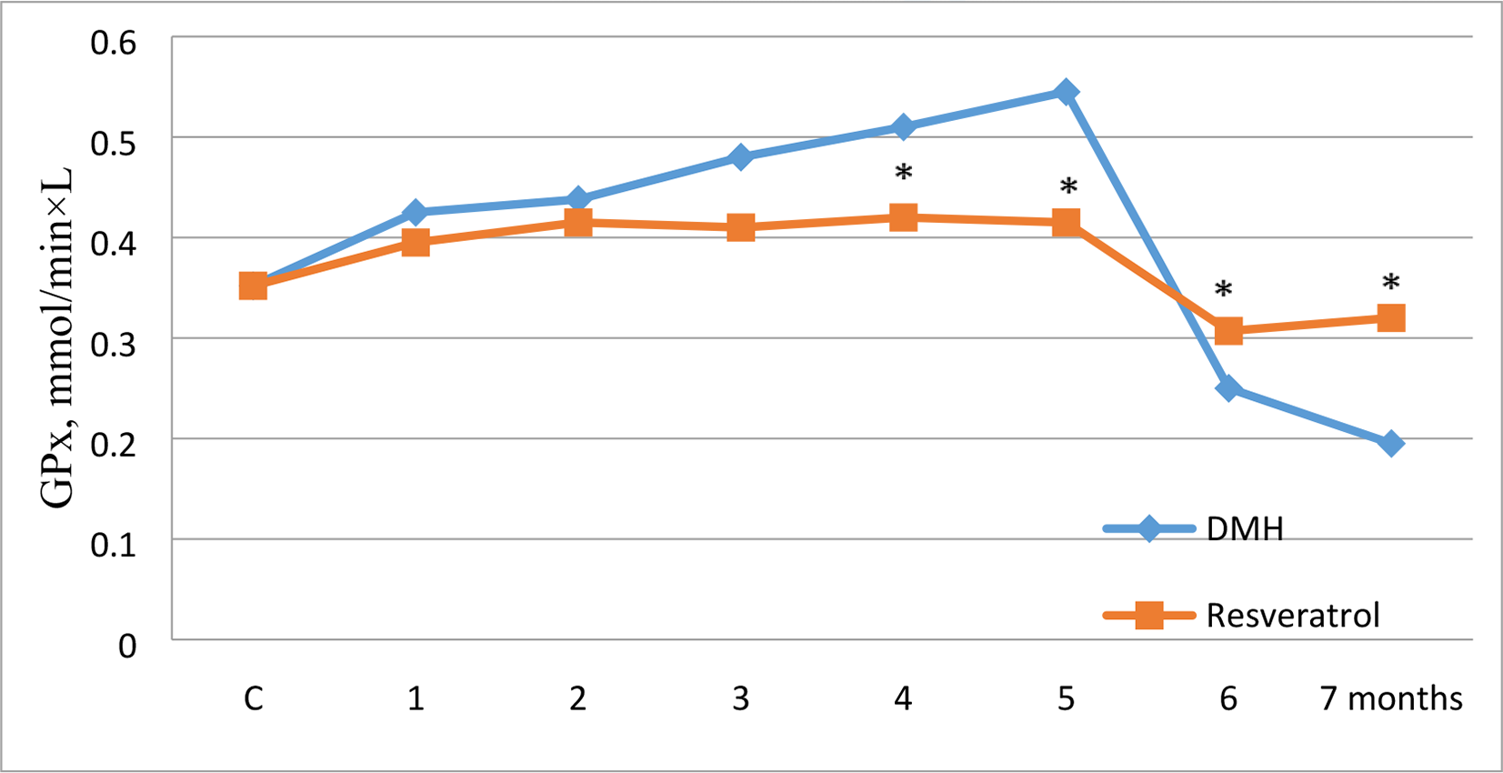
Changes in GPx concentration under the conditions of 1,2-dimethylhydrazine and resveratrol + 1,2-dimethylhydrazine administration in the dynamics of the experiment (n = 10 in each group of analysis except the 1,2-dimethylhydrazine-only treated animals on the sixth and seventh months, where n = 7).
In the fifth month of the development of DMH-induced carcinogenesis, we noticed that the GPx activity in blood serum was 1.54 times higher than that in the control. During the next phases of the experiment, this indicator decreased. In the seventh month, the usage of resveratrol led to restitution of GPx activity in the serum, which was decreased in rats in the sixth and seventh months of cancer development. From our point of view, the studied drug, due to its antioxidant properties, caused a slowdown in the oxidative activity. This led to a significant reduction in endogenous toxin levels in the body, thus decreasing their toxic effects on the liver. Under these conditions, the protein synthesizing function of the liver was able to recover. This induced the production of enzymes of the glutathione system. Thus, the positive effect of resveratrol usage on the activity of GR was established (Figure 5). This was manifested in the fourth month of the research and lasted until the end of the experiment (in the seventh month, this indicator increased by 2 times, compared with the group of rats affected by DMH).

Changes in glutathione reductase concentration under the conditions of 1,2-dimethylhydrazine and resveratrol + 1,2-dimethylhydrazine administration in the dynamics of the experiment (n = 10 in each group of analysis except the 1,2-dimethylhydrazine-only treated animals on the sixth and seventh months, where n = 7).
Colon Tissue
A characteristic feature of histological restructuring of the colon mucosa as a result of resveratrol + DMH administration was the presence of dystrophic changes in the epithelial cells of the glands. The nuclei were predominantly oval in shape with an enlightened nucleoplasm containing 1 nucleus. The goblet cells were small. The cells lining the gland had a single-row arrangement. The lumen of the glands contained mucus with desquamated epithelium. The stroma were well developed with edema and lymphoplasmacytic infiltration. The submucosal, muscular membranes and the adventitia showed slight swelling and mild lymphocytic infiltration (Figure 6).
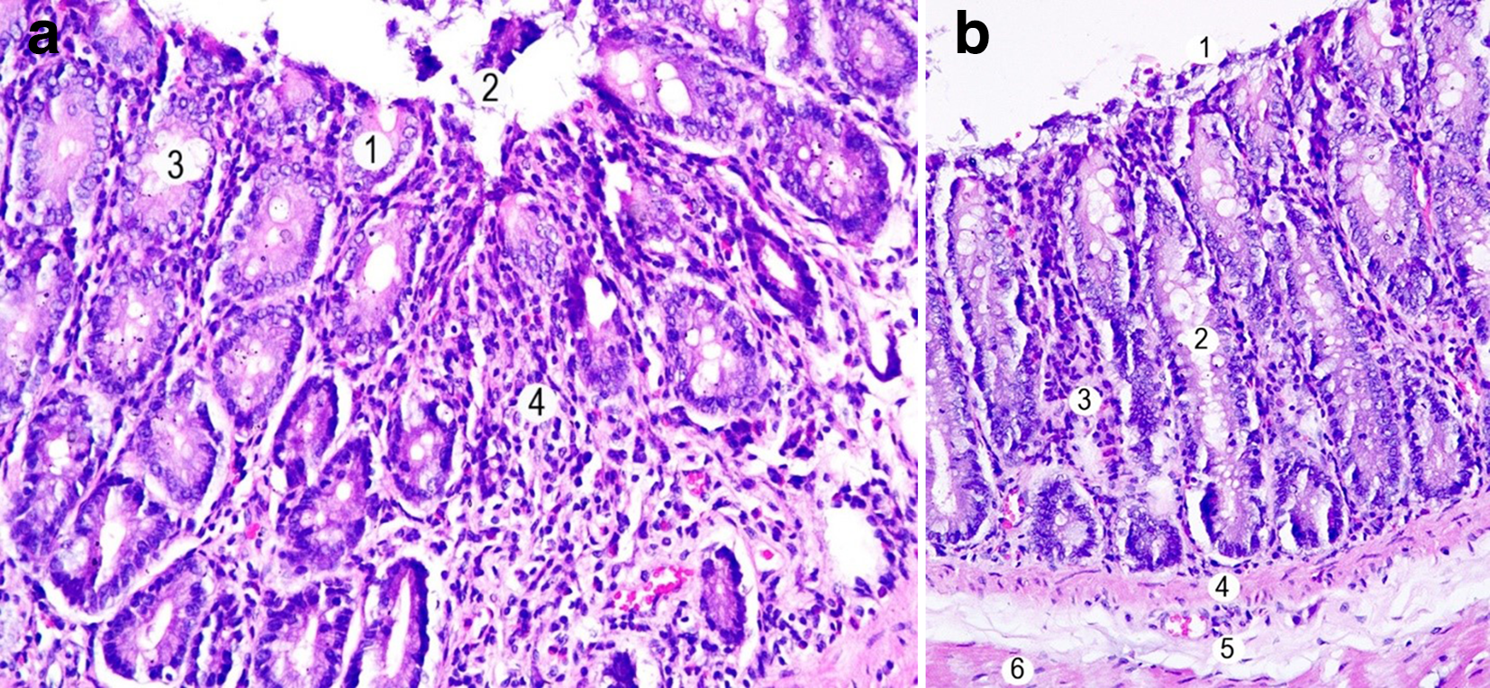
Colon tissue histological structure after 30 weeks of 1,2-dimethylhydrazine + resveratrol administration. (a) Enterocytes (1), desquamated fragments of the epithelium (2), goblet cells (3), and lymphocytic infiltration (4). (b) Desquamated fragments of the epithelium (1), crypt (2), lymphocytic infiltration (3), muscularis mucosa (4), submucosal layer (5), and tunica muscularis (6). Hematoxylin & Eosin, ×200.
Conclusion
Resveratrol, a constituent of grapes and red wine, had a positive effect on the oxidation processes in rats under the condition of induced carcinogenesis. The balance of pro- and antioxidants normalized, increasing the defense systems’ abilities of the affected organism. The effectiveness of the studied drug manifested in the recovery of glutathione system activity, which plays an important role in the realization of antiradical and antiperoxidase protection of cells. All these led to the inhibition of oxidative activity, in particular, LPO.
Prolonged administration of DMH leads to the development of intraepithelial neoplasia in the colon tissue. At the same time, the combined use of resveratrol during the 30 weeks of the experiment slowed down this process, and morphological changes in the colon were characterized as manifestations of mucous colitis.
The obtained results are very promising and they testify to a great perspective for the future usage of resveratrol for the prevention of CRC development and to the possibility of including it in the chemopreventing recommendations on tumors of the gastrointestinal tract.
Footnotes
Acknowledgment
We are greatly thankful to the staff of the Central Research Laboratory of I. Horbachevsky Ternopil National Medical University, Ukraine.
Declaration of Conflicting Interests
The author(s) declared no potential conflicts of interest with respect to the research, authorship, and/or publication of this article.
Funding
The author(s) disclosed receipt of the following financial support for the research, authorship, and/or publication of this article: This work was financially supported by the Ministry of Health of Ukraine (Grant no. 0119U002307).