
Abstract
Nowadays, the incidence of Alzheimer’s disease (AD) and type 2 diabetes (T2D) is increasing at an alarming rate. More and more studies have been investigating the relationship between these two diseases and are trying to find an effective treatment. According to amyloid hypothesis, it is very necessary to find phenolic compounds with catechol moieties, which would inhibit the aggregation of amyloid β (Aβ) and human islet amyloid polypeptide (hIAPP), while also exhibiting antioxidant activity and protective effect. We isolated rosmarinic acid (RA) from the plant Isodon japonicus (Burm.f.) H. Hara. Thioflavin T assay and transmission electron microscopy observation were carried out to evaluate the inhibitory effect of RA, caffeic acid, and 3,4-dihydroxyphenyllactic acid, which are the substructures of RA, on both Aβ and hIAPP fibrillization. 2,2-Diphenyl-1-picrylhydrazyl assay was applied to test the antioxidant activity. RA showed inhibitory effect on both peptides and strong antioxidant activity. These results suggest that the existence of catechol units plays an important role on the inhibitory activity. Therefore, RA will be a promising strategy to prevent AD and T2D.
Alzheimer’s disease (AD) and diabetes are parallel chronic diseases arising from aging, and the incidence is at an alarming rate, thereby becoming a highly concerning global health issue. Insulin resistance is considered as a core feature of type 2 diabetes (T2D), and emerging as a potentially important feature of AD. 1 Some environmental factors and susceptibility genes lead to insulin resistance, which lets diabetes uncontrolled. Too much sugar remaining in the blood may also damage the vessels and organs including the brain, furtherly cause AD. 2 T2D and AD can both lead to cognitive decline, as well as brain hypofunction, memory disturbance, blindness, and so on.
Although the exact pathological defects in AD and T2D remain unknown, the prevailing theories implicate that the aggregation of amyloid β (Aβ) and human islet amyloid polypeptide (hIAPP) leads to such diseases. Aβ is a peptide containing 36 to 43 amino acids, which are formed from cleavage of amyloid precursor protein. hIAPP is a 37-residue regulatory peptide, also the major secretory product of β-cells. Both peptides show a similar aggregation principle, and the aggregation of Aβ causes the extreme shrinkage of cerebral cortex and hippocampus in the brain, while hIAPP aggregation leads to the β-cell destruction.
Isodon japonicus (Burm.f.) H. Hara (Labiatae) is often applied in traditional Chinese and Japanese medicine, and it mainly grows in Northeast Asia. The aerial part of the plant is often used for the treatment of gastrointestinal disorders, 3 while its dried leaves show strong antioxidant activity, 4 antiallergic activity, 5 tanning activity, 6 and melanogenic inhibitory activity. 7 Besides diterpenoids, I. japonicus is also a rich source of phenolic compounds. Rosmarinic acid (RA) is one of the dominant compounds in the plant, which is the dimer of caffeic acid (CA) and with two catechol moieties.
In our previous studies, we reported that 4,5-di-O-caffeoylquinic acid and 3,4,5-tri-O-caffeoylquinic acid strongly inhibited the aggregation of Aβ42 in a dose-dependent manner. 8 Acteoside and oraposide were also found to exhibit strong activities for inhibition of Aβ aggregation and antioxidant activity. 9,10 Some phenolic compounds with catechol moieties were proved possessing the inhibitory activity on the aggregation of Aβ. Recently, we found that glucuronosylated flavonoids with a catechol moiety exhibited potent inhibitory activity of Aβ and hIAPP aggregations. 11 On the other hand, RA shows strong antioxidant activity, and it can be expected to possess more biological activities also. In this study, we aimed on investigating the protective effect of RA, CA, and 3,4-dihydroxyphenyllactic acid (DPA) (Figure 1), which are the substructures of RA, on both AD and T2D. We successfully found that RA displayed inhibitory activity on amyloid aggregation related to both diseases.

Chemical structures of RA, CA, and DPA. CA, caffeic acid; DPA, 3,4-dihydroxyphenyllactic acid; RA, rosmarinic acid. CA, caffeic acid; DPA, 3,4-dihydroxyphenyllactic acid; RA, rosmarinic acid.
To determine the inhibitory activity of RA, CA, and DPA against Aβ and hIAPP fibrillizations, Thioflavin T (Th-T) assay was used to measure the efficiency of these compounds. Among all the samples dealt with different concentration, these compounds expressed the concentration-dependent effect on protein fibrillization. As shown in Figure 2(a)-(c), RA showed the most significant inhibitory activity on Aβ42 aggregation, whose IC50 value was 4.8 µM, while CA exhibited a stronger inhibitory activity (IC50 17.9 µM) than that of DPA on Aβ42 (IC50 31.2 µM). CA is also used as a positive control in our previous reports. 8,10 Figure 2(d)–(f) depicts the inhibitory activity on hIAPP aggregation of RA, CA, and DPA. RA and CA showed the inhibitory effect on hIAPP fibrillization at IC50 3.1 and 57.6 µM, respectively. The effect of DPA was not significantly detected (IC50 >100 µM). But different from Aβ, the formation of hIAPP fibril mostly happened in the first 4-hour incubation, after reaching the maximum of Th-T fluorescence intensity at 4 hours, RA, CA, and DPA started exhibiting the effect on the fibril formation of hIAPP relatively, and after 8 hours the hIAPP entered the stable phase.

Effect of RA, CA, and DPA on the Th-T fluorescence from Aβ and hIAPP. (a) Aβ42 with RA; (b) Aβ42 with CA; (c) Aβ42 with DPA; (d) hIAPP with RA; (e) hIAPP with CA; (f) hIAPP with DPA. Aβ, amyloid β; CA, caffeic acid; DPA, 3,4-dihydroxyphenyllactic acid; hIAPP, human islet amyloid polypeptide; RA, rosmarinic acid; Th-T, Thioflavin T.
Transmission electron microscopy (TEM) images indicated the typical fibrillization of Aβ and hIAPP incubated with or without RA, CA, and DPA. Proteins incubated without these compounds showed more intensive fibril formation than those incubated with the compounds. Figure 3(b) , (f) shows the ability of RA on inhibiting the fibril formation of Aβ42 and hIAPP which was the most significant with a sparse fibrillization image. CA (Figure 3(c) and (g)) and DPA (Figure 3(d) and (h)) also affect the aggregation of both protein fibrils, but not that strong as RA.
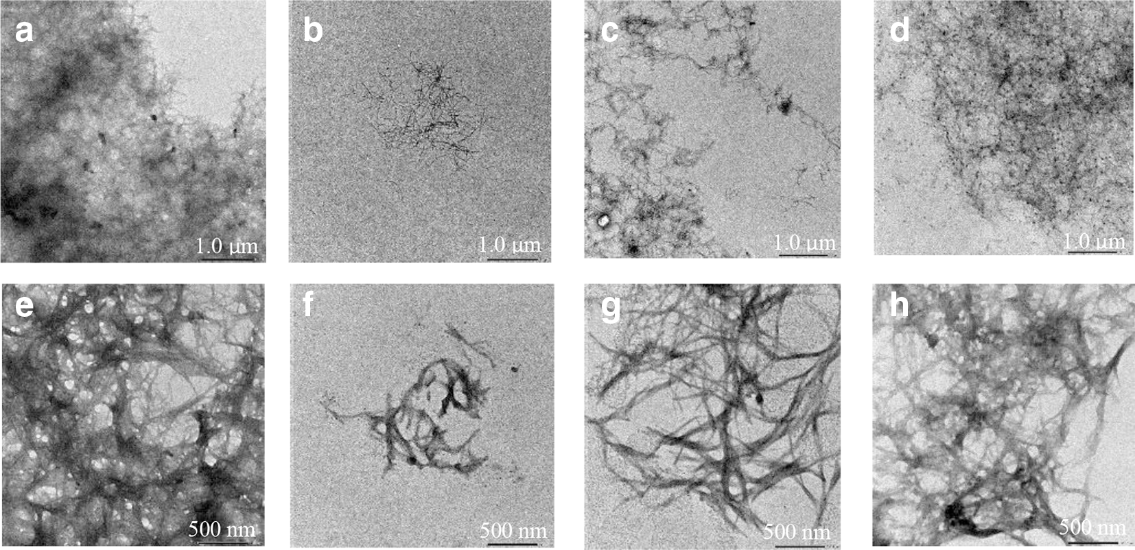
TEM observation of Aβ and hIAPP. (a) 25 µM Aβ42; (b) 25 µM Aβ42 with 100 µM RA; (c) 25 µM Aβ42 with 100 µM CA; (d) 25 µM Aβ42 with 100 µM DPA; (e) 25 µM hIAPP; (f) 25 µM hIAPP with 100 µM RA; (g) 25 µM hIAPP with 100 µM CA; (h) 25 µM hIAPP with 100 µM DPA. Aβ, amyloid β; CA, caffeic acid; DPA, 3,4-dihydroxyphenyllactic acid; hIAPP, human islet amyloid polypeptide; RA, rosmarinic acid; TEM, transmission electron microscopy.
Polyphenols affect the aggregation of Aβ and hIAPP due to the block of the aromatic ring on π–π stacking 12 and the connection of quinone (auto-oxidized from catechol unit) and amyloid monomer, due to Michael addition or the imine formation. 13 In the structure of RA, two catechol moieties are involved, which indicate that more blocks and connection exist between RA and protein chains. Comparing DPA with CA containing a carbon double bond means that is more easily auto-oxidized, possessing a stronger ability on connecting with amyloid monomers.
Besides inhibiting the aggregation of Aβ and hIAPP, the compound with antioxidant activity can also prevent the cell destruction related to AD and T2D. Reactive oxygen species (ROS) is always produced by living organisms because of the cellular metabolism and environmental factors. 14 In the AD and T2D, ROS can lead to the death of neuronal cells in the brain and also to β-cell destruction, which is related to insulin production. Oxidative stress, the shift of the balance between oxidants and antioxidants, contributes to many diseases including neurological disorders, diabetes, and so on.
2,2-Diphenyl-1-picrylhydrazyl (DPPH) radical scavenging assay was carried out to evaluate the antioxidant activity of RA, CA, and DPA, and all these compounds express the concentration-dependent antioxidant activity. As shown in Figure 4, RA shows the strongest antioxidant activity, the antioxidant rate reaching more than 80% with concentrations of 25 and 50 µM, even more significant than the positive control. DPA also shows the strong antioxidant activity (65% at 50 µM), while the antioxidant activity of CA is not significant (25% at 50 µM).

Antioxidant activity of RA, CA, and DPA. CA, caffeic acid; DPA, 3,4-dihydroxyphenyllactic acid; EGCG, epigallocatechin gallate; RA, rosmarinic acid.
Bendary et al. found that the antioxidant activity is related to the compound structure, which in turn depends on the number and position of the functional groups such as hydroxyl group, amino group, and so on. 15 In the structures of RA, CA, and DPA, RA has the most OH groups, and DPA includes more hydroxyl groups than CA. This may be the reason that the antioxidant intensity is ranked in RA, DPA, and CA.
In the present study, some relationship between AD and T2D has been found. Because Aβ and hIAPP play an important role in triggering AD and T2D, we suggested that it be necessary to find the compounds that can inhibit the protein fibrillization and show a strong antioxidant activity. In the structure of RA, CA, and DPA, the catechol moieties showed a significant influence on the inhibition of amyloid aggregation, while the hydroxyl groups expressed strong antioxidant activities. Both inhibitory activity and antioxidant activity may show the protective effect and lead to efficient therapy related on AD and T2D.
Also, as per the several reports on RA about its inhibitory activity toward Aβ and hIAPP aggregation, 16,17 we believed that applying RA in medicine will be a promising strategy to treat and prevent AD and diabetes. The findings have inspired us to conduct more investigation on various biological activities of phenolic compounds from other medicinal plants apart from I. japonicus to get more useful guidelines in relation to search for novel RA-related inhibitors against protein aggregation associated with chronic diseases.
Experimental
Isolation of RA
Isodon japonicus (Burm.f.) H. Hara was purchased from Kanai Co., Ltd. (Tokyo, Japan). After smashing, the dried I. japonicus (100 g) was extracted with MeOH (700 mL ×2) at room temperature for 24 hours. After filtration and evaporation, the MeOH extract was mixed with EtOAc (400 mL ×3) and H2O (400 mL). A portion (2 g) of EtOAc-soluble material (RJ-EA, 4.37 g) was chromatographed over octadecylsilyl (ODS) column (Cosmosil 75C18-PREP, ϕ2.2 × 30 cm, acetone:H2O = 3:7→1:0). RJ-EA-4 (260.5 mg) was separated using ODS column (Cosmosil 75C18-PREP, ϕ1.0 × 30 cm, MeOH:H2O = 3:7→1:0) to afford RJ-EA-4–1~4-8. RJ-EA-4–3 (23.5 mg) was purified by TSKgel ODS-120A, MeOH-1% AcOH in water (30:70) to obtain RA (11.1 mg) and confirmed by 1H NMR spectrum (CD3OD, 500 MHz).
CA and DPA were purchased from FUJIFILM Wako Pure Chemical Corporation, Japan.
Th-T Fluorescence Assay
Aβ42 was dissolved in 0.1% NH4OH and hIAPP (KareBay Biochem Inc., USA) was dissolved in 1,1,1,3,3,3-hexafluoro-2-propanol. The amyloid solution was diluted 10-fold with 50 mM phosphate-buffered saline (pH 7.4). Both peptides were pretreated with 1, 10, and 100 µM of RA, CA, and DPA, in the 96-well microtiter plate (Thermo Scientific), respectively. All the samples were incubated for 24 hours at 37°C. Th-T fluorescence intensity was evaluated at 0, 4, 8, and 24 hours, and found to show an excitation wavelength of 420 nm and an emission wavelength of 485 nm.
Transmission Electronic Microscopy
Aβ42 and hIAPP (each 25 µM) treated with equimolar RA, CA, and DPA were spotted onto a carbon-coated Formvar grid and incubated for 2 minutes at room temperature and air-dried for 5 minutes after washing by H2O twice and 0.4% silicotungstic solution twice. Samples were observed under the JEOL JEM-1400 electron microscope.
DPPH Radical Scavenging Assay
DPPH radical scavenging assay was applied to evaluate the antioxidant activity of RA, CA, and DPA. The positive control epigallocatechin gallate and the three compounds were pretreated to 5, 25, and 50 µM, and 10 µL of each sample was injected into a 96-well plate. Half were treated with 190 µL of DPPH solution [combined DPPH (1.9 mg)/12.0 mL EtOH, 3.0 mL of 2-(N-morpholino)ethanesulfonic acid (MES), and 9.0 mL of H2O], and half were treated without DPPH (combined EtOH 1.0 mL, 3.0 mL of MES, and 9.0 mL of H2O). After a 15 min reaction at room temperature, both samples were examined under the wavelength of 490 nm.
Footnotes
Acknowledgments
We thank Professor Kazuhiro Irie, Associate Professor Kazuma Murakami, and Dr Mizuho Hanaki, Graduate School of Agriculture, Kyoto University for preparing Aβ42.
Declaration of Conflicting Interests
The author(s) declared no potential conflicts of interest with respect to the research, authorship, and/or publication of this article.
Declaration of Conflicting Interests
The author(s) declared no potential conflicts of interest with respect to the research, authorship, and/or publication of this article.
Funding
The author(s) disclosed receipt of the following financial support for the research, authorship, and/or publication of this article: this work was partially supported by JSPS KAKENHI Grant Number JP24580156.