
Abstract
Numerous pharmacological studies on Panax plants have been performed. However, these studies were limited to ginsenosides, which are typical constituents in Panax plants. In our research program to discover novel agents to prevent dementia and improve dementia symptoms, especially Alzheimer’s disease (AD), we investigated the inhibitory activities of essential oil (EO) extracts from 4 Panax plants against β-secretase, amyloid β (Aβ) aggregation, acetylcholinesterase (AChE), and butyrylcholinesterase (BChE). An EO extract of Panax japonicus showed the most potent activity with 51.3% inhibition at 500 μg/mL against β-secretase. Panax ginseng showed the most potent inhibitory activity against AChE and BChE with 70.4% and 84.4% inhibition at 50 μg/mL, respectively. Panax notoginseng extract showed the most potent activity with 57.3% inhibition at 500 μg/mL against Aβ aggregation. From these results, an EO extract of P. ginseng could be an effective agent to improve AD symptoms, while EO extracts of P. japonicus and P. notoginseng could be suitable for AD prevention.
Araliaceae family plants, especially Panax genus plants, are recognized as an important source of crude drugs in traditional Chinese medicine and “Kampo” medicine in Japan. In fact, the genus “panax” means panacea (“cure all”) in Greek. 1 There are 13 species of Panax plants with various pharmacological effects and their active principles have been reported. Unfortunately, active principles have been limited to triterpenoid saponin and ginsenosides, and studies on other constituents have not been reported extensively. In our research program to discover effective agents to improve patients’ quality of life, we focused on aromatic compounds in Panax plants as a new source of pharmacologically active compounds.
Cho et al 2 investigated the constituents of 3 Panax plants, Panax ginseng, Panax notoginseng, and Panax quinquefolius, and found that sesquiterepene was the main constituent. Panax ginseng and P. notoginseng showed high similarity, while P. quinquefolius showed low similarity than the other 2 plants. Various pharmacological activities of essential oil (EO) extracts of red ginseng (steamed and dried roots of P. ginseng) have been reported. Bak et al 3 showed anti-inflammatory and anti-oxidative activities, and Reyes et al 4 showed inhibitory activity against Brucella infection. In addition, our research group found that EO extracts from the roots of P. ginseng inhibited acetylcholinesterase (AChE), butyrylcholinesterase (BChE), and β-secretase, which are enzymes related to treatment and prevention of Alzheimer’s disease (AD). 5 Studies on EOs from Panax plants are largely limited to P. ginseng. Therefore, in this study, we focused on EO extracts from Panax japonicus, P. notoginseng, and P. quinquefolius, as well as P. ginseng.
Dementia is a serious disease that seriously affects the quality life of patients. The number of patients is increasing, reaching over 46.8 million worldwide in 2015, and is expected to reach 131.5 million in 2050. 6 Dementia is categorized into 5 or 6 types. 7 Alzheimer’s disease is the major type and it accounts for up to 75% of dementia patients. 8 The pathogenesis of AD is well recognized based on the amyloid β (Aβ) oligomer hypothesis. 9 The Aβ oligomer hypothesis refers to the production of Aβ. Amyloid β is the major factor in AD pathogenesis and is highly toxic to brain nerve cells. In addition, Aβ is known to self-assemble into senile plaque via small molecules such as Aβ dimers and trimers. These are liberated by 2 aspartic proteases, β-secretase and γ-secretase, from amyloid precursor protein on the cell membrane of brain nerve cells. There are 2 subtypes of Aβ that differ in the degradation site of γ-secretase: Aβ1-40 and Aβ1-42. The liberated Aβs can be self-assembled to form dimers and trimers. These small aggregates are known to induce toxicity in nerve cells in the brain. 10 In addition, Aβ1-42 can be self-assembled faster than Aβ1-40, resulting in a difference in the toxicity levels of these complexes. 11 From these facts, inhibitors of β-secretase and Aβ aggregation are promising target agents to prevent AD and some drugs have already been developed.
In addition, inhibitory activities against AChE and BChE were also investigated from a therapeutic point of view. In the cholinergic hypothesis, choline transferase activity is decreased in the cerebral cortex of AD patients 12 and a loss of cognitive function is observed. In the clinical stage, AChE and BChE inhibitors, such as donepezil, galantamine, and rivastigmine, have been used as therapeutic options. Among them, galantamine is widely found in various Amarylidaceae plants. 13 Therefore, natural plant resources possessing cholinesterase inhibitory activity may have the potential to improve AD symptoms.
In our research program to discover novel agents to prevent and improve the symptoms of dementia, especially AD, we investigated the inhibitory activities of EO extracts from 4 Panax plants against β-secretase, Aβ aggregation, AChE, and BChE.
We already determined spathulenol (8.82%), bicyclogermacrene (6.23%), and β-elemene (3.94%) as major components of P. ginseng extract. 5 The gas chromatography mass spectrometry (GC/MS) analysis of the EO extract from P. japonicus allowed us to identify palmitic acid (21.85%), methyl linoleate (8.51%), and methyl palmitate (7.07%) (Table 1; Figure 1). Similarly, the P. notoginseng extract contained palmitic acid (12.41%), ethyl linoleate (7.68%), and spathulenol (7.42%) (Table 2; Figure 2) and the P. quinquefolius extract contained palmitic acid (21.20%), methyl palmitate (15.53%), and methyl linoleate (15.11%) (Table 3; Figure 3). The main contents of Panax plant extracts consisted of palmitic acid and its ester derivatives. This is the first report to identify the contents of an EO extract from P. japonicus.

Total ion chromatogram of the essential oil from Panax japonicus. A: Methyl palmitate, B: methyl linoleate, and C: palmitic acid.

Total ion chromatogram of the essential oil from Panax notoginseng. A: Spatulenol, B: ethyl linoleate, and C: palmitic acid.

Total ion chromatogram of the essential oil from Panax quinquefolius. A: Methyl palmitate, B: methyl linoleate, and C: palmitic acid.
Ten Major Components of Essential Oil From Panax japonicus Determined by Gas Chromatography Mass Spectrometry.

Rt, retention time.
aWiley 7th and NIST08.
bNot available.
cNot determined on databases.
dMass spectrum data shown in supplemental material.
Ten Major Components of Essential Oil From Panax notoginseng Determined by Gas Chromatography Mass Spectrometry.

Rt, retention time.
aWiley 7th and NIST08.
bNot available.
cNot determined on databases.
dMass spectrum data shown in supplemental material.
Ten Major Components of Essential Oil From Panax quinquefolius Determined by Gas Chromatography Mass Spectrometry.

Rt, retention time.
aWiley 7th and NIST08.
bNot available.
cNot determined on databases.
dMass spectrum data shown in supplemental material.
All extracts showed inhibitory activities against β-secretase (Figure 4). The P. japonicus extract showed the most potent activity among the extracts tested with 24.2% and 51.3% of inhibition at 100 and 500 µg/mL, respectively.
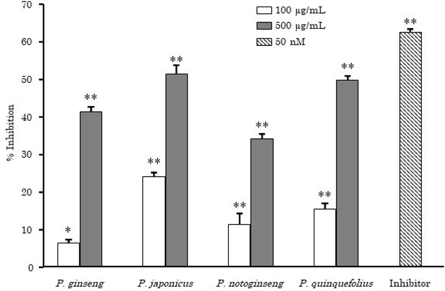
Inhibitory activities of Panax species on β-secretase. Data are shown as the mean with standard deviation as error bars. Significantly different from the control group, *P < 0.05 and **P < 0.01.
All extracts showed inhibitory activities against AChE (Figure 5). The P. ginseng extract showed the most potent activity among the extracts tested with 70.4% and 81.7% of inhibition at 50 and 250 µg/mL, respectively. From the previous reports, diterpenes, ferruginol, nezukol, and kaur-16-ene showed potent activities. 14 Terpenoids contained in the P. ginseng extract may play an important role in expressing the activities.

Inhibitory activities of Panax species on acetylcholinesterase. Data are shown as the mean with standard deviation as error bars. Significantly different from the control group, **P < 0.01.
All extracts showed inhibitory activities against BChE (Figure 6) at lower concentrations compared to those in the β-secretase and AChE inhibitory assays. The P. ginseng and P. quinquefolius extracts showed the most potent inhibitory activities of 84.4% and 79.5% at 50 µg/mL, respectively.

Inhibitory activities of Panax species on butyrylcholinesterase. Data are shown as the mean with standard deviation as error bars. Significantly different from the control group, **P < 0.01. TIP, tetraisopropylpyrophosphoramide.
Then, the fatty acids which were determined as major components in EO extracts of Panax plants were evaluated for their inhibitory activities against AChE and BChE. Methyl linoleate, palmitic acid, and methyl palmitate were selected. Among them, methyl linoleate showed the most potent activity against AChE and its IC50 value was 68.8 µM, while palmitic acid showed the most potent activity against BChE and its IC50 value was 11.8 µM (Table 4). Methyl palmitate showed weak activity as IC50 value was over 500 µM.
Fifty Percent Inhibitory Concentration Values of Compounds on Inhibition of Cholinesterases.

AChE, acetylcholinesterase; BChE, butyrylcholinesterase; IC50, 50% inhibitory concentration.
aPositive control of acetylcholinesterase.
bNot determined.
cPositive control of BChE.
Panax japonicus and P. notoginseng extracts showed inhibitory activities against amyloid aggregation of 29.8% and 57.3% at 500 µg/mL, respectively (Figure 7). The negative effect observed in P. ginseng might be due to the fluorescent effect of matrix contained in the extract.

Inhibitory activities of Panax species on amyloid aggregation. Data are shown as the mean with standard deviation as error bars. Significantly different from the control group, *P < 0.05 and **P < 0.01. Myr, myricetin.
Inhibition of β-secretase and Aβ aggregation have been recognized as therapeutic targets for AD. 15,16 β-Secretase catalyzes the degradation of Aβ peptide from a precursor protein in the first step of AD pathogenesis. This is the main reason why β-secretase and Aβ aggregation inhibitors have been rarely proven to cure AD. Therefore, we focused on the preventive effect of β-secretase and Aβ aggregation inhibitors. Aggregation and pigmentation of Aβ to form senile plaque takes many years from adolescence onward. If a subject took β-secretase and Aβ aggregation inhibitors continuously from adolescence, AD would be prevented in old age. We selected a natural plant material for the β-secretase assay as screened in the previous studies. 17 In this study, EO extracts of Panax plants were selected. Essential oil extracts can be absorbed and pass through the blood-brain barrier easily, 18 and a high level of translocation to the brain is expected. In addition, EO extracts are potential target agents for AD therapy. 19 Furthermore, the Panax plants used in this study have been consumed for a very long time and their safety features have already been proven. Therefore, EO extracts of Panax plants are ideal target agents to prevent AD.
In this study, we revealed that EO extracts from Panax plants have inhibitory activities against β-secretase, AChE, BChE, and Aβ aggregation. In particular, EO extracts of P. japonicus and P. notoginseng showed potent inhibitory activities against β-secretase and Aβ aggregation, while the EO extract of P. ginseng showed the most potent inhibitory activities against AChE and BChE among the samples tested. From these results, an EO extract of P. ginseng could be an effective agent to improve AD symptoms rather than preventing AD, while EO extracts of P. japonicus and P. notoginseng may be effective for AD prevention.
In the previous reports, P. notoginseng extract showed reduced Aβ pigmentation 20 and P. quinquefolius extract alleviated Aβ1-42 cytotoxicity 21 ; the active principles were identified as ginsenosides. In addition, ginsenosides showed inhibitory activities against cholinesterases and β-secretase. 22 However, the bio-availabilities of ginsenosides were too low when administered orally due to their complex metabolisms. 22
In this report, we revealed novel pharmacological effects of EO extracts from Panax plants. Extensive studies have been performed on ginsenosides, which are high polar compounds. Novel pharmacological activities are yet to be discovered in Panax plants in nonginsenoside fractions.
Experimental
Materials
Panax ginseng roots were purchased in Gyeondong Market in Korea. Panax japonicus rhizome, P. notoginseng roots, and P. quinquefolius roots were purchased from Tochimoto Tenkaido (Osaka, Japan). Fast Blue B Salt (FBB) and Triton X-100 were purchased from MP Biomedicals (Santa Ana, CA, United States). Sodium dodecyl sulfate (SDS) was purchased from GE Healthcare (Tokyo, Japan). Acetylcholinesterase (from Electrophorus electricus) and BChE (from equine serum) were purchased from Sigma-Aldrich (St Louis, MI, United States). Galantamine hydrobromide and myricetin were purchased from Tokyo Chemical Industry (Tokyo, Japan). Fluorescence-quenching substrate for β-secretase [MOCAc-Ser-Glu-Val-Asn-Leu-Asp-Ala-Glu-Phe-Arg-Lys(Dnp)-Arg-Arg-NH2], β-secretase inhibitor [Lys-Thr-Glu-Glu-Ile-Ser-Glu-Val-Asn-Sta-Val-Ala-Glu-Phe], and amyloid β-protein (Human, 1-42) were purchased from Peptide Institute, Inc. (Osaka, Japan). All other reagents were purchased from FUJIFILM Wako Pure Chemical Corp. (Osaka, Japan) or Nacalai Tesque (Kyoto, Japan).
Preparation of EOs From Panax Species
Dried samples were pulverized and extracted with a 10 times amount of distilled water (w/v) using EO quantifying apparatus (Japanese Pharmacopeia 17th edition) by the steam distillation method (5 hours, 100°C). 5 Result on the yield of Panax species by steam distillation method is shown in Table 5.
Samples of Panax Species Used in This Study.

Gas Chromatography Mass Spectrometry Analysis
Contents of EO were analyzed using GC/MS under the following conditions. GC Conditions: column, Inertcap Pure Wax (GL science, Tokyo, Japan, 0.25 µm, 0.25 mm i.d. × 60 m); oven temperature, 0 minute (50°C), 2 minutes (50°C), 78 minutes (240°C), and 98 minutes (240°C, postrun); injection temperature, 250°C; split ratio, 1/80; injection volume, 1.0 µL; detector, flame ionization detector, MS Conditions: EI source, electron energy of 70 eV; ionization temperature, 230°C; quadrupole temperature, 150°C; scanning range, 25 to 350 amu. The components were identified by matching their mass fragments and retention times with databases (Wiley 7th and NIST08).
Assay for β-Secretase Inhibition
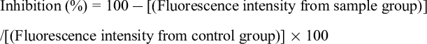
Assays were performed according to the method reported previously with modification. 23 Samples in a dimethylsulfoxide (DMSO) solution (2 µL) at an appropriate concentration were diluted with 78 µL of assay buffer (20 mM acetate buffer, pH = 4.5 containing 0.1% Triton X-100) in a 96 well-microtiter plate. Ten microliters of β-secretase solution in assay buffer (100 U/mL) were added to the sample solution and incubated at 37°C for 10 minutes. After incubation, 10 µL of substrate solution dissolved in assay buffer (0.1 mM) was added and incubated at 37°C for 1 hour. After incubation, 50 µL of 2.5 M sodium acetate solution was added to the reaction solution. The reaction solution (100 µL) was moved to a glass vial and diluted with 900 µL of water. The glass vial was incubated at 80°C for 10 minutes to terminate the reaction. The reaction solution was analyzed by high performance liquid chromatography (HPLC) under these conditions. Column: Cadenza CD-C18 (Imtakt Co., Kyoto, Japan, 3 µm, 4.6 mm i.d. × 150 mm); mobile phase, 0.1% (v/v) formic acid/acetonitrile with 0.1% (v/v) formic acid: 0 minute (9:1), 18.0 minutes (1:1), 18.1 minutes (5:95), and 23.0 minutes (5:95); column temperature, 40°C; flow rate, 1 mL/min; detection, fluorescent of excitation at 325 nm and emission at 395 nm; injection volume, 10 µL. The peak area of the degradative fluorescent fragment (R t 15.4 minutes) was integrated. Inhibitory activity of the sample was calculated using the following equation.
The β-secretase inhibitor was used as a reference compound.

Assay for AChE inhibition
Assays were performed according to a method reported previously 24,25 with modifications in order to introduce a microtiter plate for higher throughput. Samples in a DMSO solution (5 µL) at an appropriate concentration were diluted with 180 µL of assay buffer (50 mM Tris-HCl buffer, pH = 7.8) in a 96-well microtiter plate. Ten microliters of enzyme solution (2.0 U/mL) and 5 µL of 1-naphthylacetate (18 mM) were added to the mixture and incubated at 37°C for 1 hour. After incubation, 25 µL of 5% (w/v) SDS solution and 25 µL of FBB (2 mM) were added. Absorbance at 600 nm was measured and the inhibition was calculated using the equation below. Moreover, the absorbance from each sample was subtracted from that of the nonenzyme group in order to reduce the interference from absorbance of the sample itself.
Galantamine hydrobromide was used as a reference drug.

Assay for BChE Inhibition
Assays were performed according to the same method used for AChE with BChE as an enzyme instead. Tetraisopropylpyrophosphoramide was used as a reference drug.
Assay for Amyloid Aggregation Inhibition Using the Thioflavin T Test
Assays were performed according to a method reported previously 26 with modifications. Aβ1-42 peptide was dissolved in DMSO to make a 1 mM Aβ1-42 stock solution and stored at −20°C until use. Aβ1-42 stock was diluted 5-fold with assay buffer (50 mM sodium phosphate buffer, pH = 7.4). Samples in a DMSO solution (2 µL) and 88 µL of assay buffer were mixed in a white 96-well microtiter plate. Ten microliters of Aβ1-42 solution (200 µM) were added to the mixture and incubated with shaking for 1 hour (37°C, 500 rpm). After incubation, 100 µL of 5 µM thioflavin T solution (50 mM glycine-NaOH buffer, pH = 8.5) was added and incubated at 37°C for 30 minutes. Thioflavin T fluorescence intensity associated with Aβ fibrils was measured at 440/486 nm (excitation/emission) using a Perkin Elmer 2030 multilabel reader (Perkin Elmer, MA, United States). The inhibition was calculated using the following equation. Moreover, the fluorescence intensity from each sample was subtracted from that of the non-Aβ1-42 group in order to reduce the interference from the fluorescence intensity of the sample itself.
Myricetin was used as a reference drug.
Statistical Analysis
All data were analyzed with Statcel3 (The Publisher OMS, Tokorozawa, Japan), add-in software for Excel, using one-way analysis of variance. Significant difference was analyzed by Dunnet’s algorithm at P < 0.01 or P < 0.05.
Supplemental Material
Supplementary material - Supplemental material for Inhibitory Effects of Essential Oil Extracts From Panax Plants Against β-Secretase, Cholinesterase, and Amyloid Aggregation
Supplemental material, Supplementary material, for Inhibitory Effects of Essential Oil Extracts From Panax Plants Against β-Secretase, Cholinesterase, and Amyloid Aggregation by Hirokazu Kawamoto, Fumiaki Takeshita and Kazuya Murata in Natural Product Communications
Footnotes
Acknowledgment
We are grateful to Shinichi Matsumura and Yuri Yoshioka at INABATA KORYO CO., LTD. for GC-MS analysis.
Declaration of Conflicting Interests
The author(s) declared no potential conflicts of interest with respect to the research, authorship, and/or publication of this article.
Funding
The author(s) received no financial support for the research, authorship, and/or publication of this article.
References
Supplementary Material
Please find the following supplemental material available below.
For Open Access articles published under a Creative Commons License, all supplemental material carries the same license as the article it is associated with.
For non-Open Access articles published, all supplemental material carries a non-exclusive license, and permission requests for re-use of supplemental material or any part of supplemental material shall be sent directly to the copyright owner as specified in the copyright notice associated with the article.