
Abstract
Baicalin is a natural product isolated from Scutellaria genus and has been reported to have many different pharmacological activities, which include anti-viral, anti-inflammatory, and anti-allergic effects. We investigated the effects of baicalin administered after I/R-induced brain injury in a mouse model. I/R-induced brain injury was induced by MCAO for 2 h. Baicalin was orally administered to mice once daily for 2 consecutive days after 2 h of reperfusion. Single daily oral administrations of baicalin at 10, 30, or 100 mg/kg/day for 2 consecutively days after I/R significantly reduced infarct volumes, edema indices, and water contents in mouse brains, serum levels of AQP-4. IL-1β, TNF-α, and ROS, and lipid peroxidation levels in brain ipsilateral hemispheres were suppressed by baicalin. This result is believed to be due to the anti-inflammatory effect of baicalin and its downregulation of AQP-4 protein expression in affected brain tissues after I/R-induced brain injury in mice.
Approximately 60% of cardiovascular mortalities including around a half of total coronary deaths and two thirds of stroke-attributed deaths occurs in low and middle income countries. 87% of all strokes are ischemic, 10% are intracerebral hemorrhages, and 3% are subarachnoid hemorrhages, thus, ischemic stroke is a devastating disease that causes much morbidity and disability, and poses an enormous burden on healthcare systems and on patients that need rehabilitation and long-term care. 1,2 To date, r-tPA is the only thrombolytic agent approved by the US FDA for acute ischemic stroke, but its clinical applications are limited by an extremely small treatment window after stroke onset. 3–6 Accordingly, there is an urgent need for preclinical research aimed at developing new therapeutic agents for ischemic stroke.
BBB leakage in brain diseases, such as ischemic stroke, exacerbates disease progression. 7,8 BBB leakage has also been reported to be correlated with AQP-4 induction in perivascular astrocytes. 9 Interestingly, Tu et al. reported that baicalin inhibited MMP-9 expression and MMP-9-mediated occludin degradation and reduced MCAO-induced brain edema and BBB permeability in rats. 10 Recent preliminary studies conducted in our laboratory showed that baicalin reduces brain edema associated with ischemic brain damage and reduces total infarction volumes in ipsi-lateral hemispheres. In the present study, we explored the anti-inflammatory effects of baicalin on I/R-induced inflammation and its protective effect on BBB leakage in MCAO-induced brain injured mice.
Scutellaria Radix, the dried root of Scutellaria baicalensis Georgi, contains flavonoids such as baicalin, baicalein, and wogonin, and baicalin content is used to gage the quality of medicinal Scutellaria Radix in Korea. 11 Baicalin has been reported to have potential beneficial effects in many diseases, for example, it has been shown to control inflammation and to possess antioxidant activity. 12–14 Furthermore, several researchers have proposed baicalin is likely to inhibit brain damage caused by ischemia and suggested that its anti-inflammatory, anti-oxidative, and anti-apoptotic effects contribute to its mode of action. 10,12–19 Thus, baicalin appears to have considerable potential for the treatment of ischemic stroke, although its neuroprotective properties when administered orally after ischemic stroke onset, have not been explained. Accordingly, the action mechanism responsible for its effects needs further exploration.
This study was conducted to investigate the effects of baicalin post-treatment on I/R-induced brain injury in a mouse model. Body weights showed a nonsignificant decreasing tendency after I/R (Supplementary Figure 1SA), which was presumed to be due to dehydration and food intake difficulties after surgery. Baicalin treatments did not affect body weights. Mean serum electrolyte levels in the MCAO and sham control groups were similar (Supplementary Figure 1SB), indicating the absence of any physiological change that might affect outcome after I/R. Representative sections of a brain stained with TTC are shown in Figure 1A.
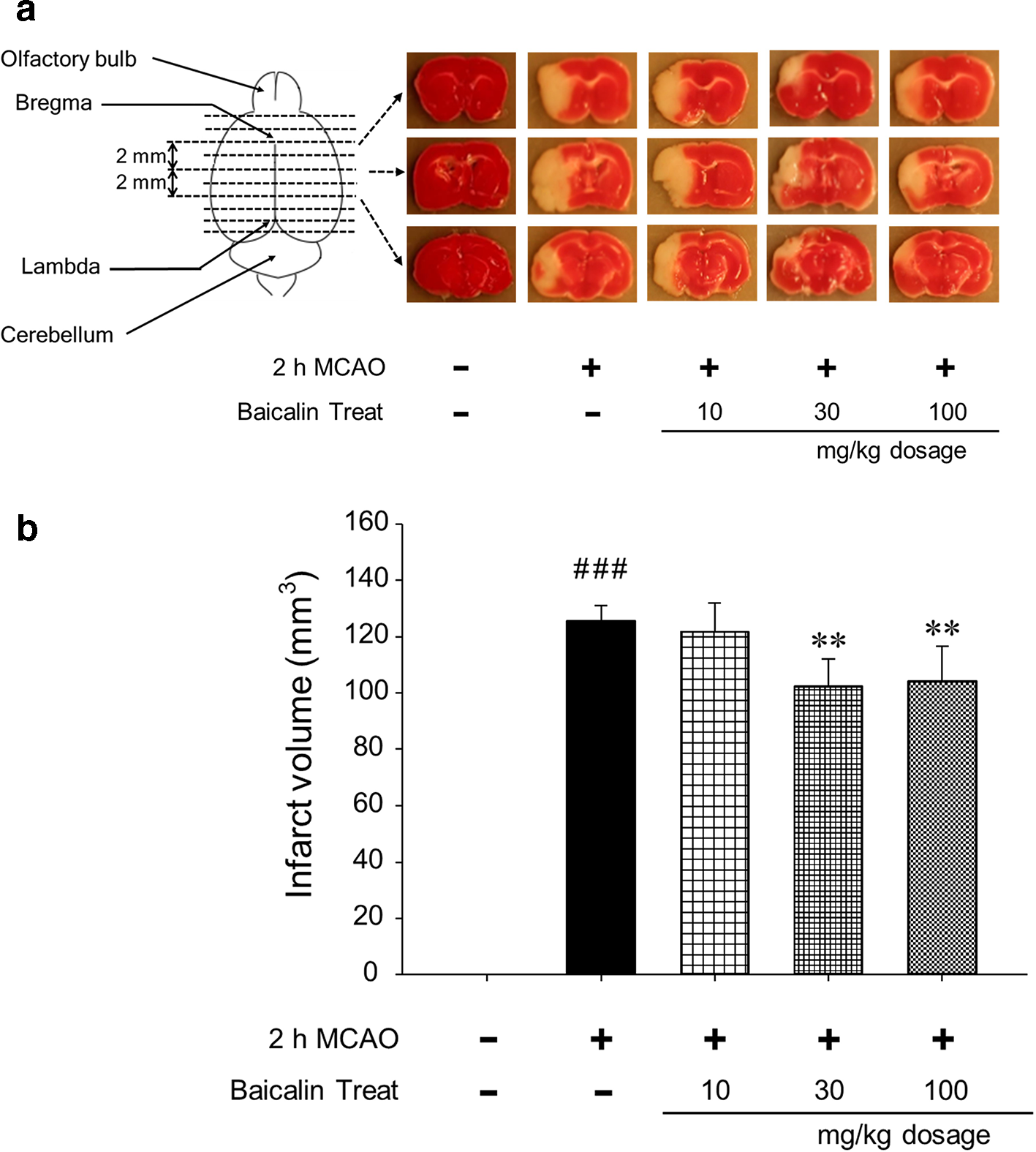
Representative images (A) of brain sections and the effect of baicalin post-treatment on infarct volumes (B) in the brains of MCAO-injured mice. The treatment with 30 or 100 mg/kg of baicalin daily for 2 days after MCAO significantly decreased infarct volumes. results are presented as means ± SDs. ### P < 0.001 vs sham controls, **P < 0.01 vs MCAO controls; n = 6 per group.
Sham operation did not cause brain damage, but I/R caused extensive damage to left ipsilateral hemispheres (mean ± SD; 125 ± 5.5 mm3). However, after treatment with 30 or 100 mg/kg baicalin for two consecutive days after I/R, infarction volumes were significantly reduced (102 ± 9.6 mm3 and 104 ± 12.4 mm3, respectively) as compared with those of the MCAO control group (Figure 1B). CEIs were calculated by dividing brains into three parts, as shown in Figure 2A, and mean CEIs in the 30 and 100 mg/kg baicalin groups were significantly lower than that in the MCAO control group (Figure 2B), which was considered to be associated with total infarct volumes (Figure 1B). Brain water contents were also significantly diminished by baicalin administration (Figure 2C).
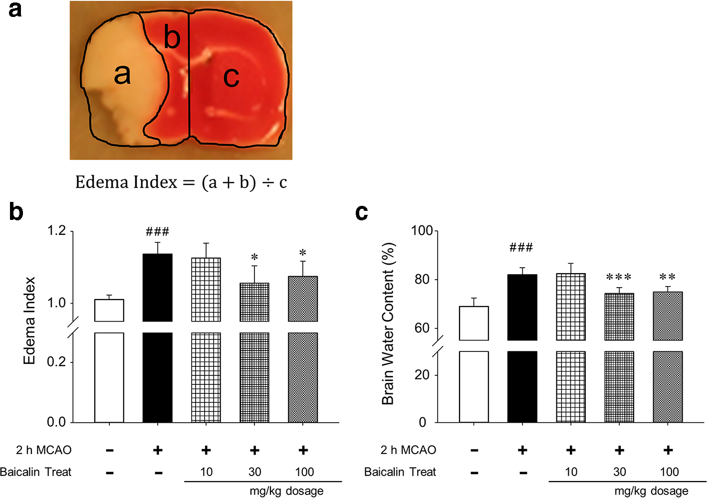
Calculation of edema index (A) and the effects of baicalin post-treatment on edema indices (B) and water contents (C) in the brains of MCAO-injured mice. Post-treatment with baicalin at 30 or 100 mg/kg daily significantly suppressed MCAO-induced increases in whole brain edema indices (B) and water contents (c). Results are presented as means ± SDs. ### P < 0.001 vs sham controls, *P < 0.05, **P < 0.01, ***P < 0.001 vs MCAO controls; n = 6 per group.
Interestingly, brain water contents showed more significant changes than CEIs, possibly because difficulties associated with sectioning brain tissues for TTC staining. NDSs increased significantly after I/R, but no improvements were observed in the three baicalin groups (Supplementary Figure 2S).
The effects of baicalin on brain inflammation after I/R were assessed by measuring pro-inflammatory cytokine levels in brain tissue homogenates. Pro-inflammatory cytokines, such as IL-1β and TNF-α, stimulate leukocytes attached to activated vascular endothelium during the early stage of focal cerebral ischemia, 20 and ROS generated during inflammation are believed to play critical roles in many diseases. 21 IL-1β levels were higher in the MCAO control group than in the sham control group (648.7 ± 77.3 pg/ml and 182.8 ± 34.8 pg/ml, respectively), and the same tendency was observed for TNF-α levels (434.7 ± 59.3 pg/ml and 143.2 ± 32.8 pg/ml, respectively). Baicalin treatment post-I/R inhibited both IL-1β (30 mg/kg, 541.3 ± 40.4 pg/ml; 100 mg/kg, 551.7 ± 69.1 pg/ml) and TNF-α (30 mg/kg, 339.0 ± 72.2 pg/ml; 100 mg/kg, 327.5 ± 61.8 pg/ml) increases (Figure 3A , B). Based on published results and our own, we believe baicalin reduces infarction in stroke by inhibiting inflammatory responses to ischemic brain injury. ROS and MDA contents in brain tissue homogenates were used as surrogates of antioxidant activity. The levels of both were significantly higher in the MCAO control group (ROS, 232.2 ± 29.2%; MDA, 1.39 ± 0.13 nmol/mg) than in the sham control group (ROS, 123.5 ± 13.2%; MDA, 0.63 ± 0.13 nmol/mg). However, ROS and MDA levels in the 100 mg/kg baicalin group were significantly lower than in the MCAO control group (ROS, 186.0 ± 22.3%; MDA, 1.04 ± 0.24 nmol/mg) (Figure 3C , D). Thus, it was assumed that baicalin administration significantly suppressed the MCAO-induced upregulations of IL-1β and TNF-α in ipsi-lateral hemispheric brain tissues, resulting in inhibition of ROS production and lipid peroxidation.

Effects of baicalin post-treatment on IL-1β, TNF-α, ROS, and MDA protein levels in the brains of MCAO-injured mice. Baicalin post-treatment significantly suppressed I/R-induced increases in IL-1β (A), TNF-α (B), ROS (C), and MDA (D) levels. Results are presented as means ± SDs. ### P < 0.001 vs sham controls, *P < 0.05 vs MCAO controls; n = 6 per group.
AQP-4 is an aquaporin derived from major endogenous proteins, and its deregulation may lead to water accumulation, 22,23 and thus, AQP-4 is important for the maintenance of brain water balance. 7,8 To assess the role played by AQP-4 after I/R, we measured protein levels in ipsi-lateral hemispheres. AQP-4 levels were higher in the MCAO than in the sham control group, indicating I/R-induced brain injury resulted in partial blood-brain barrier (BBB) disruption. However, AQP-4 expression was significantly lower in the 100 mg/kg baicalin group than in the MCAO group (Figure 4).

Effects of baicalin post-treatment on AQP-4 protein levels in the ipsi-lateral hemispheric brains of MCAO-injured mice. Baicalin post-treatment significantly inhibited MCAO-induced increases in AQP-4 protein levels. Representative western blot analysis of AQP-4 protein expression showing the effect of baicalin on AQP-4 levels in brain tissue. Results are presented as mean ± SDs. ### P < 0.001 vs sham controls, **P < 0.01 vs MCAO controls; n = 6 per group.
Many mechanisms may be responsible for the effects of baicalin on ischemic brain injury, though the present study indicates the inhibitions of AQP-4 overexpression and of inflammatory processes reduced infarction sizes after I/R. A brief scheme of possible mechanisms is provided in Figure 5.
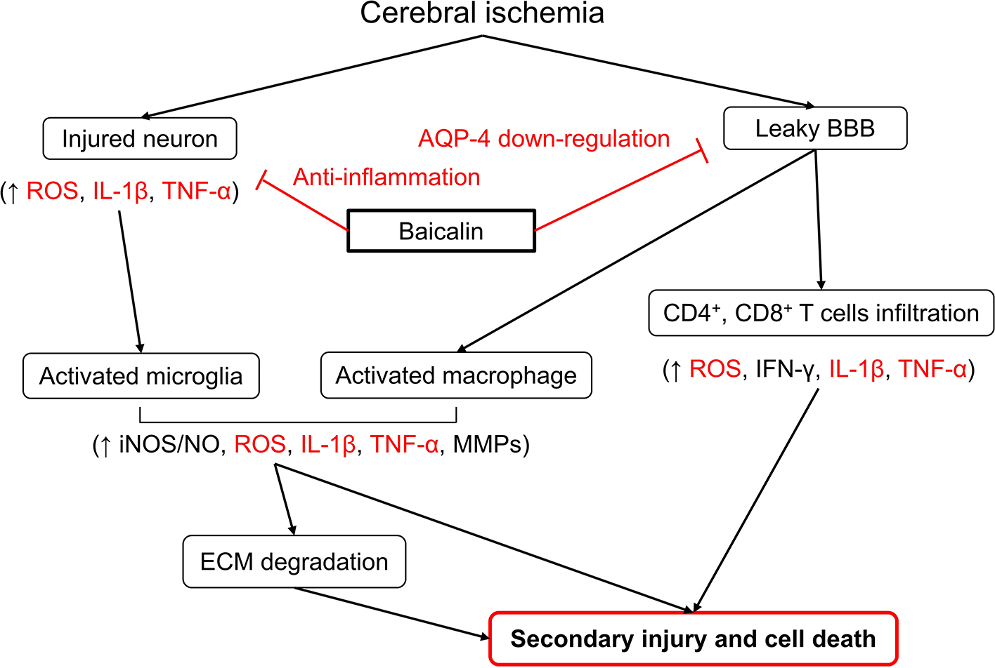
Schematic of the proposed neuroprotective mechanism of baicalin in our murine MCAO model. Red arrows indicated proposed effects of baicalin on I/R-induced brain damage.
Taken together, our results demonstrate baicalin suppressed I/R-induced brain injury in our murine model, probably by reducing inflammation and brain edema. In particular, stroke-related increases in AQP-4 protein levels in ipsi-lateral hemispheres of injured brain tissues were suppressed by oral baicalin administration after stroke onset, which suggests AQP-4 be considered a target for the treatment of brain injury. In addition, our findings highlight the importance of traditional herbal medicines and their components as resources for novel compounds with therapeutic potential, and suggest baicalin be considered a natural product that might be useful for the treatment of ischemic brain injury.
Experimental
Ischemic Stroke Model and Baicalin Treatment
Adult SPF C57BL/6 male mice (22, 25 g) were obtained from Daehan Biolink Co. (Chungbuk, Korea) and housed in temperature and humidity-controlled animal facilities under a 12 hours light/dark cycle. All animal experiments were approved beforehand and regulated by the ethics committee of Pusan National University (PNU) (approval number, PNU-2016-1087). Ischemic stroke was induced in mice by MCAO, as previously described. 24 Baicalin was purchased from Sigma-Aldrich (MO) and dissolved in physiological saline. In order to verify the quality of baicalin, fingerprinting data were obtained by HPLC (Supplementary Figure 4S). Mice were divided into 5 groups containing six mice per group, as follows; a sham operated control group (the sham control group), a MCAO-operated but not baicalin treated group (the MCAO control group), and three MCAO-operated and baicalin treated groups, in which baicalin was administered at 10, 30, or 100 mg/kg daily (the 10, 30, and 100 mg/kg baicalin groups). Animals in the three baicalin-treated groups were administered 0.5 ml of each concentration orally at 2 and 26 hours I/R (Supplementary Figure 3S). Mice in the sham and MCAO control groups received the same amount of physiological saline as the animals in the three treatment groups.
Body Weights and Physiological Parameters
Mice were weighed daily during the experiment. Blood samples were collected 2 days after I/R and serum was obtained by centrifuging at 1,500 × g for 15 minutes at 4 ℃. Serum levels of sodium (Na+), potassium (K+), and chloride (Cl−) were measured to monitor potential imbalances that may have influence brain edema data using an electrolyte analyzer (Dri-Chem 3500i, Fuji, Japan).
Infarct Volume Measurements and Brain Edema Calculation
Forty-8 hours after I/R, mice were euthanized by CO2 inhalation and brains were excised and sliced into ten coronal sections (1 mm thick). Sections were stained 2% TTC for 17 minutes at room temperature (25 ℃) and then immersed in 10% NBF for 2 hours. Total infarct volumes were calculated using lesion areas, as described by Jung et al. 23 using ImageJ software (NIH, MA). Cerebral edema indices were calculated by dividing areas in damaged ipsilateral hemispheres by the areas of corresponding normal contralateral hemisphere areas using TTC-stained brain slice images, as described by Sebastiani et al. 24 In order to evaluate the effects of baicalin treatment on edema development after MCAO, wet weight/dry weight ratios of whole brains were determined using a reliable speed-vacuum drying method. 25
Neurological Deficit Scores
NDSs were blindly quantified using a 5-point scale, as described by Jung et al. 23 at 2, 26, and 50 hours after I/R, as follows. Grade 0, no neurological deficit; Grade 1, incomplete extension or failure of the right foreleg; Grade 2, on pulling the tail, spontaneous movement in all directions noticeable decreased and the animal turned to its right; Grade 3, highly sensitive to pain induced by tail stimulation, and the animal walked and circled to its right; Grade 4, no response to tail stimulation or brain injury-related death.
Inflammatory Cytokine Analysis
Brains were carefully separated from skulls, and ischemic ipsilateral hemispheres were homogenized in PBS (pH 7.4, 5% w/v). Resulting homogenates were clarified at 10,000 × g for 20 minutes at 4 ℃ and supernatants were subjected to ELISA. Levels of IL-1β and TNF-α in brain tissues were measured by ELISA using a commercially available kit (Abcam, Cambridge, MA). The absorbances of reaction products were measured at 450 nm using a microplate reader.
Determinations of ROS and Lipid Peroxidation Levels
ROS levels in tissue homogenates were determined using DCFH-DA, as previously described. 24 Tissue homogenates were incubated with 1 mM DCFH-DA at 37 ℃ for 30 minutes, and absorbances were measured using a fluorescent microplate reader at an excitation wavelength of 485 nm and an emission wavelength of 535 nm. In order to assess oxidative stress in damaged brain tissues, we measured MDA, a biomarker of lipid peroxidation levels in ischemic hemispheres was using a TBARS assay kit (Cayman Chemical, MI). 26 ODs were read at 540 nm using a spectrophotometer and results are presented as μM/μg on wet tissue.
Western Blot Analysis
AQP-4 levels in mouse brains were evaluated by western blotting, which was performed using a modification of a method we previously described, 24 Briefly, 2 days after I/R, mice were euthanized by CO2 inhalation, brains were excised and placed on ice cold glass. Ischemia-induced left ipsi-lateral hemispheres were dissected, homogenized in modified PBS containing 150 mM NaCl, 1 mM EDTA, 50 mM Tris, and 1: 100 (v/v) proteinase inhibitor, centrifuged (13,250 × g) at 4 ℃ for 10 minutes, and total protein were obtained from supernatants using protein extract solution (Pro-prep, iNtRON, Gyeonggi-do, Korea). Total protein contents of lysates were measured using a protein assay kit (Quick Start, Bio-Rad, Hercules, CA, USA). In addition, same amounts of protein extracted from brain tissue lysates were separated by electrophoresis on 10% SDS-PAGE gels and transferred to PVDF membranes (Millipore, Darmstadt, Germany), which were then blocked with 5% skim milk in TBST (Sigma-Aldrich) for 1 hours at 25 ℃, incubated with anti AQP-4 (1:500) or anti β-actin (1:1000) primary antibody (Cell Signaling, MA), and washed with TBST. Membranes were then incubated with a secondary antibody conjugated with HRP (goat anti rabbit, 1: 5000) (Enzo Life Sciences, NY), and incubated at 25 ℃ for 2 hours. After washing three times with TBST, membranes were treated using an ECL kit (GenDEPOT, TX) and transferred to a light emitting analyzer system (Amersham™ Imager 600, UK). Blot signal intensities were analyzed using ImageJ (NIH, MD), and amounts of proteins were determined relative to AQP-4 or β-actin.
Statistical Analysis
Results are expressed as mean ± SDs, and the significances of intergroup differences were determined by one-way ANOVA and Tukey’s post hoc analysis. The Shapiro-Wilk test was used to determine the normality of data distributions. The analysis was conducted using Sigmaplot v12.0 software, and statistical significance was accepted for P values < 0.05.
Footnotes
Declaration of Conflicting Interests
The author(s) declared no potential conflicts of interest with respect to the research, authorship, and/or publication of this article.
Funding
The author(s) received no financial support for the research, authorship, and/or publication of this article.
Supplemental Material
References
Supplementary Material
Please find the following supplemental material available below.
For Open Access articles published under a Creative Commons License, all supplemental material carries the same license as the article it is associated with.
For non-Open Access articles published, all supplemental material carries a non-exclusive license, and permission requests for re-use of supplemental material or any part of supplemental material shall be sent directly to the copyright owner as specified in the copyright notice associated with the article.