
Abstract
Background:
A baseline level of lipofuscin in the retinal pigment epithelium (RPE) is inevitable with age, but increased levels due to increased oxidative stress can result in deleterious vision loss at older ages. As earlier detection of differences in levels can lead to superior preventative management, we studied the relationship between lipofuscin accumulation and dietary lifestyle (vegetarian vs. nonvegetarian) in the younger, healthy South Asian population using retinal fundus autofluorescence (FAF) imaging.
Methods:
In this pilot study, we examined 37 healthy subjects (average age 23 years ± 1) all undergoing similar stress levels as medical students at Rutgers New Jersey Medical School. Levels of lipofuscin concentrations were imaged using a FAF retinal camera (Canon CX-1). Two images (color and FAF) were captured of the left eye and included in the analysis. FAF quantitative scoring was measured in 2 regions of the captured image, the papillo-macular region (P) and the macula (M), by determining the grayscale score of a 35.5 mm2 rectangle in the respective regions. Standardized scores (corrected to remove baseline fluorescence) were then obtained. Means, standard deviations, and t tests were performed for comparisons.
Results:
Fundus autofluorescence scores of regions P and M were significantly different (P < .05) between groups. Region P was further standardized and results remained significant.
Conclusions:
Our preliminary results show that in this cohort, vegetarians had statistically significant lower levels of autofluorescence. These findings can have potential implications regarding long-term retinal health and risk for developing certain diseases over decades in subjects at risk for vision-threatening diseases.
Lipofuscin is a marker of oxidative stress and cellular aging that accumulates in postmitotic cells in the retinal pigment epithelium (RPE). These pigmented, autoflorescent molecules found in the eye are unique from lipofuscin in other body tissues as they aggregate secondary to the incomplete digestion of photoreceptor outer segments and can potentially be phototoxic to the RPE. 1 Individuals are not born with this ocular pigment, as young eyes show few or no lipofuscin granules. 2 These granules slowly accumulate over time and thus, a baseline level of lipofuscin is expected in the healthy eye. An overall increase in lipofuscin can be seen from the third decade as these granules start to appear as clusters, concentrating significantly in the posterior pole of the retina, which encompasses the area between the optic nerve and the macula. 3
Although the creation of lipofuscin is a natural process that allows for normal retinal health, severely increased levels of lipofuscin can be devastating leading to gradual visual impairment, as seen in age-related macular degeneration.4,5 The rate of its accumulation varies between individuals and can be accelerated in conditions of high oxidative stress created either by abnormally high levels of free radical production (retinal oxygenation and light exposure) or decreased antioxidant protection levels (cigarette smoke exposure).1,6 There is also increasing evidence that suggests that oxidative stress has a heavy impact in the pathogenesis of both types of diabetes mellitus. The creation of reactive oxidative species is disproportionally elevated in diabetes due to the high levels of glucose that under go various oxidative processes. 7 These unfavorable conditions can gradually lead to the complications of diabetes, including diabetic retinopathy.
Studies have also suggested an association between nutrition, lipofuscin, and oxidative stress, but this has only been briefly explored. The LAST (Lutein Antioxidant Supplementation Trial) study suggested that eating an equivalent to 10 mg of lutein, a yellow pigment found in dark green leafy vegetables such as spinach and kale, 3 to 4 times a week, can lead to a modest improvement in visual acuity in patients who have age-related macular degeneration. 8 Furthermore, the first reported human clinical case of lipofuscin reversal was through a nutraceutical intervention using an oral polyphenolic mixture containing resveratrol, commonly found in red grapes and red vines. 9 Its ability to act as an antioxidant by directly eliminating free radicals led to the improvement of the 80-year-old subject’s visual function and visible clearing of the pigment inside the cells within 5 months. 9
To expand on existing theories of nutrition, oxidative stress, and eye health, this study investigates the relationship between dietary lifestyles and lipofuscin accumulation as a result of oxidative stress, specifically comparing the impact of a vegetarian and a nonvegetarian diet on the RPE. A number of individuals around the world practice a vegetarian diet, but the vegetarian population in India is exceptionally high and accounts for nearly 35% of the entire population. 10 Furthermore, many Indians, even after emigrating from the homeland, have upheld their dietary traditions and maintained a vegetarian lifestyle that typically tends to be lower in saturated fat and cholesterol and higher in fiber, magnesium and potassium, vitamins C and E, folate, carotenoids, flavonoids, and other phytochemicals. 11 The nutritional differences observed in a vegetarian diet could yield lower levels of oxidative stress and may carry the potential to benefit those with certain diseases, such as diabetes in which a nutrient rich diet is a major factor in achieving proper glycemic control to reduce progression of complications. 7 With this knowledge, we aimed to explore the effects of diet on oxidative stress in the RPE by measuring lipofuscin levels amongst vegetarians and nonvegetarians within the South Asian population.
Methods
This prospective pilot imaging study was approved by the Rutgers University Internal Review Board and was fully HIPAA compliant. All imaging was conducted in the Telemedicine Laboratory at University Hospital in Newark, New Jersey, during the month of August 2014. A total of 37 healthy subjects (average age 23 years ± 1) all undergoing similar stress levels as medical students were enrolled in the study. Inclusion criteria for imaging were ages between 20 to 25 years, South Asian descent, and student enrollment in Rutgers New Jersey Medical School. Exclusion criteria were preexisting ocular conditions (cataracts, ocular opacities, and retinal findings) or ocular surgeries and a history of smoking or current smoking practices. Prior to imaging, participants completed a preliminary survey about their pertinent medical and social history. Subjects were categorized as either vegetarians or nonvegetarians based on their self-reported dietary habits. A vegetarian diet was one that did not consist of meat, poultry, or fish. The consumption of eggs, dairy, and other animal products was classified under a vegetarian diet. Those subjects who practiced both lifestyles at some point in their lives were categorized as vegetarians if they practiced a vegetarian lifestyle for more than half their life. Those that did not meet these criteria were grouped as nonvegetarians. No further dietary specifications were made. The intent of the study and the imaging protocol was explained in full detail to all participants. No mydriatic agents were used prior to imaging. All participants remained in a dimly light room (125 Candela) for 3 minutes to allow for natural pupillary dilation. All participants were screened for preexisting ocular conditions (cataracts, ocular opacities and retinal findings) via the written survey and a color digital 45
Lipofuscin is best imaged with fundus autofluorescence imaging which relies on stimulating light emissions from the molecules in the RPE using wavelengths based on previously established standards centered on autofluorescence properties of the retinal-pigmented epithelial cells.12,13 The fundus autofluorescence (FAF) image illustrates the spatial distribution of the lipofuscin with dark (hypoflorescent) regions representing lower levels of lipofuscin and bright (hyperflorescent) regions representing higher levels of lipofuscin. Levels of lipofuscin accumulation were imaged using a FAF retinal camera (Canon CX-1 16 megapixel camera with CMOS sensor, Tokyo, Japan) and a minimum natural pupillary dilation of 3.2 mm. The Canon CX-1 was equipped with optical filters for autofluorescence, with an exciter filter of 535-585 nm (wide band), and barrier filter with a bandwidth range of 605-715 nm (narrow band). All images were captured at an angle of 45
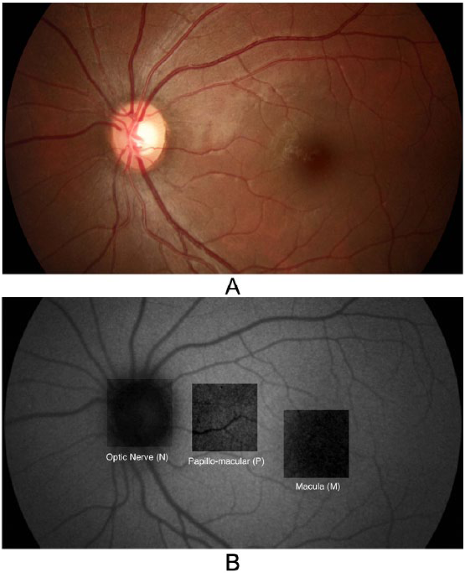
(A) Color nonvegetarian. (B) FAF nonvegetarian.
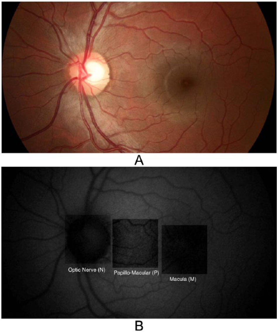
(A) Color vegetarian. (B) FAF vegetarian.
Autofluorescence quantitative scoring was obtained in 2 regions: the macula (M) and the papillo-macular zone (P), the zone between the temporal edge of the optic nerve and the macula. A grayscale pixel score of a 35.5 mm2 rectangle, approximately 1 disc diameter in length, in the respective regions was determined for each image (Figures 1B, 2B). One disc diameter was used as the standard measurement as it covered the appropriate desired area without overlapping on adjacent structures. The same images were then “standardized,” or corrected to remove baseline fluorescence by leveling autofluorescence at the optic nerve using Adobe Photoshop 7.0.1. 15 All nonmydriatic color and FAF images were viewed on SRS monitor Rx23.C-6. Statistical analysis was performed for comparisons using Microsoft Excel (12.2.7). Descriptive analyses included mean, standard deviation, and t tests, which was used to compare means. A P value less than .05 was considered statically significant.
Results
Out of 43 subjects that presented for imaging, 42 met inclusion criteria. One subject with significant smoking history was excluded from the analysis. In addition, 5 eyes did not meet the camera’s minimum pupillary dilation criteria (3.2 mm) and were excluded from the analysis due to poor FAF images. In total, 37 participants and thus 37 left eyes were included in the final analysis. Table 1 lists demographic and clinical characteristics of our study population. There were 21 nonvegetarians and 16 vegetarians and all participants were non-smokers. The mean age and body mass index (BMI) of both groups were not significantly different (p >0.05). Autofluorescence scores of regions P and M were significantly different (P < .05) between groups. Region P was further standardized and results remained significant. All FAF scores are listed in Table 2.
Screening Participants’ Demographics and Clinical Characteristics.
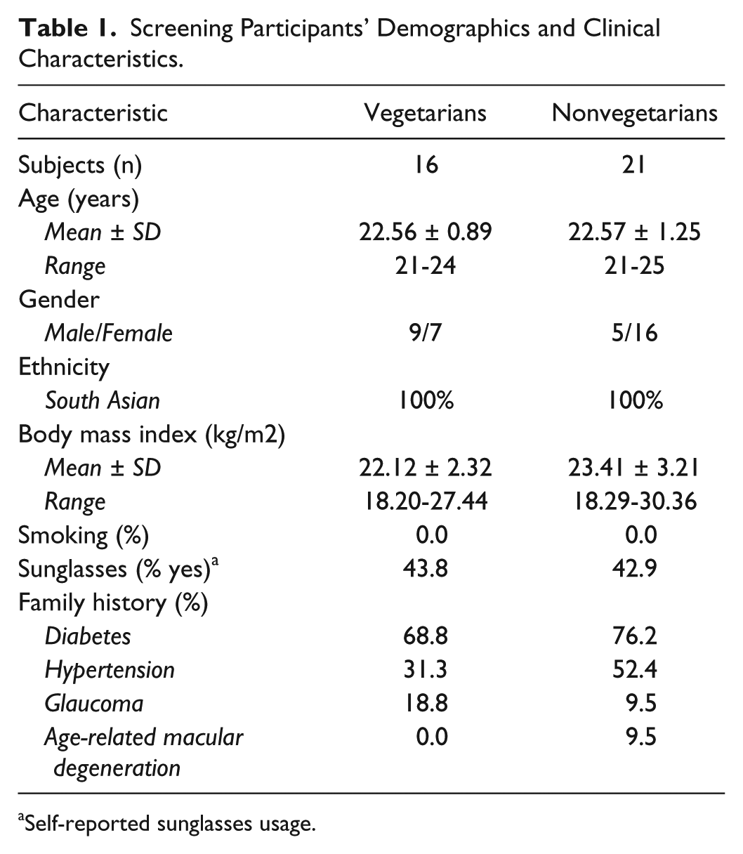
Self-reported sunglasses usage.
Comparing Levels of FAF Scores in Vegetarians Versus Nonvegetarians.
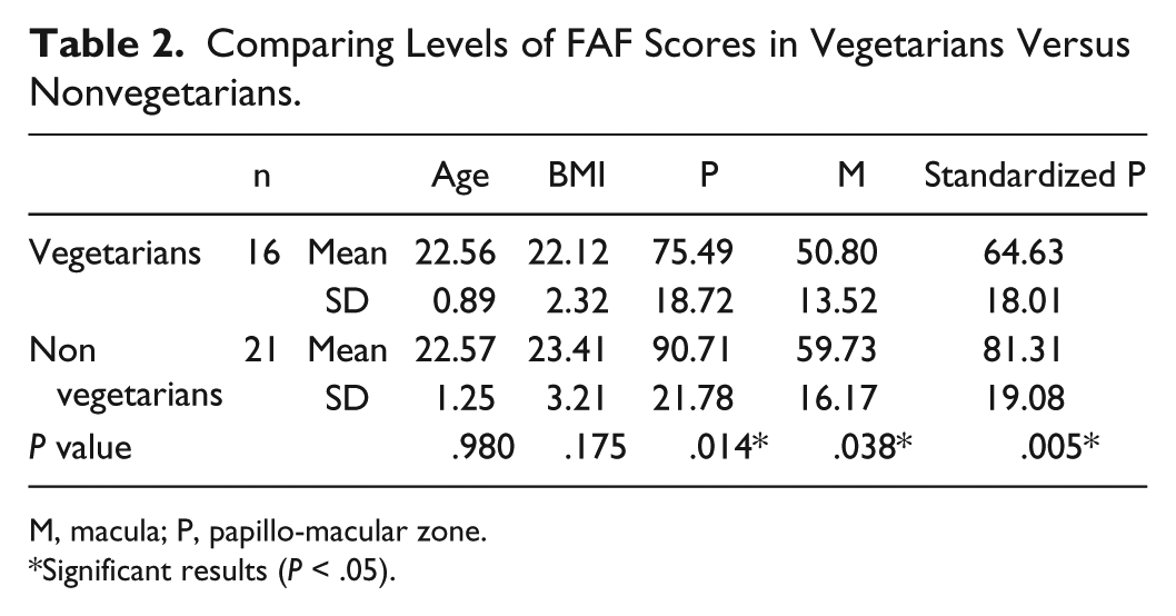
M, macula; P, papillo-macular zone.
Significant results (P < .05).
The color images were used to screen for any pathology or abnormalities prior to autofluorescence analysis. Small, hard drusens were observed in a total of 5 eyes (4 nonvegetarians and 1 vegetarian). However, they were all located peripheral to the inferior and superior vascular arcades and were not in the area used for analysis. Furthermore, a few subjects had tilted disks, which is not unusual for the South Asian population. No other anterior or posterior pole findings were noted when reading the color images.
Discussion
In reading the literature, our pilot study proves to be the first prospective imaging study that seeks to characterize any differences in lipofuscin levels within the RPE between vegetarians and nonvegetarians in the South Asian population. We hypothesized that a vegetarian diet would lead to higher rates of clearance of lipofuscin and thus less overall deposition of lipofuscin in the RPE. Our results support the original hypothesis and show a statistically significant difference (P < .05) in lipofuscin levels between the region P of both groups. Compared to region P, region M had lower levels of pigment due the region M’s distinctive characteristics. The M region has minimal florescence at the fovea due to the increased absorption by macular pigments, lutein and zeaxanthin, derived from diet.4,16 Since most of the lipofuscin accumulates in the posterior pole of the retina, we expected the P region to show the most difference between groups and the results confirmed our hypothesis.
What are the differences in dietary intake between a vegetarian and a nonvegetarian diet that could explain the differences observed in RPE lipofuscin levels? Perhaps there is something about a vegetarian diet that enables the extended ability to “discard” lipofuscin. As previously mentioned, a balanced vegetarian diet includes greater amounts of eye healthy nutrients such as vitamin C and E, B-carotene, zinc, lutein, and zeaxanthin. Furthermore, the eye is particularly susceptible to oxidative damage by direct, light exposure and high rates of metabolism. Thus, it requires greater levels of antioxidant protection. Vitamin C, mainly found in fruits and vegetables, is a highly effective antioxidant and protects vital molecules in the body from free radical and reactive oxygen species damage and may play a role in the maintenance of vitamin E levels in the eye. Vitamin E describes a group of fat-soluble antioxidants that mainly block free radicals from triggering lipid oxidation. The retina is highly saturated with fatty acids, further establishing the role of vitamin E as essential to good eye health. 17 Main sources of vitamin E include nuts, vegetable oils, and legumes. B-carotene is an orange pigment, mostly found in carrots and sweet potatoes, that when taken with other vitamins has proven to decrease the risk of development of AMD. 18 Zinc, found in nuts, legumes, and diary, plays a role in the structure of many enzymes and exhibits both antioxidant and immune functions. Lutein and zeaxanthin are carotenoids found in the lens and concentrated in the macula. In addition to their antioxidant properties, these macular pigments absorb incoming blue light and block ROS formation, thus limiting retinal oxidative damage. 17 Two foods that are found to have the highest amounts of lutein and zeaxanthin are kale and spinach. All the antioxidant vitamin and minerals previously discussed can be easily acquired from a vegetarian diet. Thus, a well-balanced vegetarian lifestyle could naturally involve higher intake of each nutrient. This promotes higher levels of antioxidants in the retina, leading to greater clearance of lipofuscin in vegetarians.
Although younger eyes do not have significant pathology, older eyes could have pathology that leads to altered levels of fluorescence and thus must be considered in following studies. Areas of severely decreased autofluorescence can be directly related to lipofuscin levels, but could also be due to areas of severe retinal damage and atrophy, increased luteal pigment in areas such as the fovea, and the presence of fibrotic scars. Increased melanin content, either due to pathology such as RPE hypertrophy or due to racial differences in melanin levels, can also cause a decrease. In addition, the presence of opacities in the cornea, anterior chamber, lens or the vitreous humor can affect the detected autofluorescence. 4 On the other hand, an increased autofluorescence signal can be caused by ocular conditions including Stargardt’s disease and Best disease; where the genetic disposition to rapid accumulation of lipofuscin leads to early vision loss. Pathologic conditions such as age-related macular degeneration, where the idiopathic buildup of lipofuscin over time leads to decreased vision, show areas of increased FAF and have displayed localized functional deficits. 4 Eventually, abnormal concentrated accumulations of lipid and protein in the extracellular epithelial space, known as drusens can cause localized hyperflorescent regions. Drusens are classified as hard or soft; however, the large, soft drusens are more greatly associated with RPE atrophy and vision loss, therefore remaining one of the greatest risk factors for AMD, after age. 19
When discussing our findings, some limitations concerning the methodology of the present study should be mentioned, as these could have led to the inaccurate measurement of lipofuscin levels. The total number of subjects included was low, dietary history was entirely self-reported, and the criteria for a vegetarian and a nonvegetarian diet was not heavily standardized, which may have allowed for human bias. For a pilot study, we felt that variability in diet would not detract from the main goal of the project but subsequent studies should include a more detailed dietary questionnaire with a greater focus on specific eating practices and methods of food preparation (smoked, grilled, etc). We would also survey and include a larger number of subjects. Although AMD and greater levels of lipofuscin are dangerous to an older age group than we studied in this pilot project, our goal was to understand if differences could be detected from a younger age. As previously mentioned, lipofuscin is not seen in very young eyes and only significantly increases in levels around the third decade. Thus, we chose subjects in the early twenties to reduce the confounding effect of age-related lipofuscin accumulation. In this way, any differences detected could be attributed to lifestyle choices such as diet.
Although our study did show promising results, further studies are necessary to assess the affect of a vegetarian diet on retinal health and the potential changes over a lifetime. This preliminary study is the first of a 2-phase study, in which second phase will investigate lipofuscin levels in older groups of individuals. A follow-up study will be conducted amongst a similar ethnic population but involving an older age group, specifically the 40- to 55-year-old population. This age group allows for lipofuscin accumulating for a greater period of time, while avoiding the age (>60s) where confounding effects of age-related pathology such as cataracts could alter measured FAF levels. Our goal is to expand this study to include subjects of various ethnic groups, ultimately allowing these findings to be applied to the entire population. We hope the tracing of lipofuscin differences between individuals with different dietary practices over decades may unfold the dietary secrets to maintaining a healthy retina from a younger age.
Conclusions
Lipofuscin is an autoflorescent pigment that accumulates with age and is a part of normal retinal health in limited aggregates. However, rates of its accumulation are greatly dependent on factors such as smoking and as our study further suggests, diet. Compared to nonvegetarians, vegetarians had statistically significant lower levels of FAF in the RPE. These findings can have potential implications regarding long-term retinal health and risk for developing certain diseases over decades in subjects at risk for vision-threatening diseases.
Footnotes
Abbreviations
FAF, fundus autofluorescence; ROS, reactive oxidative species; RPE, retinal pigment epithelium.
Declaration of Conflicting Interests
The author(s) declared no potential conflicts of interest with respect to the research, authorship, and/or publication of this article.
Funding
The author(s) received no financial support for the research, authorship, and/or publication of this article.