
Abstract
This article reviews research efforts on developing single-walled carbon nanotube (SWNT)-based near-infrared (NIR) optical glucose sensors toward long-term in vivo continuous glucose monitoring (CGM). We first discuss the unique optical properties of SWNTs and compare SWNTs with traditional organic and nanoparticle fluorophores regarding in vivo glucose-sensing applications. We then present our development of SWNT-based glucose sensors that use glucose-binding proteins and boronic acids as a high-affinity molecular receptor for glucose and transduce binding events on the receptors to modulate SWNT fluorescence. Finally, we discuss opportunities and challenges in translating the emerging technology of SWNT-based NIR optical glucose sensors into in vivo CGM for practical clinical use.
Introduction
Diabetes affects 346 million people worldwide.1,2 Frequent hyperglycemia can cause serious complications, including heart disease, stroke, blindness, nervous system disease, and kidney disease.3–6 In order to avoid such complications, diabetes patients rely on glucose sensors to maintain target blood glucose levels. 7 As a consequence, effective management of diabetes requires continuous monitoring of blood glucose levels. 8 An easy-to-use and accurate continuous glucose monitoring (CGM) sensor would thus improve the quality of patients' lives and help prevent and/or reduce complications that may arise from diabetes.5,6,8–10 The next generation of glucose sensor technology therefore focuses on in vivo CGM.8,10–18
The current standard for in vivo CGM is the electrochemical method that uses enzymatic reactions.8,10,19,20 Several CGM electrochemical glucose sensors have been approved by the Food and Drug Administration and are commercially available.2,8,21 However, these sensors still have several limitations:8,10,16,22 they have a short lifetime (2–7 days in vivo), they require frequent recalibration (typically at least once a day), they use a needle-like transdermal electrode and thus leave an open wound, they often overestimate hypoglycemia [which can limit their use with a “closed-loop” insulin pump (i.e., artificial pancreas)], and they are intrinsically susceptible to biofouling because they measure the flux of glucose rather than the concentration.
Because of their advantages over electrochemical sensors for in vivo continuous monitoring, noninvasive or minimally invasive optical glucose sensors are of great interest.16,23,24 A key advantage is that optical glucose sensors do not require an electrode that physically penetrates the skin as in commercial electrochemical CGM sensors; rather these implanted sensors can be optically interrogated into the skin. Optical glucose sensors include minimally invasive, implantable fluorescence- or surface-plasmon-resonance-based sensors and completely noninvasive spectroscopic sensors.16,23–25 Fluorescence-based sensors typically use glucose-responsive fluorophores and fluorescence resonance transfer pairs of fluorophores that emit fluorescence at visible wavelengths. However, such fluorophores often photobleach, which is intrinsically unfavorable for long-term in vivo glucose sensing; furthermore, fluorescence at visible wavelengths does not penetrate well through the optically turbid biological tissue.16,23,26 On the other hand, current noninvasive spectroscopic-based sensors lack specificity and dynamic range for glucose detection.11,12,21,24,25 For example, a noninvasive CGM sensor based on a multisensor concept, embedding dielectric spectroscopy and optical sensors, and a mathematical model is only valid for real-time monitoring of glycemic variation. 24
Single-walled carbon nanotubes (SWNTs) have unique physical and chemical properties for applications in life sciences and medicine, including in vivo sensing and imaging.27,28 Particularly, nonphotobleaching, near-infrared (NIR) fluorescence of SWNTs, and the capability to modulate SWNT fluorescence selectively in response to specific analytes offer great opportunities to develop NIR optical biomedical sensors. For example, we and others have demonstrated the selective modulation of SWNT fluorescence in response to target analytes, including glucose,29–32 and have developed various SWNT-based NIR optical sensors,29–37 which even operate at the single-molecule level.38–40 Here we review research efforts on developing completely passive, implantable SWNT-based NIR optical sensors toward long-term in vivo CGM. We first describe the unique optical properties of SWNTs and compare SWNTs with traditional organic and nanoparticle fluorophores regarding in vivo optical glucose sensing. We then present our development of SWNT-based glucose sensors that use glucose-binding proteins (GBPs) and boronic acids (BAs) as high-affinity molecular receptor for glucose. Finally, we discuss challenges and opportunities in translating the emerging technology of SWNT-based NIR optical glucose sensing into in vivo CGM for practical clinical use.
Optical Properties of Single-Walled Carbon Nanotubes for In Vivo Sensing
Carbon nanotubes are a high-aspect ratio nanostructure, conceptually constructed by rolling graphene sheets into a seamless cylinder. Single-walled carbon nanotubes consist of a single layer of graphene whereas multiwalled carbon nanotubes (MWNTs) consist of multiple layers of graphene. The quasi-one-dimensionality of SWNTs imparts sharp electronic state densities at Van Hove singularities (
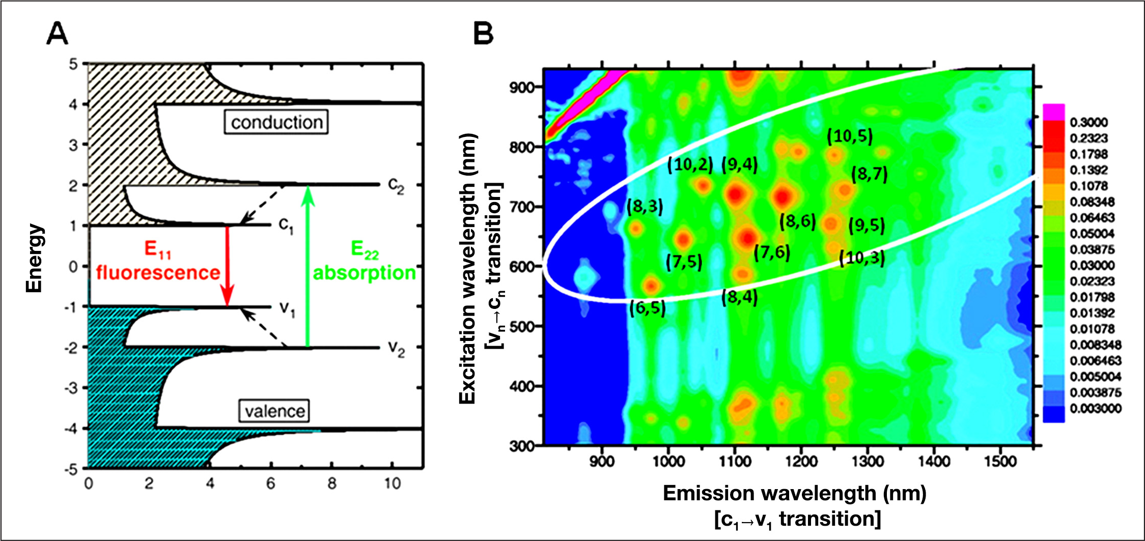
Optical properties of SWNTs. (
Conventional fluorophores, including fluorescent quantum dots, typically emit fluorescence at visible wavelengths and often photobleach rapidly, whereas SWNTs emit fluorescence at NIR wavelengths (900 to 1600 nm), within the so-called biological “tissue transparency window,” in which biological tissues are optically transparent (
Fluorescence properties of SWNTs and absorption and scattering in biological tissues. (
Next we compare the quantum yield, optical tissue transparency, tissue penetration depth, and photostability of SWNTs with those of conventional organic and nanoparticle fluorescence probes, regarding in vivo sensing applications.
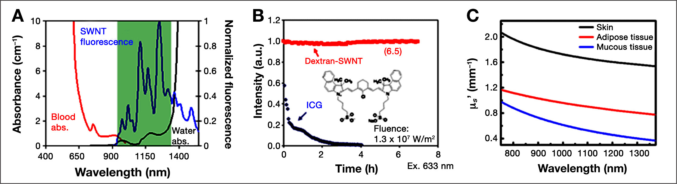
The intensity of the signal emitted by optical sensors is an important design constraint. For in vivo optical sensing, the optical signal emitted by optical sensors depends both on the quantum yield and the absorption in tissues. The quantum yield is the efficiency of energy transfer from incident light to emitted fluorescence. The absorption in biological tissues strongly depends on the wavelength (
Quantum Yield, Absorbance in Human Whole Blood (μ), and φe−2μd of Common Visible and Near-Infrared Organic and Nanoparticle Fluorescent Probes a
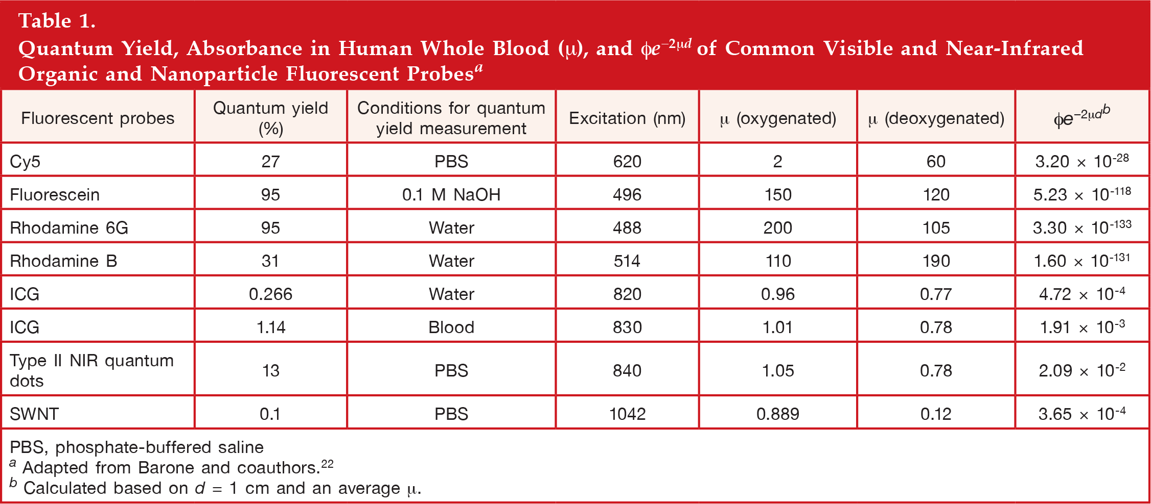
PBS, phosphate-buffered saline
Adapted from Barone and coauthors. 22
Calculated based on d = 1 cm and an average μ.
In addition to absorption, scattering also attenuates the fluorescent signal from optical sensors (
Experimental and simulation modeling studies of the effect of tissue scattering on the tissue penetration depth showed that the maximum penetration depth can be obtained at the wavelength range 1000–1400 nm, called the second NIR widow (NIR II).44–47 Single-walled carbon nanotubes emit fluorescence within the NIR II, whereas most commercially available NIR fluorophores emit fluorescence in the NIR I (700 to 900 nm), including indocyanine green (ICG) and NIR fluorescent quantum dots.44,48 The lower scattering and autofluorescence in the NIR II may compensate for higher absorption in NIR II (mostly due to water) than NIR I and make NIR II-emitting SWNTs outperform conventional NIR I-emitting fluorophores for in vivo applications (
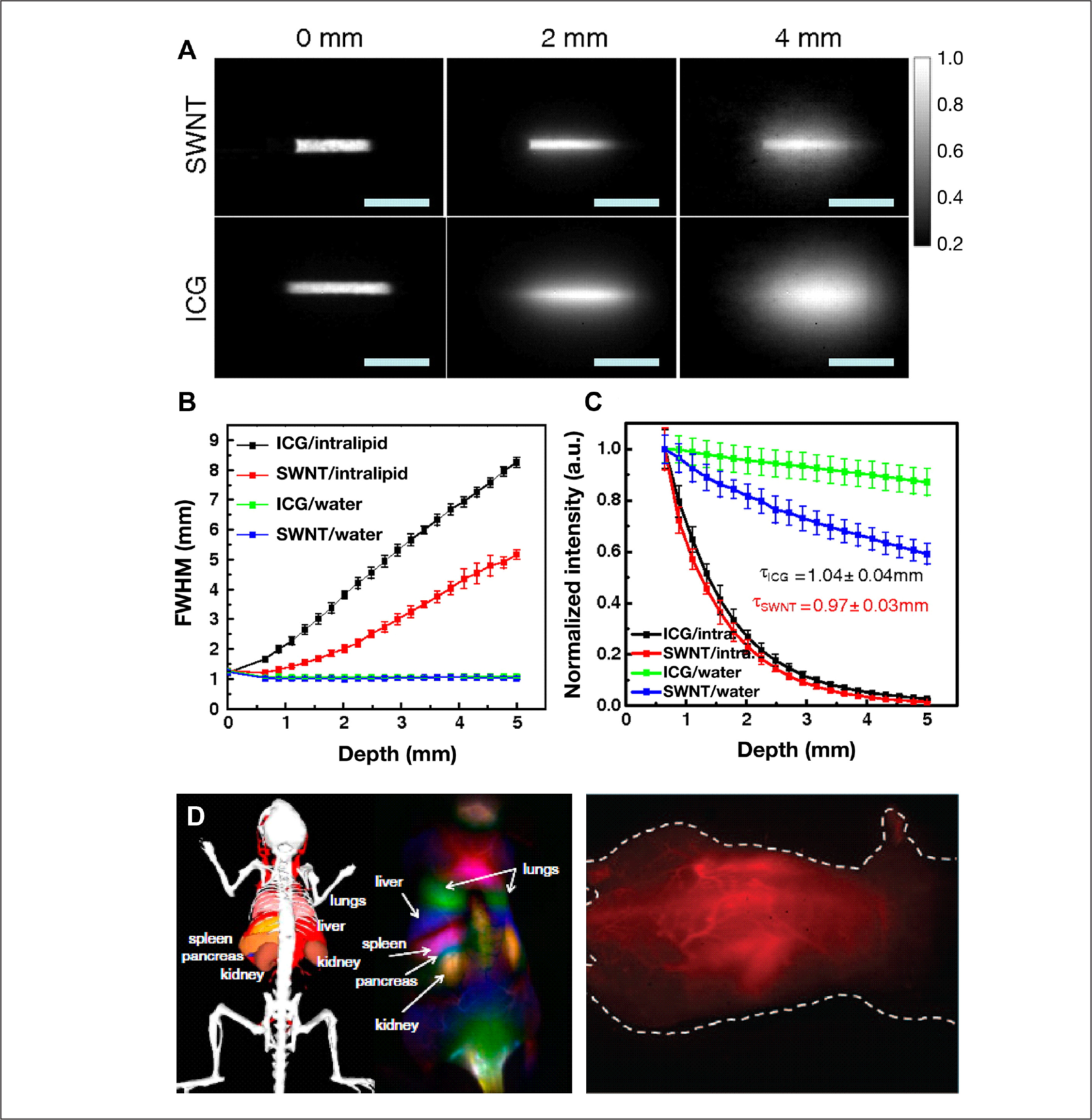
Optical penetration depth of NIR II-emitting SWNTs and NIR-I-emitting ICG in tissue phantoms. (
The photostability of fluorophores determines the lifetime of optical sensors. Assuming the maximum tolerable error of viable in vivo glucose sensors of 20% for clinical applications,
5
a simple calculation from the attenuation factor of e−kτ in the fluorescent signal intensity IS estimates the lifetime τ of optical sensors as τ = -ln(0.8/k).
Photobleaching and Sensor Lifetime of Common Organic and Nanoparticle Fluorescent Probes a
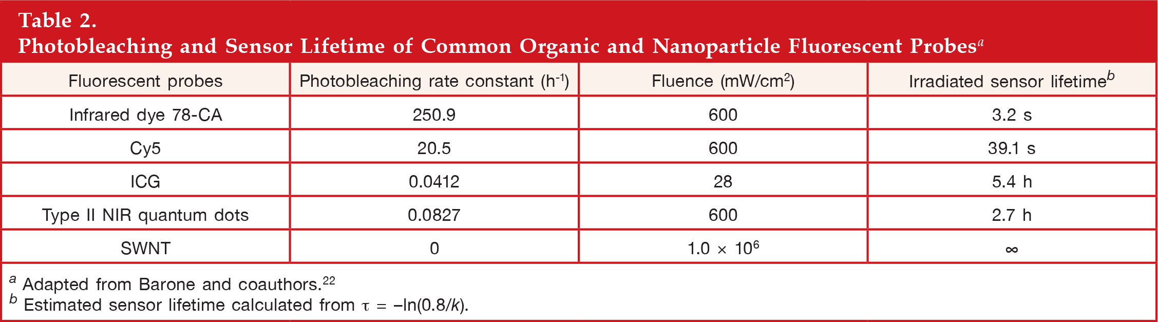
Adapted from Barone and coauthors. 22
Estimated sensor lifetime calculated from τ = -ln(0.8/k).
Selective Modulation of Single-Walled Carbon Nanotube Fluorescence
The fluorescence of SWNTs is highly responsive to the local physical and chemical environment (e.g., local dielectric environment), making SWNTs a unique nanoscale optical sensing platform.53–55
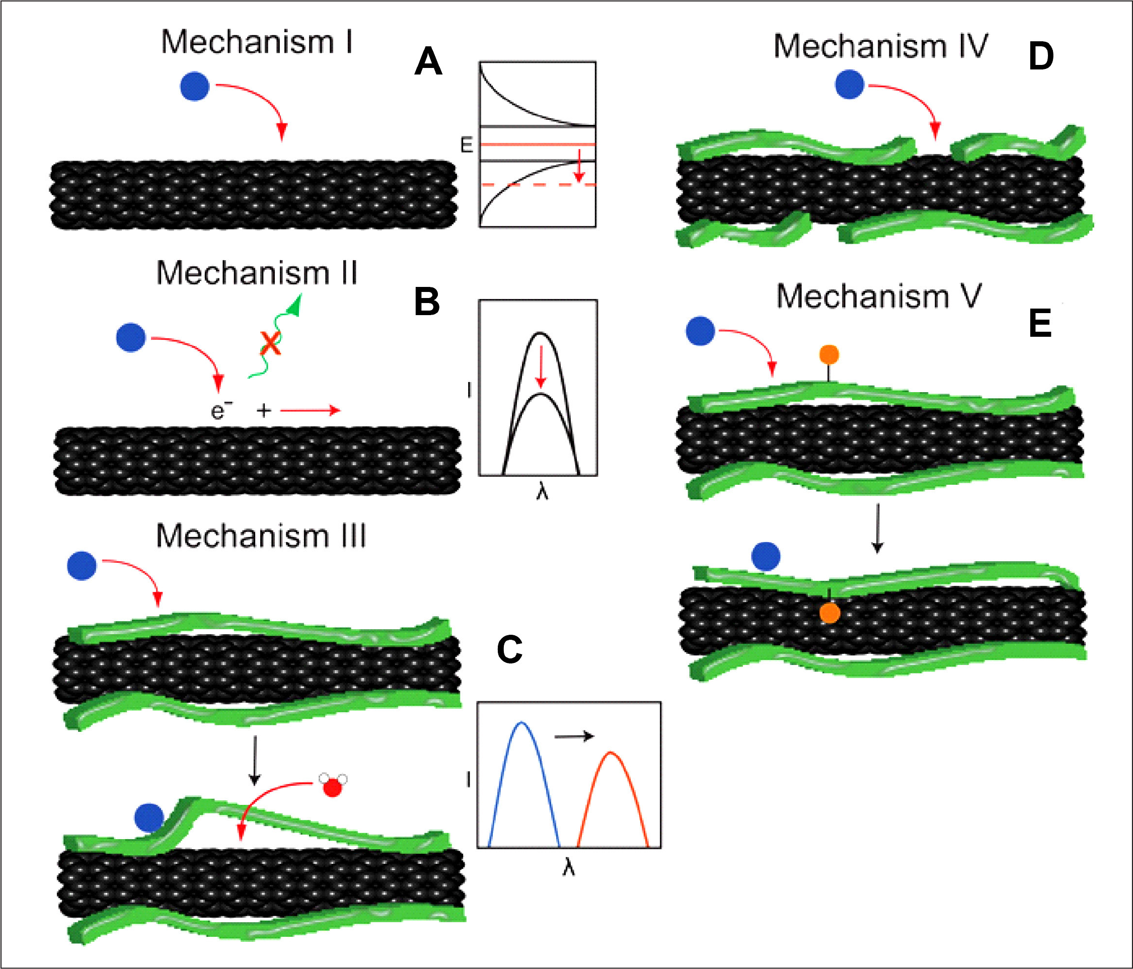
Representative mechanisms of SWNT fluorescence modulation.
Single-Walled Carbon Nanotube-Based Near-Infrared Optical Glucose Sensors
We have demonstrated a proof-of-concept for modulating the NIR fluorescence of SWNTs in response to glucose and developed prototype SWNT-based glucose sensors.29,30 A central challenge in designing SWNT-based sensors is to engineer the nanotube interface to be selective to glucose and effectively modulate SWNT fluorescence, while maintaining the colloidal stability of SWNT complexes. Because direct covalent functionalization of the surface of SWNTs disrupts SWNT fluorescent emission, noncovalent functionalization is required. Here we describe two SWNT-based glucose sensors: glucose-binding-protein-based and BA-based SWNT optical glucose sensors. Our earlier sensors have been reviewed previously. 7
Glucose Binding Protein-Based Single-Walled Carbon Nanotube Glucose Sensors
Glucose-binding protein is a periplasmic-binding protein with high affinity for glucose that changes its conformation upon binding of glucose (from the open to the closed form;
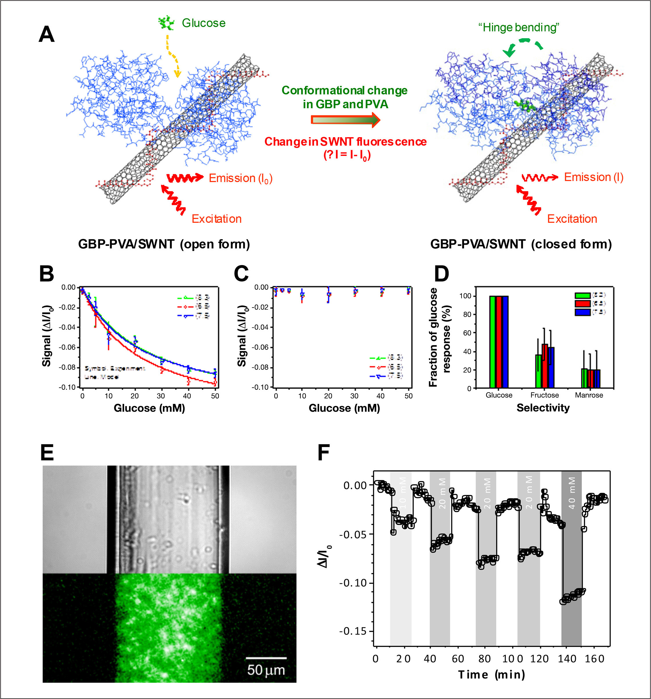
Glucose binding protein-based SWNT optical glucose sensors.
Boronic-Acid-Based Single-Walled Carbon Nanotube Glucose Sensors
Boronic acids are an excellent molecular receptor for glucose.62,63 The reversible complexation of glucose with aromatic BAs produces a stable boronate anion, changing the electronic properties of the BAs.64–67 This alternation in the electronic properties of aromatic BAs upon binding of glucose has been a basic scheme for various BA-based glucose-sensing approaches.63–66,68–70 It was hypothesized that the complexation of glucose with aromatic BAs conjugated on the surface of SWNTs could modulate the SWNT fluorescence signal in response to binding of glucose
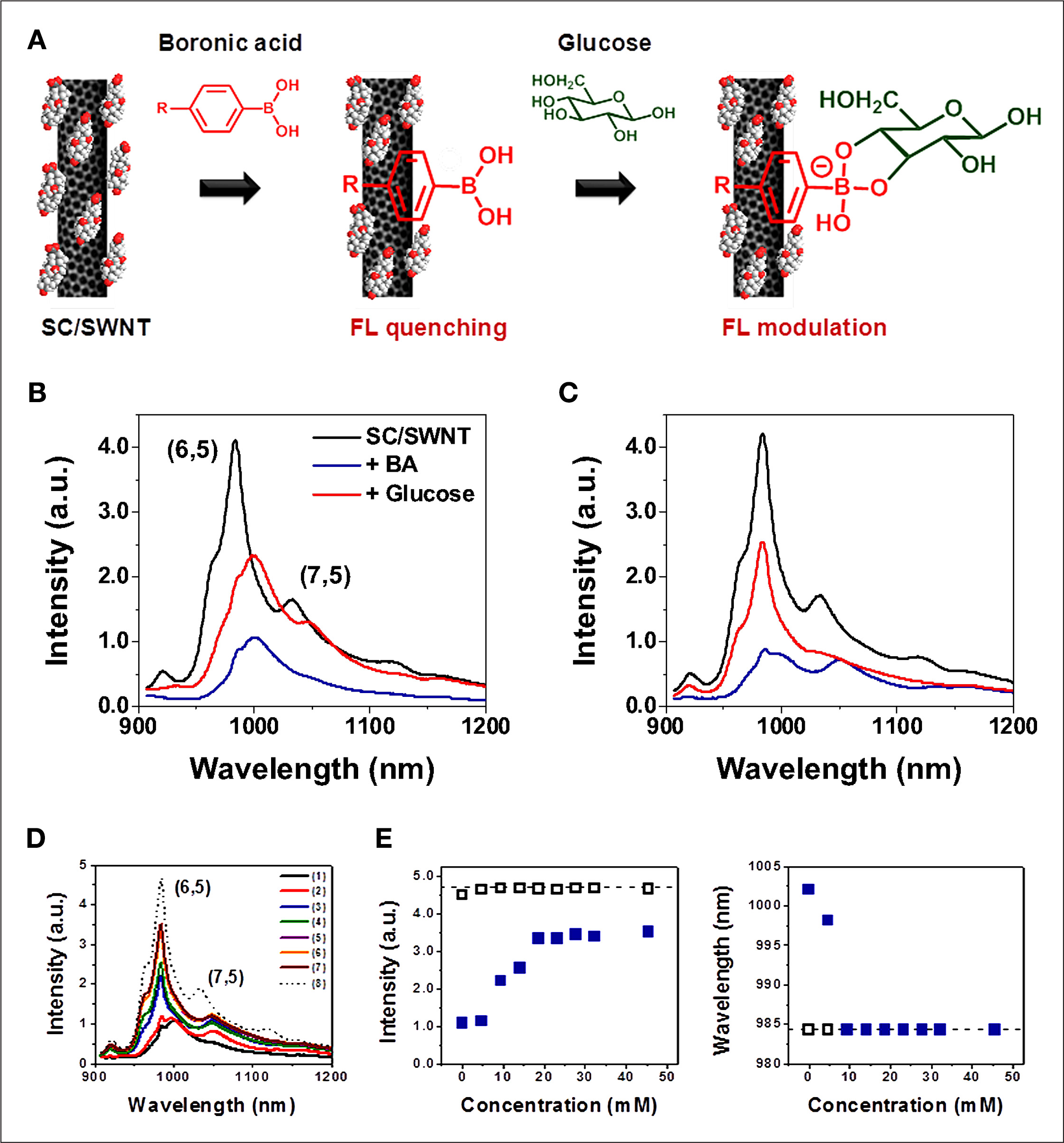
Reversible fluorescence quenching of BA-SWNT complexes for glucose detection.
This “turn-on” sensing scheme that uses the reversible fluorescence quenching and wavelength shift of the BA-SWNT complex has a potential to be applied to develop a highly stable and sensitive NIR optical glucose sensor. However, the current form of the BA-based SWNT sensors requires surfactant in the nanotube sensor solution to disperse the nanotubes in aqueous solutions.
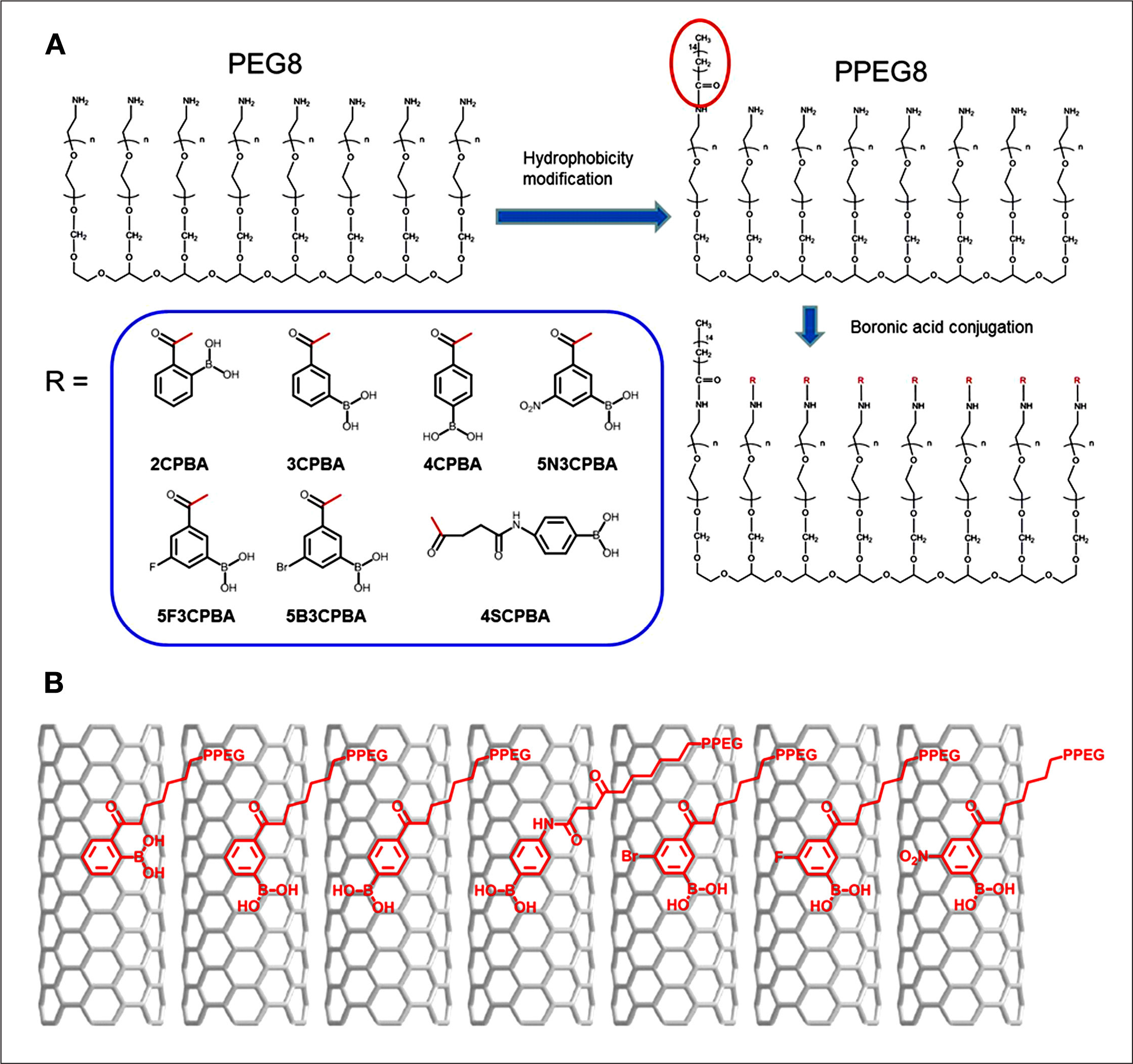
Polymer complexes that can wrap SWNTs and disperse them in aqueous solutions and contain BA receptors to specifically detect not only glucose, but also other saccharides are thus under active research, including BA-conjugated polyethylene glycol-based polymer complexes.
71
In this work, we demonstrated both functions using a homologous series of seven phenyl BAs conjugated to a polyethylene glycol, eight-member, branched polymer (PPEG8) that allows for aqueous dispersion of SWNT and quenching of the NIR fluorescence in response to saccharide binding. We compared the 2-carboxyphenylboronic acid (CPBA), 3CPBA, 4CPBA, N-(4-Phenylboronic)succinamic acid, 5-Bromo-3-carboxy, 3-Carboxy-5-fluoro, and 3-carboxy-5-nitro, demonstrating a clear link between SWNT photoluminescence quantum yield and BA structure
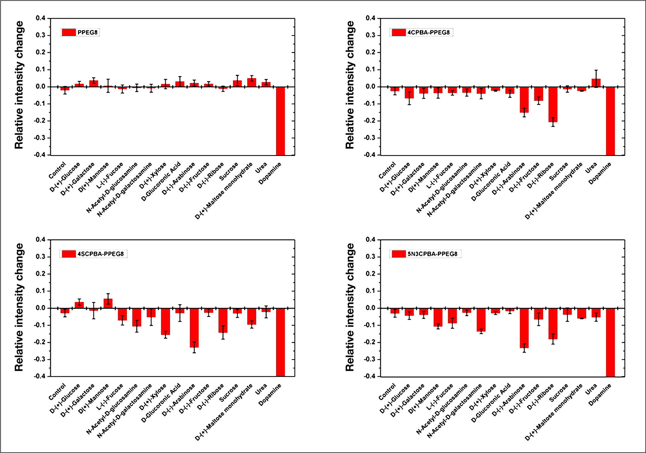
Response of saccharides in different phenylboronic acid-PPEG8 polymer dispersed SWNT solutions. All polymer-wrapped SWNT solutions were diluted using phosphate-buffered saline buffer to a final SWNT concentration of 2 mg/liter. The concentration of SWNT was determined using absorbance at 632 nm with c = 0.036 (mg/liter) 1 cm−1. All analytes were dissolved in water at 1 M concentration, and 2 μl analyte solutions were added to 200 μl SWNT, such that the final analyte concentration was 10 mM. The mixture solution was incubated for 1 h before the NIR fluorescence measurement. The intensity change is calculated based on the fluorescence of SWNT (9,4). Error bar is given by repeating three times for each measurement. Dopamine completely quenched fluorescence of SWNTs as a positive control. Reprinted with permission from Journal of the American Chemical Society. 71
Opportunities and Challenges for In Vivo Continuous Glucose Monitoring
Testing in vivo responses of the SWNT glucose sensors is critical for their clinical applications. A challenge is to improve the sensitivity, selectivity, and lifetime of the sensors in physiologically relevant conditions, including varying oxygen levels, pH, temperature, and potential interferents. An approach to achieve this is to use an internal reference by using two or more different types of SWNTs that emit fluorescence at different wavelengths for sensors and references, respectively. Another task to consider for in vivo applications is to construct microscale structures that encapsulate SWNT-based sensors. For in vivo sensing applications, the direct injection of nanoscale water-soluble SWNT sensors would not be practical, because the nanoscale SWNT sensors can circulate and distribute through the body;
44
instead, microscale glucose-responsive structure that are small enough to be injected or implemented in the body yet large enough to remain at the implantation site for a long period of time would be required for stable in vivo sensing.72,73 Because the SWNT sensor is in aqueous suspension, the final sensor architecture is flexible, including dialysis capillary (
Another challenge is that sensors implanted in the body often fail because of biofouling of the sensor surface, inflammation, and fibrosis-induced vessel regression at the implantation site.74–78 An approach to reduce the problems is to coat the surface of the glucose-sensing structure (e.g., hydrogel microparticles encapsulating SWNT glucose sensors) with the antifouling coating and incorporate anti-inflammatory agents, such as dexamethasone (DX), and angiogenic factors, such as vascular endothelial growth factor (VEGF), into the glucose-sensing structure.74–78 For instance, delivering VEGF-producing cells to the site of glucose sensor implantation using a tissue-interactive fibrin biohydrogel induced significant neovascularization and improved the glucose sensor function in vivo. 76 The surface coating of hydrogels and microcapillaries with DX and VEGF and the release of DX and VEGF in the tissue reduced the biofouling and developed local environments favorable for long-term, reliable in vivo sensing.74,79
Testing the toxicity of SWNT sensors is an important issue for in vivo applications. Many studies have investigated the potential toxicity of carbon nanotubes to in vitro cell cultures and in vivo animal models and have shown various results, depending on the type and geometry of nanotubes, nanotube materials preparation, and surface functionalization. 27 Properly surface-functionalized carbon nanotubes have not induced obvious toxicity in cell culture experiments and in vivo studies,80–83 whereas raw carbon has induced pulmonary toxicity and mechanical blockage of the upper airways in animal models.84–88 Long MWNTs introduced into the abdominal cavity of mice also developed asbestos-like, length-dependent pathogenicity, including inflammation and formation of granulomas. 88 For the proposed SWNT-based in vivo glucose sensing, the SWNT sensor-encapsulating microstructures, such as hydrogel microparticles and microcapillaries, provide another protective layer (i.e., biocompatible hydrogels) other than biocompatible functionalization layers on SWNTs, which reduce the potential toxicity resulting from the direct contact of SWNTs with biological entities and the needle-like shape of SWNTs. 88 Considering the growing interest and use of SWNTs for biomedical application, further investigations are required to fully address the SWNT toxicology.27,88
Conclusions
SWNT-based NIR optical glucose sensors with their extraordinary NIR, nonphotobleaching fluorescence have a great potential for long-term in vivo CGM. A key challenge in designing SWNT-based glucose sensors is to identify and engineer molecular receptors for glucose that will work effectively in a SWNT sensing system and to understand how to effectively transfer glucose-binding events into the modulation of SWNT fluorescence to achieve the selectivity, sensitivity, and dynamic range of the sensors for in vivo glucose monitoring. Advancement of this technology also requires simultaneous research efforts toward translating the laboratory development of SWNT-based NIR optical glucose sensors into in vivo glucose sensing for practical clinical uses. The ability to sense glucose with nanoscale SWNT-based NIR optical sensors would also allow for new ways to answer important biological questions regarding glucose metabolism. The emerging technology of SWNT-based NIR optical glucose sensors can also be extended to various in vivo health monitoring systems.
Footnotes
Abbreviations:
This work is funded by a grant from Sanofi-Aventis.