
Abstract
Using zinc nitrate hexahydrate as the precursor and Rhus vulgaris fruit extract as a natural reducing and capping agent, this study describes the environmentally friendly synthesis of zinc oxide nanoparticles (RV-ZnONPs). The presence of bioactive substances like phenols, flavonoids, tannins, alkaloids, saponins, terpenoids, steroids and glycosides responsible for the reduction of Zn2+ ions nucleation and stabilization of ZnONPs. UV-Vis spectroscopy, X-ray diffraction (XRD), zeta potential analysis (XRD), transmission electron microscopy (TEM) and Fourier-transform infrared (FT-IR) spectroscopy were used to characterize the biosynthesized RV-ZnONPs. A distinctive absorption peak with an estimated band gap energy of 3.20 eV was found by UV–Vis analysis at approximately 371 nm. While TEM images revealed primarily irregular nanoparticles with an average size of ∼23. 5 nm, XRD confirmed the formation of highly crystalline ZnONPs with a hexagonal wurtzite structure and an average crystallite size of ∼22.7 nm. Excellent colloidal stability was demonstrated by the high negative zeta potential value (−35.1 mV) and FT-IR analysis verified the role of functional groups derived from plants in capping and stabilizing nanoparticles. The photocatalytic, antioxidant, and antibacterial properties of RV-ZnONPs were also assessed. Under UV irradiation, the RV-ZnONPs demonstrated effective photocatalytic degradation with maximum degradation rate of 65.4%. The degradation efficiency rose with nanoparticle concentration and reaction time. Antioxidant activity assessed by the DPPH assay demonstrated concentration-dependent free-radical scavenging with RV-ZnONPs showing a significantly lower IC50 value (74.46 µg/mL) compared to the extract from crude fruit. Furthermore RV-ZnONPs demonstrated significant antibacterial activity against Staphylococcus aureus and Escherichia coli which was ascribed to membrane disruption, reactive oxygen species generation, and Zn2+ ion release. This study highlights the promising potential of Rhus vulgaris fruit extract for environmental remediation antioxidant applications and antimicrobial treatments by demonstrating that it offers an efficient green pathway for the synthesis of stable and multifunctional ZnONPs.
1. Introduction
Nanotechnology and nanopharmacology, which focus on the manipulation and study of matter at the atomic scale (typically 1 to 100 nm), have emerged as critical areas of research and development in the 20th century.1–4 When materials are reduced to the nanoscale, their surface-to-volume ratios increase significantly, enhancing surface effects and altering physicochemical properties.5,6 When compared to their bulk counterparts nanomaterials with larger surface areas show better reactivity and biological properties creating a high opportunities in various scientific Fields. 7 These enhanced features, particularly high reactivity and surface functionality, make nanoparticles especially suitable for pharmacological and biomedical applications.8,9 These findings suggest the possibility of surface engineering for enabling spatially controlled modulation of the electronic structure of low-dimensional nanocrystals on a scalable fashion. 10
Among the various types of nanoparticles, metal oxide (MO) nanoparticles are particularly promising due to their unique biological, physical, and chemical characteristics, including solubility, chemical stability, and adhesiveness. 11 However, traditional techniques for preparing MO nanoparticles often rely on hazardous chemicals that are harmful to the environment, animals, and plants. Conventional physical and chemical synthesis methods frequently require high temperatures, excessive energy consumption, and toxic organic solvents or reducing agents such as hydrazine and sodium borohydride. 10 Consequently, there is a growing shift towards green chemistry. Synthesizing MO nanoparticles using green methods, such as plant extracts, offers a simple, affordable, eco-friendly, and non-toxic alternative. 11 This approach addresses the limitations of current ZnO nanoparticle research, where the toxicity of chemical reductants and the environmental cost of physical methods remain significant barriers to sustainable industrial application.
ZnO, MgO, Fe2O3, CuO, Ag2O, CaO, NiO, and ZrO2 are examples of metal oxide nanoparticles that have garnered significant interest due to their potential applications in biomedicine and environmental remediation.12–16 However, the synthesis of complex architectures and the management of interfacial properties in these challenging compounds based on them remain significant hurdles. 17 ZnO nanoparticles, in particular, are promising candidates in biomedicine, demonstrating anti-cancer, antibacterial, anti-inflammatory, antioxidant, and anti-diabetic properties. 18 Specifically, ZnONPs possess versatile properties investigated for electrical conductivity, catalytic functions, and use in the cosmetic industry, 19 However, the selection of applications in this study is driven by specific global challenges. Photocatalytic activity is targeted to address environmental pollution, specifically the degradation of persistent organic dyes like methylene blue in wastewater, which poses a threat to aquatic ecosystems. Antibacterial activity is selected to combat the rising crisis of antibiotic-resistant bacterial infections in healthcare. Antioxidant activity is crucial for mitigating oxidative stress-related conditions in biological systems. Green manufacturing of ZnO nanoparticles using plant extracts can enhance these properties while minimizing environmental impact. 20
Rhus vulgaris is a wild edible fruit-producing plant under the family Anacardiaceae, primarily found in temperate, subtropical, and tropical regions, particularly in North America and Africa (including Tanzania, Uganda, Kenya, Ethiopia, Mozambique, Malawi, Zambia, and Zimbabwe). 21 Phytochemical studies on R. vulgaris have revealed a high concentration of bioactive compounds, including phenols, glycosides, alkaloids, saponins, tannins, and terpenoids.22,23 The specific advantage of using R. vulgaris fruit extract lies in its rich phytochemical composition. The phenolic hydroxyl and carboxyl groups present in these compounds act as potent electron donors, effectively reducing metal ions (Zn2+) to metallic ZnO, while simultaneously acting as capping agents to stabilize the nanoparticles and prevent agglomeration.11–13 While R. vulgaris possesses known biological characteristics, including antibacterial, anti-inflammatory, wound-healing, antiulcerogenic, and anti-cancer effects, 24 there is a notable research gap regarding its application in nanotechnology. Although R. vulgaris has been utilized for therapeutic applications, ZnO nanoparticles mediated by R. vulgaris fruit extracts and their potential for combined photocatalytic, antioxidant, and antibacterial activities have not been thoroughly investigated. Most existing studies focus on other plant species, leaving the unique potential of R. vulgaris underutilized.
Therefore, the current study utilizes Zinc Nitrate Hexahydrate as a starting material and R. vulgaris fruit extracts as a reducing and stabilizing agent to synthesize ZnONPs. This work aims to evaluate the synthesized nanoparticles for photocatalytic degradation of pollutants such as methylene blue and biomedical applications such as combating bacterial infections and oxidative conditions. By leveraging the eco-friendly properties of R. vulgaris, this research seeks to contribute to the development of sustainable, multifunctional nanomaterials for wastewater treatment and healthcare.
2. Material and methods
2.1. Plant collection
Rhus vulgaris Fruits were collated from the Amhara region, East Gojam zone, Mertulemariam Wereda, Ethiopia, located about 364 km from the capital city of Addis Ababa, Ethiopia. The collection of this plant material was carried out in accordance with current institutional, national and international directives and legislation. For a collection of Rhus vulgaris fruit, permissions or licenses were obtained from the Agricultural Center of Mertulemariam Wereda. The collected plant materials were taxonomically identified by Dr. Melkamu Wondaferah, Department of Botany, and Addis Ababa University. National guidelines and legislation were strictly followed for the sample collection of plant sample. The voucher specimens of the collected plant materials were deposited at the National Herbarium with voucher number 3074. The plant samples will be collected when ready for consumption according to traditional usage. After gathering the healthy Fruits, they were adequately cleaned using tap water to remove any remaining soil. After washing, the Fruits were allowed to air dry for 15 to 20 days. Using a mortar and pestle, the dried fruit components were ground into a fine powder and placed in a container without air for later usage at room temperature (23 oC).
2.2. Rhus vulgaris fruit extraction procedures
The chemical composition of the Rhus vulgaris fruit extract including major phytochemicals like flavonoids, phenolics, alkaloids, and terpenoids, has been detailed in previous studies.25,26 The plant extraction from the Rhus vulgaris fruit was carried out based on the methods used by Shankar et al. (2004). 27 The extraction temperature of 60 °C and duration of 10 minutes were selected based on previous studies indicating that this range optimizes the yield of phenolic and flavonoid compounds while minimizing thermal degradation of heat-sensitive phytochemicals.28,29 100 ml of distilled water were added to a conical flask containing 8 grams of finely ground Rhus vulgaris fruit. The flask was then heated continuously at 60 °C on a hotplate for 10 minutes to facilitate the extraction of bioactive compounds. To prevent contamination and evaporation during heating, the mouth of the flask was tightly sealed with aluminum foil. The mixture was heated and then left to cool naturally to room temperature (23 oC). To get rid of particles and create a clean solution, the cooled extract was filtered using Whatman filter paper number 1. The filtrate was stored at room temperature in a sterile, airtight container for further laboratory activities. These settings were used to retain the stability and bioactivity of this aqueous extract for use in pharmacological testing. Studies showed that the aqueous extraction of bioactive compounds from Rhus vulgaris fruit can enhance biological functions. 30
2.3. Biosynthesis of zinc oxide nanoparticles
RV-ZnONPs were prepared utilizing green synthesis method by means of Rhus vulgaris fruit extract. After preparation of the plant extract as described previously, 30 mL of this extract was put into a flask and heated gradually. When the temperature reached 60 °C, 3 g of zinc nitrate hexahydrate were added to this extract. After that the mixture was continuously stirred, maintaining the temperature at 60 °C, at pH value of 10 until the mixture converted into a yellowish paste after 1 h. It is obvious that, the temperature of reaction played important role in producing nanoparticles (NPs), the optimal yield of NPs were achieved at 60 °C. The calcination temperature of 400 °C was chosen based on literature reports which suggest that this temperature is sufficient to convert zinc hydroxide precursors into crystalline ZnO without causing significant particle agglomeration or excessive energy consumption.31,32 Afterward the paste was blazed in a furnace at 400 °C for about 2 h then the residual was washed by ethanol and distilled water several times. The powder was then heated at 100 °C to dry the samples. Then zinc oxide nanoparticles were obtained and they were ready for characterization. Figure 4 shows the procedure of synthesizing zinc oxide nanoparticles using Rhus vulgaris fruit extract and zinc nitrate hexahydrate as a precursor.
The mechanism and the responsible biomolecules for the nanoparticle’s formation via plant extracts have not yet been entirely documented.
33
The plausible mechanism of RV- ZnONPs formation, using fruit extracts, is described as the following


The biomolecules such as natural phenolic compounds (Ar–(OH)n) of Rhus vulgaris fruit extract, responsible for formation RV- ZnONPs, could lose their electron for the efficient reduction of Zn2+ ions to Zn0. This might cause the Zn0-phenolate complex by chelating effect causing nucleation and growth of NPs at 60 °C. This complex undergoes direct decomposition at higher temperature of 100 °C in air and lead to RV- ZnNPs. Thus, the natural phenolic compounds had favorable effects on RV-ZnO NPs synthesis.
2.4. Characterization of nanoparticles
The UV-Vis absorbance spectrum of the RV-ZnONPs was recorded using a UV-Vis spectrophotometer (JASCO V-670). The measurements showed a strong absorption peak in the 300–400 nm range which was a crucial sign that RV-ZnONPs had been successfully synthesized and offered crucial information about the process of nanoparticle formation.
34
Visual cues like a discernible color shift in the reaction mixture were used to confirm the nanoparticle formation. The powder particles were subjected to TEM analysis at an accelerating voltage of 200 kV yielding a resolution of about 1 Å. To achieve this a probe sonicator was used to disperse the RV-ZnONPs sample in triple distilled water (TDW). Before performing the TEM characterization, a droplet of the resultant suspension was placed onto a carbon-coated copper grid and allowed to air-dry at room temperature. Using Fourier Transform Infrared (FTIR) Spectroscopy the crucial functional group utilized as an RV-ZnONP reducing and capping agent was characterized. An FTIR spectrometer was used to gather the RV-ZnONPs FTIR spectra within a wavenumber range of 400–4000 cm-1. An IS50 FTIR spectrometer was used to perform the FTIR analysis.
35
Using an XRD diffractometer with monochromatic Cu Kα radiation (λ = 1. 54056 Å) over a 2θ range of 5°–80°. The crystallite size and morphology of the RV-ZnONPs produced from zinc nitrate hexahydrate were assessed. Equation (3) the Scherrer equation was used to calculate the crystallite size.
36

2.5. Antioxidant activity
The antioxidant activities of RV-ZnONPs were performed using DPPH (2,2-diphenyl-1-picrylhydrazyl) free radical assay using a methods used Blois by Blois et al. (1958)
37
and Desmarchelier et al.
38
A 0.1 mM methanolic DPPH solution was prepared fresh daily. RV-ZnONPs stock solutions were prepared at 1000 µg/mL in methanol and serially diluted to working concentrations of 25.0, 75.0, 100.0, and 125.0 µg/mL. Upon addition of a hydrogen-donating substance (antioxidant material from Rhus vulgaris Fruit extracts) to the alcoholic DPPH solution, the DPPH radical is reduced, resulting in a color change from purple to pale yellow. Accordingly, 2 mL of a freshly prepared 0.1 mM methanolic DPPH solution was mixed with 2 mL of RV-ZnONPs solution at different concentrations of 25.0, 75.0, 100.0, and 125.0 μg/mL. After that, the test tubes were placed in the dark for 30 minutes. After incubation, the absorbance was measured at 517 nm. A concentration of ascorbic acid (AA) ranging from 25 to 125 μg was utilized as a reference or control. Through measuring the absorbance of the reaction mixture during measurement, a higher degree of free radical scavenging activities can be identifies.
39
Equation (4) was used to determine the DPPH radical scavenging properties of the samples using AA as a reference in order to evaluate their antioxidant capabilities.

2.6. Antibacterial activity
Using the agar well diffusion method, the antibacterial susceptibility of RV-ZnONPs was evaluated against two different bacteria species obtained from Gondar university which are isolated clinically such as S. aureus, and E. coli. Bacterial cultures were maintained on nutrient agar slants and sub-cultured in nutrient broth to obtain a standard turbidity of 0.5 McFarland standard (approx. 1.5 x 10^8 CFU/mL). The bacterial culture was evenly distributed across the hardened nutrient agar plates using a sterilized glass spreader dipped in the inoculum suspension. After that, a 6 mm well was made on the agar plates’ surface. RV-ZnONPs stock solution (1000 µg/mL) was diluted to 500 µg/mL in sterile distilled water for the assay. After that, a 6 mm well was made on the agar plates’ surface. After adding of the produced RV-ZnONPs solution to the wells, the Petri plates were incubated for 24 hours at 37°C in order to assess the zone of inhibition. 40 In this experiment, streptomycin (10 µg/ml) served as the reference. Millimeters were used to measure the inhibitory zone’s diameter and compare it to the control.
2.7. Photocatalytic activity
UV irradiation was used to analyze the photocatalytic activity of RV-ZnONPs using previously reported procedures done by Fallah et al.
41
and Zhai et al.
42
depending on the photodegradation response of methylene blue (MB) dye. A 10-ppm stock solution of MB was prepared in deionized water. RV-ZnONPs were dispersed in deionized water to create working concentrations of 100, 200, 300, 400, and 500 µg/mL. To investigate the photocatalytic activities of RV-ZnO NPs, MB was subjected to high-intensity (365 nm, 115V-60 Hz, 2.5A, 100 W) Lamp UV irradiation. After forming a 10 ppm stock solution of MB in 500 ml of deionized water, 2 mg of different samples such as RV-ZnONPs1, RV-ZnONPs2, RV-ZnONPs3, RV-ZnONPs4 and RV-ZnONPs5 were added to 60 ml of a 10 ppm dye solution. Before photoirradiation, the solution was agitated with a magnetic stirrer for 15 minutes in the dark to form adsorption-desorption equilibrium in the system. The samples were then exposed to UV light generated by a high-intensity lamp (365 nm, 115V-60Hz, 2.5A, 100 W) for 25 minutes. The absorbance of each sample was measured to identify the one with the highest photocatalytic activity. Subsequently, the effect of reaction time on dye degradation was investigated at intervals of 25, 50, 75, 100, and 125 minutes. After the designated irradiation periods, the samples were removed from the colloidal mixture. The supernatant was then separated by centrifugation at 5,000 rpm for 15 minutes to remove any residual nanoparticles. The absorbance spectra of the supernatant were recorded across the wavelength range of 350 to 850 nm using a PerkinElmer UV-Vis spectrophotometer (USA). The concentration of the dye during degradation was calculated at 660 nm using Equation (5).

2.8. Statistical analysis
Origin 2025 software was utilized to evaluate variance using a one-way ANOVA. The standard deviation (SD) and average of the three runs of each measurement, which were performed three times (n=3), are displayed. Significance was determined using P ≤ 0.05.
3. Result and discussion
The aqueous extract of Rhus vulgaris fruit contains Saponins, Tannins, Phenols, Alkaloid, Flavonoids, Terpenoids, Steroids and Glycoside from our previous research work. 43 These phytochemicals possess Rhus vulgaris fruit to have different pharmaceutical activities such as lipid peroxidation, metal ion reduction abilities, antibacterial, free radical scavenging or antioxidant, and stabilization abilities.25,26 In this study, phytochemicals obtained from aqueous fruit extract of Rhus vulgaris was utilized to cap Zn2+ ions, thereby promoting the nucleation of ZnONPs. The synthesized RV-ZnONPs were then characterized employing various analytical techniques and evaluated for their potential photocatalytic, antioxidant, and antibacterial activities. Based on their size and functionalization, nanoparticles have the rare capacity to change the properties of larger materials. 44 As observed in FTIR peaks (Figure 5) at 3400 cm-1 indicates the presence of O-H, at 1625 cm-1 the presence of C=O functional group to specific phytochemicals like phenols and flavonoids. The bioactive compounds from fruit extract of Rhus vulgaris such as flavonoids and phenolic compounds exhibit versatile chelating and reducing properties, enabling them to cap Zn2+ ions and facilitate ZnO nucleation. Phenolic compounds form several chelating bonds that stabilize the ZnONPs after nucleation while flavonoid groups initiate the capping process on Zn2+ ions resulting in the formation of nanoparticles with a variety of size distributions. The chemical as well as the physical properties of ZnONPs can be enhanced by green preparation of nanoparticles.
3.1. UV-vis spectrum
The biosynthesis of RV-ZnONPs was verified by a discernible color shift in the reaction mixture, which turned from white at the start to yellow at the end, signifying the synthesis of ZnONPs. The UV-Vis absorption spectrum (Figure 1), obtained using zinc nitrate hexahydrate as the zinc source, displays a distinct absorption peak at a maximum wavelength of 371.3 nm, which is characteristic of ZnO nanoparticles.
35
The absence of additional peaks confirms the purity of the synthesized RV-ZnONPs. This absorption peak at 371.3 nm indicates electron transfer from the valence band to the conduction band, resulting in intrinsic band gap absorption.
45
The optical band gap energy was determined by constructing a Tauc plot of (αhv)2 versus photon energy (hv) was generated, and the linear portion of the curve was extrapolated to the energy axis (Figure 1(b)). This extrapolation yielded an optical band gap of 3.20 eV, which falls within the typical range of 3.1 to 3.4 eV characteristic of ZnO.
46
This band gap represents electronic transitions between the valence band, largely composed of oxygen 2p orbitals (O2p), and the conduction band, primarily composed of zinc 4s orbitals (Zn 4s). These results are consistent with earlier research; for instance, Sharmila et al. (2019),
47
examined the capacity of T. castanifolia to synthesize ZnONPs, and Abdelbaky et al. (2022),
48
reported a green synthesis of ZnONPs using P. odoratissimum as a mediator. In both studies, a characteristic UV-Vis spectrophotometry peak was observed at approximately 370 nm. Additionally, Nozipho et al. (2024),
7
found a high absorption peak at 350 nm confirming the formation of ZnONPs via M. pruriens extracts as a capping or reducing agent. The absorption property at 350 nm is in line with earlier findings regarding ZnONP synthesis.49,50 (a) UV-visible absorption spectrum of RV- ZnONPs). (b) Tauc plot (αhv)2 versus photon energy (hv) used to determine the optical band gap. The extrapolation of the linear portion of the curve yields a band gap of 3.20 eV.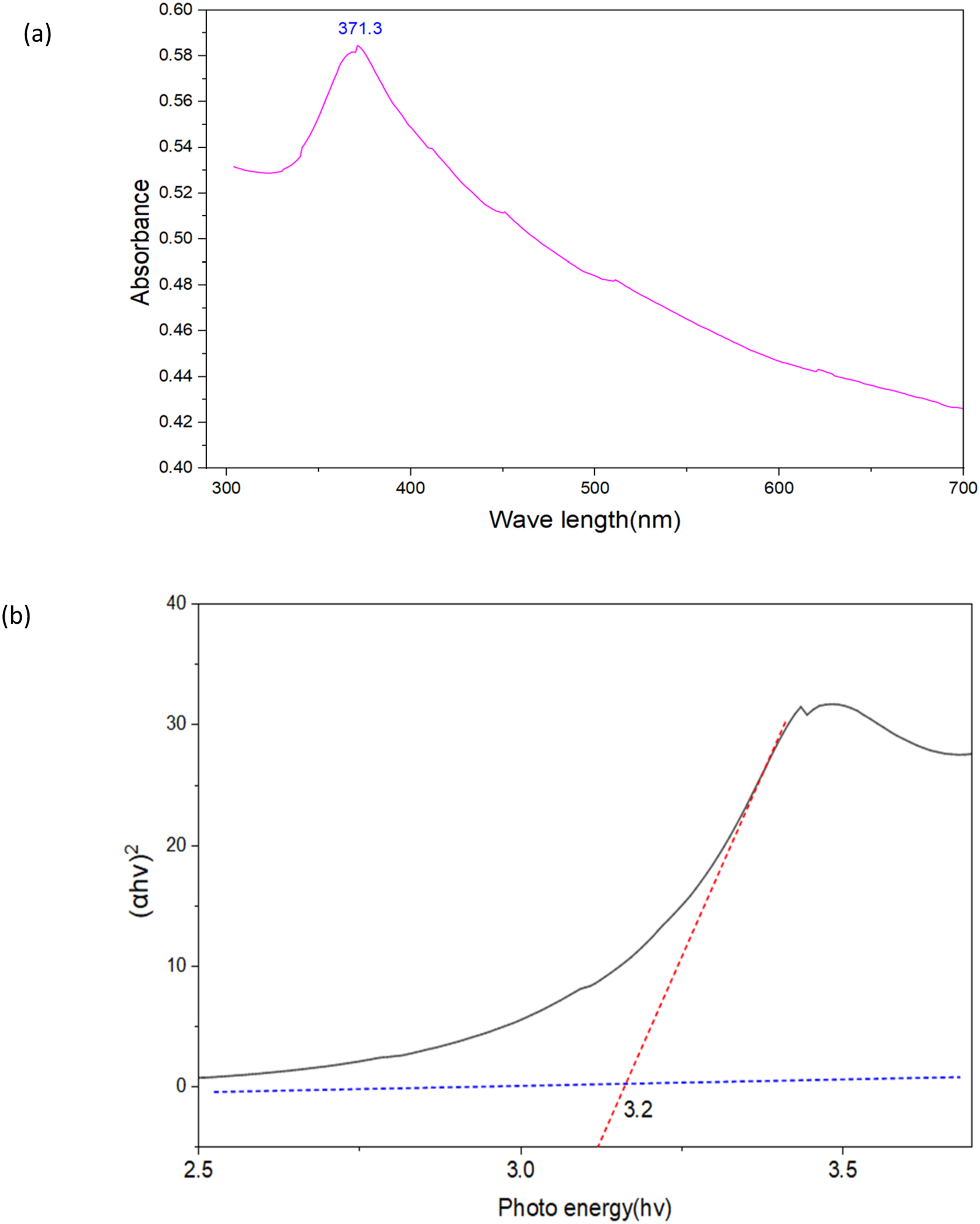
3.2. X-ray diffraction (XRD) analysis
By measuring interatomic distances, bond angles and atomic arrangements within crystalline solids, XRD is a potent method for determining a materials crystal structure.
51
The crystalline nature of the fruit extract of Rhus vulgaris mediated ZnONPs made using zinc nitrate as the precursor is evident in the XRD pattern of the synthesized RV-ZnONPs (Figure 2). Six distinct diffraction peaks are observed at 2θ values of 32.52°, 34.38°, 36.49°, 47.81°, 56.74°, and 62.73° (Figure 2). These diffraction peaks which correspond to the lattice planes (hkl) of (100), (002), (101), (102), (110), (103) and (112) respectively were indexed to the hexagonal wurtzite crystal structure of RV-ZnONPs. This indexing verifies the successful synthesis of crystalline ZnONPs with the anticipated crystal structure and is in line with the standard reference pattern from the Joint Committee on Powder Diffraction Standards (JCPDS) card number 36-1451.
52
The lattice parameters (a and c) were calculated using the Bragg’s law and the relationship for hexagonal systems. The calculated lattice parameters were found to be a = 3.25 Å and c = 5.21 Å, which are consistent with the standard JCPDS card 36-1451. X-ray diffraction (XRD) pattern of RV-ZnONPs. Peaks correspond to the hexagonal wurtzite structure of ZnO (JCPDS 36-1451), confirming crystallinity and phase purity.
The average crystallite size of the ZnONPs was calculated using the Debye-Scherrer equation (equation (3)), utilizing the strongest diffraction signal at the (101) plane.53,54 In this equation, K is the shape factor (typically 0.9), lambda (λ) is the X-ray wavelength, beta (β) is the full width at half maximum (FWHM) of the peak in radians, and theta (θ) is the Bragg angle. The FWHM is a critical parameter for assessing nanoparticle size distribution; narrower peak widths indicate a more uniform crystallite size distribution, whereas broader peaks suggest a wider distribution or the presence of microstrain. Based on the FWHM of the (101) peak, the synthesized ZnONPs exhibited an average crystallite size of 22.7 nm. This size is consistent with findings from earlier studies on ZnONP synthesis conducted by Elumalai 49 and Zak. 50 The XRD data confirms the hexagonal wurtzite structure and suggests a relatively narrow size distribution due to the sharpness of the diffraction peaks. This demonstrates the efficacy of the green synthesis method employing Rhus vulgaris fruit extract in generating well-defined RV-ZnONPs.
3.3. Zeta potential
A zeta potential of −35.1 mV was observed at pH value of 10 indicating strong colloidal stability because of the high negative surface charge. The current value is comparable to or marginally better than previously published results when compared to reports on other Rhus species used for nanoparticle synthesis. For example ZnO nanoparticles made with extracts from Rhus coriaria have demonstrated zeta potential values between −25 and −30 mV indicating moderate but sufficient stability for colloidal systems. 55 In studies on Rhus typhina-mediated ZnONPs have reported values of approximately −28 mV, which indicate stable nanoparticle suspensions with lower electrostatic repulsion than the −35.1 mV found in this work. 55 These comparisons imply that in comparison to other Rhus species, Rhus vulgaris fruit extract may offer more effective capping and stabilization of ZnONPs leading to improved colloidal stability. Different research indicated that, higher zeta potential value of ZnONPs possess high stability for long period of time at room temperature with no significant change in zeta potential or particle size.56,57
The zeta potential values of RV-ZnONPs (-35.1 mV) effectively indicate their stability and surface charge (Figure 3). The negative charge which prevents aggregation and enhances dispersion stability is primarily caused by the hydroxyl groups present in bioactive compounds in the fruit extract of Rhus vulgaris. The negative charge potential of the generated ZnONPs is most likely caused by the reducing agents found in the Rhus vulgaris fruit extract such as the flavonoid and phenolic components.58,59 The stability and charge distribution of the generated nanoparticles are provided by the crucial information in Zeta potential. The present study highlights the significance of surface charge distribution and nanoparticle durability as critical elements for potential applications in biological fields environmental cleanup and catalysis. The outcome demonstrated that by ensuring stable and uniformly distributed nanoparticles the green synthesis method using Rhus vulgaris fruit extract offers a sustainable way to synthesize RV-ZnONPs with desired physicochemical characteristics. Zeta potential (mV) distribution for RV-ZnONPs, indicating a positive surface charge that contributes to colloidal stability through electrostatic repulsion.
3.4. Transmission electron microscopy (TEM)
Transmission Electron Microscopy (TEM) is used to analysis both morphology and size distribution of the synthesized ZnONPs using Rhus vulgaris fruit extract. The Transmission Electron Microscopy (TEM) analysis (Figure 4(a)) revealed that the synthesized RV-ZnONPs predominantly exhibited irregular morphologies rather than perfect spherical geometries. This morphological diversity is often characteristic of green synthesis routes where kinetic factors during nucleation and growth are influenced by the complex biological matrix. Figure 4(b) presents the particle size distribution histogram, indicating a relatively narrow size range with the lowest particle size recorded at 21.1 nm and the highest at 26.8 nm, yielding an average particle size of approximately 23.5 nm. While the size distribution is fairly monodispersed, the slight variation in dimensions is attributed to the heterogeneous nature of the phytochemical capping agents present in the Rhus vulgaris fruit extract. These bio-molecules, likely comprising flavonoids, phenolics, and terpenoids, act as dual-functioning agents: they facilitate the reduction of Zn2+ ions to ZnO nuclei and simultaneously cap the particle surface. The diversity in functional groups (e.g., -OH, -COOH) within the extract leads to varying binding affinities and steric hindrance, which modulates the growth rate of the nanoparticles, resulting in the observed irregular shapes and slight size dispersion.60,61 The average particle size of RV-ZnONPs (∼23.5 nm) is aligns well within the typical size range observed in other Rhus species-mediated syntheses. For instance, Mongy and Shalaby
55
reported ZnONPs synthesized using Rhus coriaria extract with spherical to irregular morphologies, with average sizes around 20-25 nm. In another research work, ZnONPs synthesized via Rhus chinensis extract showed sizes approximately 25-40 nm. In this finding slightly larger particles with diverse shapes, including irregular and spherical shaped are observed.
62
ZnONPs were synthesized using Rhus verniciflua with size range from 10 to 30 nm.
63
TEM characterization of zinc nitrate hexahydrate RV-ZnONPs. (a) Micrograph with scale bar showing morphology. (b) Particle size distribution histogram indicating average size.
Rhus vulgaris fruit extract contains phytochemicals that have a stabilizing effect on the size of RV-ZnONPs. Investigations in Al-Askar et al. (2023) 64 verified that variations in the phytochemical composition of the plant extract which affects the stability and reduction kinetics of nanoparticles may have contributed to the smaller size of ZnONPs. Particle size and shape can also be significantly impacted by synthesis parameters like temperature, pH and raw material concentration which may help to explain the observed discrepancies. 65 Variations in the synthesis conditions or the particular kinds of biological agents used could be the cause of the observed variation in nanoparticle characteristics. Larger particle sizes and irregular morphologies may also result from the plant extracts components ineffective capping or stabilization which could cause aggregation.66,67 This variability is largely caused by the range of phytochemicals present in different plant extracts which can act as reducing and capping agents and affect nucleation rates growth mechanisms and particle stabilization.
3.5. Fourier-transform infrared (FT-IR) spectroscopy
The synthesized RV-ZnONPs were analyzed using FT-IR spectroscopy which revealed several distinct peaks corresponding to different functional groups (Figure 5). Structural polymeric relationships are indicated by a strong absorption band at 3400 cm-1 which is attributed to O–H stretching vibrations.
68
The peaks at 2975 cm-1 are caused by the stretching vibrations of aldehydic and alkane C-H.
69
C=O stretching vibrations are indicated by the absorption at 1625 cm-1,
70
whereas methyl and methylene group bending vibrations are detected at 1405 cm-1 and 1430 cm-1,
70
respectively. Furthermore peaks at 1237 cm-1 and 1050 cm-1 are attributed to the carboxylic acid C–OH and C–C stretching vibrations.
71
The aromatic C=C vibrations are what cause the peak at 660 cm-1. Zn–O vibrations that fall between 600 and 400 cm-1 are frequently observed.
72
The presence of peaks at 557 and 453 and the lack of peaks in Rhus vulgaris Fruit Extract (RVFE) suggested that ZnONP synthesis took place on the RVFEs surface. Fransform infrared (FT-IR) spectrum of Rhus vulgaris mediated ZnO nanoparticles. The spectrum displays characteristic absorption bands corresponding to the functional groups of phytochemicals present on the surface of the nanoparticles, confirming the capping and stabilization of the ZnO NPs by plant metabolites.
The FTIR peaks demonstrated that phytochemicals including carboxylic acids polyphenols and flavonoids are crucial for the synthesis of RV-ZnONPs and act as stabilizing and capping agents. The chemical interactions and bonding that occur during the synthesis of RV-ZnONPs are essential to their stability and functionality. 73 This study provides insightful information about these processes by demonstrating how compounds derived from plants promote green synthesis and improve the characteristics of nanoparticles for possible uses in the biomedical and environmental domains.
3.6. Application of RV-ZnONPs
3.6.1. Photocatalytic activities of biosynthesis RV-ZnONPs
The solid-phase photodegradation of MB dye under UV light was used to assess the photocatalytic activity of RV-ZnONPs1, RV-ZnONPs2, RV-ZnONPs3, RV-ZnONPs4, RV-ZnONPs5 and MB (Figure 6). To establish a baseline and ascertain the degree of MB degradation brought on exclusively by UV light a blank test without any photocatalyst was carried out. The photocatalytic activities of the RV-ZnONPs samples during a 125-minute irradiation period are depicted in Figure 6(a) which demonstrates that MB degradation increases with higher concentrations of RV-ZnONPs in the order of RV-ZnONPs1 to RV-ZnONPs5. For RV-ZnONPs1, RV-ZnONPs2, RV-ZnONPs3, RV-ZnONPs4, and RV-ZnONPs5, the corresponding absorption peak of MB at 2.25 gradually drops to 2.17, 2.01, 1,80, 1.70 and 1.47, respectively. The length of UV light exposure has a major impact on MB deterioration, as seen in Figure 6(b). The UV absorbance of the RV-ZnONPs sample was monitored at 25-minute intervals for a total of 125 minutes. The absorbance of RV-ZnONPs5 after 125 minutes was significantly lower (0.79) than that after 25 minutes (2.28) (Figure 6). The result indicated that the absorbance decreased steadily with increasing irradiation time from 25 minutes to 125 minutes. The decrease in absorbance of a sample indicated the gradual degradation of the MB dye. UV-vis spectrum alterations in the MB using the RV-ZnONPs at varied concentration RV-ZnONPs1, RV-ZnONPs2, RV-ZnONPs3, RV-ZnONPs4 and RV-ZnONPs5 (a) and UV-vis light exposure time (0, 25, 50, 75, 100, and 125 min) for RV-ZnONP5(b).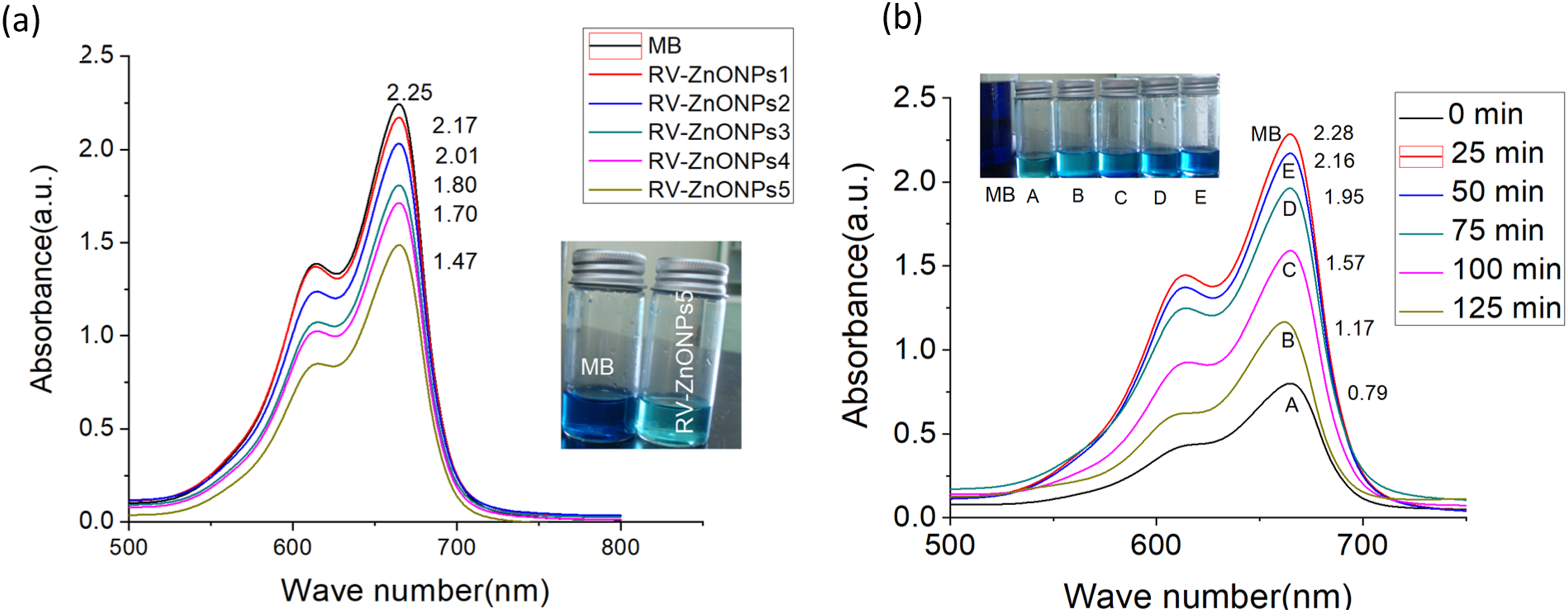
Figure 6 shows the photocatalytic activities of bio-fabricated ZnONPs as determined by UV absorption. Higher degradation is indicated by a lower absorption peak. As seen in Figure 6, when ZnO concentration rises so does the production of electron-hole pairs (e- and h+) under UV radiation. When oxygen (O2) and water (H2O) molecules are adsorbed on the catalyst surface these charge carriers interact with them to create reactive oxygen species like superoxide anions (O2-) and hydroxyl radicals (•OH) which are very effective at oxidatively breaking down MB dye molecules. Consequently, higher ZnO concentrations lead to increased production of these reactive species, resulting in more efficient photocatalytic degradation of MB.74–76 This is because the ZnONPs increased the photocatalytic activity by encourages the production of reactive oxygen species (O2- and •OH) during extended UV exposure, which results in a more effective breakdown of the MB dye. The observed increase in the degradation rate as the reaction proceeded can be explained by the high generation of reactive species. 77 These findings underscore the potential of bio-fabricated ZnONPs for efficient polluted dye degradation in wastewater treatment applications.
The rates at which RV-ZnONPs break down methylene blue (MB) dye at various concentrations from 100 µg/mL to 500 µg/mL and reaction time from 25 minute to 125 minute are displayed in Figure 7. Increasing the concentration of ZnONPs greatly increased the degradation efficiency the maximum efficiency was 34. 6 % at 500 µg/mL (RV-ZnONPs5) but it was only 3.5 % at 100 µg/mL (RV-ZnONPs1). This pattern is in line with previous studies that demonstrate that higher photocatalyst loadings encourage the generation of more reactive species accelerating dye degradation.78,79 Photocatalytic degradation efficiency of methylene blue (MB) dye using RV-ZnONPs. (a) Comparative analysis of the percentage degradation of MB using different synthesized samples (RV-ZnONPs1–5) under. (b) Time-dependent variation of MB degradation percentage using the optimized catalyst, RV-ZnONPs5, over a reaction period of 65 minute.
Furthermore, the rate of MB breakdown increased significantly with reaction time when treated with RV-ZnONPs. The dye was successfully and consistently degraded over time as seen in Figure 8 where the degradation increased from at least 5.2% at 25 minutes to 65.4% after 125 minutes. Similar kinetic trends reported in the literature indicate that prolonged exposure to UV light increases photocatalytic efficiency.
80
To further validate the performance of the biosynthesized RV-ZnONPs, a comparative analysis was conducted with other green-synthesized ZnONPs reported in the literature, specifically those derived from Rhus coriaria and Aloe vera. As summarized in Table 2, the RV-ZnONPs exhibit superior photocatalytic efficiency compared to ZnONPs mediated from both plants. The Rhus coriaria-derived ZnONPs achieved 60.0% degradation in 120 minutes Percentage inhibition of RV-ZnONPs4, RV-ZnONPs3, RV-ZnONPs2, RV-ZnONPs1 and DPPH) samples (a) and their respective color change when DPPH is added (b).
Increased concentrations of ZnONPs provide more active sites for photocatalytic reactions which boost the generation of reactive oxygen species (ROS) like hydroxyl radicals and superoxide anions which more effectively degrade the dye molecules. There is less interaction with MB molecules and less photocatalytic activity if there are fewer nanoparticles available at lower concentrations. In addition to improving light absorption and increasing the chance of electron-hole pair formation higher nanoparticle concentrations also boost degradation efficiency. By acting as a reducing and stabilizing agent the plant extract used in the synthesis improves the surface characteristics of RV-ZnONPs through biofunctionalization. This enhances the adsorption of MB molecules and makes it easier for reactive oxygen species (ROS) like hydroxyl radicals (•OH) and superoxide anions (O2•−) to be produced effectively when exposed to light. These ROS are responsible for attacking and degrading the dye molecules into less harmful substances. Extended reaction times facilitate the generation of more reactive oxygen species (ROS) and more extensive interaction between the dye and nanoparticles which increases photocatalytic activity. According to the aforementioned findings, ZnO absorbing ultraviolet light to produce photogenerated electron-hole pairs is a possible mechanism for the photocatalytic performance of RV-ZnONPs. The ensuing photogenerated electron (e-) and hole (h+) may reach the surface of the RV-ZnONPs and interact with both H2O and O2 to form O2- and •OH, which may participate in the direct oxidation that causes dye degradation.82–85 This mechanism demonstrates the biosynthesized ZnONPs superior performance in comparison to other studies indicating their potential for sophisticated environmental remediation applications.
3.6.2. Antioxidant activities
The antioxidant properties of the green-synthesized RV-ZnONPs was evaluated by measuring the decrease in DPPH absorbance at 517 nm, which reflects their capacity to scavenge free radicals generated by DPPH. 86 The observed shift from blue at the beginning to yellow at the end showed the nanoparticles’ antioxidant activities. In this study, the DPPH technique was used to measure the antioxidant activity of phytochemical-assisted biosynthesized RV-ZnONPs. 87 Methanol was used as a solvent, and DPPH was used as a free radical, which gave a violet color to methanol. When biosynthesized ZnONPs and the fruit extract are combined in an alcoholic solution, the violet color of DPPH is reduced; if the antioxidant property is high, then it changes to yellow after 30 minutes. RV-ZnONPs were synthesized with different concentrations of RV-ZnONPs (100, 200, 300, and 400 µg/ml), respectively. The resulting solutions were then labeled as RV-ZnONPs1, RV-ZnONPs2, RV-ZnONPs3, and RV-ZnONPs4, respectively (Figure 8(b)). An equal quantity of DPPH was then added for the same duration. The percentage inhibition of DPPH for RV-ZnONPs samples at different concentrations is shown in Figure 8(a). The percentage inhibition for biosynthesized RV-ZnONPs were 33.22±0.87 for p-ZnONPs1 and 81.22±0.83 for p-ZnONPs5; however, for RVFE the percentage inhibition was 21.21±1.74 at 100 µg/mL and 43.66±0.25 at 500 µg/mL, which are less than those of biosynthesized RV-ZnONPs samples. According to Figure 8, the inhibition percentages for AA at comparable concentrations were 48.23±0.23 and 87.2±0.45, respectively. The results presented in Figure 8 clearly indicate that the antioxidant activity of biosynthesized RV-ZnONPs is concentration-dependent, with higher concentrations exhibiting greater activity.
Antioxidants help prevent oxidative damage by donating electrons to free radicals, thus neutralizing their detrimental effects on biological processes. As observed in Figure 8, antioxidant chemicals transform the violet-colored DPPH radical into its yellow form by donating hydrogen atoms or electrons, resulting in DPPH-H. This color change is monitored spectrophotometrically to assess the scavenging activity of the synthesized RV-ZnONPs.
7
As shown in Figures 8 and 9, the RV-ZnONPs4 exhibit enhanced antioxidant potential compared by decolorizing the DPPH (decreasing the absorbance of DPPH) than RV-ZnONPs3, RV-ZnONPs2, and RV-ZnONPs1. This showed the effectiveness of ZnONPs synthesis in boosting antioxidant activities at higher concentration. As observed in Figure 10, there is big gap in the value of absorbance between the DPPH sample and RV-ZnONPs samples. Nanoparticles have a very large surface area relative to their size. When their concentration increases, the total surface area available for interaction with dye molecules increases, allowing more dye to be adsorbed or degraded. Higher nanoparticle concentration means more active sites. These sites interact with dye molecules, leading to enhanced adsorption or catalytic degradation.
88
The change in absorbance between methyl blue and different RV-ZnONPs samples. IC50 value for biosynthesized RV-ZnONPs.

Figure 10 shows the IC50 value for biosynthesized RV-ZnONPs samples. A lower IC50 value indicates better antioxidant activity against DPPH free radicals. Our results showed that RVFE had an IC50 value of 475.6 μg/mL, which is significantly higher than the biosynthesized RV-ZnONPs, with an IC50 value of 74.46 μg/mL, as shown in Figure 10. This means that biosynthesized RV-ZnONPs have better antioxidant properties than RVFE. In comparison, other studies found varying IC50 values for ZnONPs made with different leaf extracts. The ZnONPs made with T. heyneana Wall. Leaf extract had a much higher IC50 value (89.47–185.8 µg/ml) than biosynthesized RV-ZnONPs. 89 On the other hand, the ZnONPs made with P. lentum and A. altissima leaf extract had higher IC50 values (83.79 and 78.23 µg/mL) compared to biosynthesized ZnONPs. 90 Alghamdi et al. (2023) 91 showed a low IC50 value of C. argentea mediated ZnONPs with an IC50 value of 91.24 mg/ml, comparable to RV-ZnONPs. Compared to well-known standard chemicals like ascorbic acid (AA) (IC50 = 27.03 mg/ml), RV-ZnONPs has a higher IC50 value. The enhanced antioxidant efficacy achieved through nanoparticle formulation is highlighted by the significantly lower IC50 value of biosynthesized RV-ZnONPs (74. 46 μg/mL) compared to RVFE (475. 6 μg/mL). This is probably because of increased surface area and biofunctionalization from plant phytochemicals. According to Stan et al. 2015, 92 biological processes frequently produce nanoparticles with better radical scavenging than chemical process which is consistent with trends in green synthesis of the current findings.
3.6.3. Antibacterial activity
The zone of inhibition (mm) that p-ZnONPs against S. aureus and E. coli.

The mean value ± SD was utilized after more than three examinations of each data set. Subscriptions a, b, c and d have values in the same raw that differ significantly (P ≤ 0.05).

The zone of inhibition of RV-ZnONPs1, RV-ZnONPs2, RV-ZnONPs3 against S. aureus (a), and E. coli (b).
The antibacterial activity of the three ZnO nanoparticle samples (RV-ZnONPs1, p-ZnONPs2, and RV-ZnONPs3) showed clear variation in their effectiveness against both E. coli and S. aureus. In both bacterial species, the trend of inhibition zones followed the order RV-ZnONPs3 > p-ZnONPs2 > RV-ZnONPs1, indicating that RV-ZnONPs3 possessed the strongest antimicrobial properties with value of 23.14 ± 0.95 and 14.35± 1.00 for E. coli and S. aureus respectively. This enhanced activity is likely linked to differences in physicochemical characteristics such as particle size, surface area, crystallinity, and the presence of active functional groups. Smaller, more crystalline nanoparticles generally interact more efficiently with bacterial cells, producing higher levels of reactive oxygen species (ROS) and enabling more potent membrane disruption. As expected, the positive control streptomycin produced the largest inhibition zones (28.00 mm), reflecting its strong, targeted antibiotic mechanism compared to the broader, nonspecific mode of action of ZnO nanoparticles.
A slight difference in bacterial susceptibility was also observed, with S. aureus showing marginally higher inhibition zones than E. coli for the same nanoparticle treatments as three different samples. This variation is attributed to differences in cell wall structure: S. aureus, a Gram-positive bacterium, lacks the outer lipopolysaccharide membrane found in Gram-negative bacteria like E. coli, making it more accessible to ZnO nanoparticles. In contrast, the additional outer membrane in E. coli acts as a barrier that limits nanoparticle penetration, reducing antimicrobial sensitivity. The results demonstrate that RV-ZnONPs3 possesses the highest antibacterial efficiency, while streptomycin remains the most potent agent due to its specific antimicrobial action.
Based on the data in Table 1, RV-ZnONPs demonstrate notable antibacterial activity against E. coli and S. aureus, with inhibition zones ranging from 17.21±0.11 to 23.14 ± 0.95 mm for E. coli and 5.14± 0.30 to 14.35± 1.00 mm for S. aureus, with RV-ZnONPs3. There is a significant difference (P ≤ 0.05) between the inhibition zone of RV-ZnONPs1, RV-ZnONPs2, RV-ZnONPs3 and Streptomycin. The highest efficacy among the nanoparticles was observed in RV-ZnONPs3. However, their activity remains below that of Streptomycin, which exhibits a zone of 28.00 mm against both bacteria, indicating stronger antibacterial potency. Similar findings in previous studies, such as Zhang et al. (2020), 93 report ZnONPs with inhibition zones around 15-20 mm, aligning with these results, and highlight that nanoparticle synthesis parameters—like size and surface modifications—significantly influence antimicrobial effectiveness. The comparatively lower activity against S. aureus, a Gram-positive bacterium with a thicker peptidoglycan layer, is consistent with the general trend that Gram-positive bacteria tend to be more resistant, requiring more potent formulations.
Researchers have suggested several potential antibacterial mechanisms for the interaction of ZnO NPs with bacteria. There are hypotheses that smaller NPs’ enhanced surface reactivity and faster cell penetration cause them to release Zn2+. The release of Zn2+ from ZnONPs, which is known to impede a number of bacterial cell functions, including active transport, metabolism, and enzyme activity, is a basic idea behind antibacterial actions. 94 ZnONPs emit Zn2+ ions, which adhere to and harm the bacterial cell wall. Additionally, it disrupts the bacterial cell membrane’s layer of lipids, proteins, and carbohydrates. Additionally, DNA damage brought on by Zn2+ ion attacks may cause bacteria to die. Their attack breaks down DNA, which stops bacteria from growing and kills them. The nanoscale dimensions of the material enhance its surface area, thereby amplifying its antibacterial activity, while the small size facilitates easy entry into cells. 95 The generation of reactive oxygen species (ROS), which causes oxidative stress and cell damage or death, is the source of the other suggested antibacterial activity. ROS production is frequently used by ZnONPs as an antibacterial activities. 96 Upon accumulating on the surfaces of bacterial cell walls, the nanoparticles penetrate the bacterial interior via various protein channels, where they become activated and initiate the generation of reactive oxygen species (ROS) and metal ions. High ROS production finally destroys the integrity of the cell wall and disrupts the nucleoids of bacterial cells.97,98 The deposition of Zn2+ ions in the cytoplasm and cell membranes, as well as the interaction of ROS or hydrogen peroxides (H2O2) with bacterial cell membranes, may be the reason why ZnONPs exhibit their antibacterial activity. They generate H2O2, which is mixed with bacterial proteins and lipid bilayers. The oxidative damage in the cellular component may be caused by H2O2 in the cytoplasm.. 99 As Fan et al. (2021) 100 showed in their finding the size and composition of nanoparticles can affect their toxicity. Because of their small size, nanoparticles may be transported quickly and easily, which increases ROS generation and nanotoxicity. 101 The lethal activity of ZnONPs due to their attachment to bacterial cell membranes and accumulation inside the cytoplasm, which damages the integrity of the cell membrane and causes the leakage of cell contents, ultimately leading to cell death, is another suggested explanation. 98
3.7. Comparative analysis of physicochemical and biological properties
Comparative analysis of RV-ZnONPs with previously reported green-synthesized ZnO nanoparticles.

3.8. Challenges, limitation and future works
To guide the practical application of the developed nanomaterials, a dedicated “Future Perspectives” section has been incorporated into the revised manuscript. This section critically examines key challenges, including the scalability of the synthesis process, the necessity for standardized extraction protocols, and the imperative for comprehensive in vivo toxicity assessments to ensure environmental safety. Furthermore, while the current study utilized synthetic dye solutions to validate efficacy, future work will prioritize testing the nanoparticles in real wastewater matrices to account for complex interfering substances. Additionally, the Discussion has been updated to explicitly acknowledge that surface area analysis (BET) and elemental composition (EDX) were not performed in this study; these characterizations are reserved for subsequent investigations to provide a more complete physicochemical profile of the nanoparticles.
4. Conclusion and recommendations
The green synthesis of RV-ZnONPs using Rhus vulgaris fruit extract as a stabilizing and reducing agent is demonstrated in this work. Phenols, flavonoids, tannins and saponins are examples of phytochemicals that aid in Zn2+ reduction nucleation and capping to produce stable well-defined NPs. A 3.20 eV band gap and a UV-Vis absorption peak at 371 nm were found during characterization. TEM revealed irregular shapes with an average size of 23. 5 nm whereas XRD verified a hexagonal wurtzite structure with crystallites measuring 22.7 nm. FT-IR revealed surface functionalization by polyphenols, flavonoids and carboxylic acids while a high zeta potential (-35.1 mV) guaranteed colloidal stability. RV-ZnONPs demonstrated multifunctional properties including improved antioxidant activity (IC50 = 74.46 µg/mL), effective photocatalytic degradation of methylene blue under UV light and antibacterial effects against Staphylococcus aureus and Escherichia coli. These environmentally friendly nanoparticles outperform chemical synthesis techniques and show promise for wastewater treatment biomedical applications and sustainable nanotechnology.
Footnotes
Acknowledgments
The authors thanks Debre Tabor university to provide laboratory chemicals and equipment to preform the laboratory works.
Author contributions
Limenew Abate Worku contributed to the investigation, methodology, and software aspects of the study and writing the manuscript.
Funding
No financial support was provided to the authors for the research, authorship, or publication of this article.
Declaration of conflicting interests
The author declared no potential conflicts of interest with respect to the research, authorship, and/or publication of this article.
Data Availability Statement
Datasets generated and/or analyzed during this study are available in the paper.