
Abstract
Gold nanoparticles have many applications in the biomedical field, mainly for drug delivery, cancer therapy, and detection of pathogenic microorganisms. In this study, gold nanoparticles synthesized using Platycodon grandiflorum (Balloon flower plant) extracts were evaluated for their antibacterial potential. Gold nanoparticles were synthesized at 20–50°C using different volumes of the leaf extract. Biosynthesis of gold nanoparticles was confirmed by ultraviolet–visible spectral absorption at 545 nm by surface plasmon resonance. The morphology and size of the P. grandiflorum gold nanoparticles were further characterized as spherical in shape with an average size of 15 nm in diameter by scanning electron microscopy and transmission electron microscopy. Energy-dispersive X-ray analysis clearly displayed the presence of gold particles. The structural analysis results with face central cubic crystalline nature and elemental composition, including gold, were confirmed by X-ray diffraction and X-ray photoelectron spectroscopy, respectively. In addition, Fourier transform infrared results identified the functional group in P. grandiflorum that is involved in the reduction of metal ions to gold nanoparticles. The synthesized P. grandiflorum gold nanoparticles exhibited efficient antibacterial activity against Escherichia coli (16 mm) and Bacillus subtilis (11 mm). This report confirms the synthesis of gold nanoparticle from balloon flower plant extracts, which can be used as a reducing and stabilizing agent and demonstrates its antibacterial applications.
Introduction
Nanotechnology is a developing area of research with various applications in science and technology, including the synthesis of metal nanoparticles. 1 –3 Metal nanoparticles are extremely small and have higher surface-to-volume ratios than large particles. 4 Various types of metal nanoparticles have already been synthesized using physical, chemical, electrochemical reduction, photochemical reduction, heat evaporation, and biological approaches. Traditionally, the nanoparticles have been developed by physical and chemical approaches 5 ; however, these routes are laborious and potentially dangerous to the environment and human health. 6 In contrast, the biological approach for synthesizing various nanoparticles is safer, eco-friendly, and cost-effective. 7 –9 Many biological sources including plants, bacteria, fungi, and algae were used to synthesize of nanoparticles. Among the biological approaches, plant-mediated synthesis of nanoparticles, including gold nanoparticles (AuNPs), has greater advantages than microbial approaches because of its high reaction rate, reduced cost, and large-scale production. 10 Further, plant extract-mediated nanoparticle production drastically reduces the number of steps involved. Among the various nanoparticles, silver nanoparticles and AuNPs have been extensively used in the biomedical field due to their potential applications. AuNPs are carrying the additional advantages such as higher biocompatibility, easier to tailor with different sizes, and highly amenable to the surface chemical functionalization. AuNPs are extensively used in the biomedical field for drug delivery, cancer therapy, DNA labeling, biological sensing, and detection of pathogenic microorganisms in clinical samples. 4,11 –13 In addition, AuNPs have applications in chemical and biochemical sensing. 14,15 For the downstream applications with AuNPs, different gold (Au)-mediated nanostructures, such as Au nanourchin, nanofilm, nanostar, nanorod, and other Au-based zero–three-dimensional nanostructures, were generated.
AuNPs were prepared ecofriendly using the naturally occurring reagents (e.g. plant extract) as the reducing and stabilizing agents. 16 Similarly, AgNPs have also been synthesized from many plants extracts, 17 –22 but only a few researchers have reported AuNP synthesis from plant sources, such as Azadirachta indica, 23 Cinnamomum camphora, 24 Coleus amboinicus, 25 and Sapindus mukorossi. 26 Recently, the formation of AuNPs obtained successfully using bovine serum albumin as the reducing and stabilizing agents at room temperature. 27 . In addition, the size-controlled AuNPs were synthesized using the nonionic surface Tween-80. 28 Platycodon grandiflorum (balloon flower) contains various natural metabolites, such as alkaloids, flavonoids, saponins, steroids, tannins, terpenoids, proteins, phenolic acids, and other bioligands. 29 Recently, Choi et al. 30 reported the synthesis of nanoparticles using saponins, which are compounds from P. grandiflorum that can be used as catalysts for the reduction of 4-nitrophenol. These compounds as the secondary metabolites are playing a pivotal role in the synthesis of AuNP and act as the capping and reducing agents. These compounds may be involved in the transformation of inorganic metal substances into nanoparticles, including AuNPs. The compounds from P. grandiflorum exhibit antimicrobial, anti-inflammatory, anti-allergic, and anticancer properties. 31 Further, nanoparticles display higher potential in gram-negative bacteria rather than gram-positive bacterial species due to their differences in the peptidoglycan layer, which is acting as the barrier for nanoparticle penetration. P. grandiflorum plant is also used as an ingredient in Korean foods (e.g. Bibimbap) and is a cheap resource during the synthesis of nanoparticles. Therefore, the present study was conducted to optimize the conditions for the synthesis of AuNPs from P. grandiflorum. In addition, the biosynthesized AuNPs were evaluated for their antibacterial properties against pathogenic bacterial strains.
Materials and methods
Materials
The hydrogen tetra chloroaurate (III) hydrate (HAuCl4.3H2O) used in this study was procured from Sigma Aldrich (St. Louis, Missouri, USA). The filtrate membrane was purchased from Sartorius Stedim Biotech (Germany). For transmission electron microscopy (TEM) analysis, the carbon-coated copper grids were procured from Electron Microscopy Sciences (Hatfield, UK). Luria Bertani (LB) agar media were procured from Biopure Reagent (Korea).
Balloon flower plant leaf extract preparation
Balloon flower plant leaves were collected from Incheon Park, South Korea (Figure 1(a)). The collected leaves were thoroughly washed with sterile distilled water and dried for 5 days. About 10 g of the dried leaves were added to distilled water (100 mL) and boiled for 20 min using a water bath. Next, the boiled leaf extract was cooled and filtered through Whatman No. 1 filter paper, followed by filtration through a filter membrane (0.45 µm). Finally, the filtered extract was stored for AuNP synthesis.
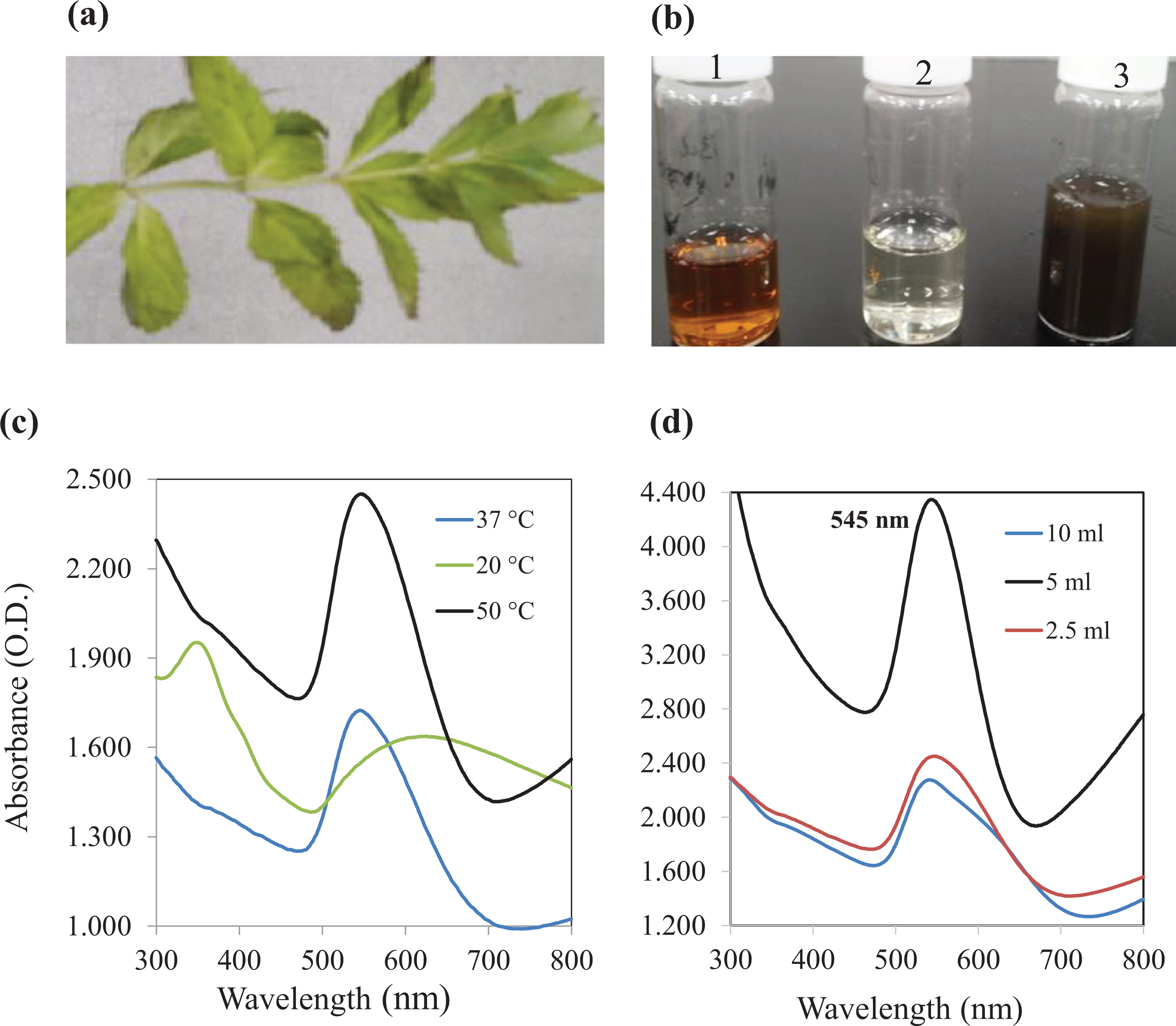
(a) Photograph of Platycodon grandiflorum plant, (b) plant extract (1), HAuCl4 solution (2), and synthesized AuNPs (3), (c) UV-Vis spectrum showing AuNP synthesis at 20, 37, and 50°C, and (d) different concentrations of the plant extract (2.5, 5, and 10 mL) at 50°C. HAuCl4: hydrogen tetra chloroaurate; AuNPs: gold nanoparticles; UV-Vis: ultraviolet–visible.
Au nanoparticle synthesis and optical absorption
Based on previous literature regarding AuNP biosynthesis, 32 approximately 30 mL of HAuCl4.3H2O (1 mM) solution was mixed with 10 mL of the leaf extract. Next, the mixed solution was incubated at different temperatures (20, 37, and 50°C). After incubation, a color change in the reaction mixture was used as an indication of AuNP formation. Furthermore, the amount of leaf extract was optimized (2.5 mL, 5 mL, and 10 mL) to effectively obtain nanoparticles at 50°C. The reaction mixture was centrifuged repeatedly at 10,000 × g (10 min) to obtain purified AuNPs. The final residue was subsequently dried and stored. A control experiment was performed without the addition of leaf extract. Purified AuNPs were used for characterization and application studies. The formation of AuNPs was confirmed by measuring the absorbance at 300–700 nm on a ultraviolet–visible (UV-Vis) spectrophotometer (Jasco V-770).
Characterization of PgAuNPs
Morphological analysis
Scanning electron microscopy
The morphology of PgAuNP was determined using a scanning electron microscopy (SEM) (Hitachi, S-4300SE, Japan). SEM was used to scan the AuNP sample with high-energy beams at 15 kV. Images were captured at 60,000× magnification. Energy-dispersive X-ray (EDX; EDAX, Mahwah, New Jersey, USA) analysis was performed to determine the elemental composition of the samples (Au and other elements). The PgAuNP samples were prepared on silicon wafer for SEM and EDX analyses.
Transmission electron microscopy analysis
The size and shape of the PgAuNPs were also examined by TEM ((JEM 2100F microscope, Jeol, Japan). The PgAuNP samples were prepared by placing a drop of AuNP solution on a carbon-coated copper grid and drying the samples using a vacuum desiccator. The images of the PgAuNPs are shown with scale bar 100 and 20 nm.
Structural analysis and size distribution
X-ray diffraction
The synthesized PgAuNPs were examined by X-ray diffraction (XRD), using a DMAX-2500 XRD system (Rigaku, Japan) equipped with a Nickel filter and a Cu Kα (1.54059 Å) radiation source. The scanning range used was from 10° to 90°, and the scanning rate was 0.5 s−1. The XRD method was performed to analyze the crystalline structure and purity of the PgAuNPs. 33
X-ray photoelectron spectroscopy
X-ray photoelectron spectroscopy (XPS; Thermo Scientific, K-Alpha, UK) was carried out to examine the elemental states of the nanoparticles. The PgAuNP samples were prepared on a silicon wafer by dropping and then drying the sample suspension.
Fourier transform infrared
FTIR spectroscopy was performed to analyze the molecular configuration of the PgAuNPs. This analysis was performed using a Vertex 80 V FTIR system (Bruker, Germany) to register the infrared spectrum of PgAuNPs by either absorption or emission of the sample. A potassium bromide pellet was made with PgAuNPs to study the FTIR spectrum (4000–400 cm−1).
Antimicrobial potential
The antimicrobial activity of the AuNPs synthesized from P. grandiflorum leaf extract was tested against Escherichia coli (gram-negative) and Bacillus subtilis (gram-positive) using a disc diffusion assay. 34 Both strains were maintained on LB agar plates. Both strains were grown in LB broth at 37°C for 24 h (180 r min−1) to obtain the bacterial suspensions. Subsequently, the bacterial suspensions (1 × 10 6) were spread on LB agar plates using a sterile glass spreader. Sterile filter paper discs (6 mm diameter) were placed on inoculated plates and different concentrations of AuNPs and control (sterile water) were loaded onto each disc. All plates were subsequently incubated at 37°C (24 h). After incubation, the inhibitory zone produced upon incubation with different concentrations of the nanoparticles was estimated.
Results and discussion
Plants were used to synthesize environmentally benign metal nanoparticles, because of their ease of availability, large-scale production, and affordability. Temperature plays an important role in the synthesis of nanoparticles by increasing the reaction rate and production. 35 Therefore, in this study, AuNPs were prepared at 20–50°C using 10 mL of P. grandiflorum leaf extract mixed with 1 mM HAuCl4 solution. The results revealed that a temperature of 50°C was the most effective for the synthesis of PgAuNPs due to excitation of plasmon resonance in AuNPs (Figure 1(b) and (c)). Various concentrations of the leaf extract (2.5–10 mL) were also analyzed to optimize the synthesis of PgAuNP at 50°C. The color changed from pale yellow to brownish red after a 5 min incubation with the extract (5 mL), indicating PgAuNP formation due to the reaction between the leaf extract and the metal ions. However, Au nanoparticle synthesis required a 15 min incubation with 2.5 mL of the extracts. Recently, other researchers have reported the synthesis of Au nanoparticles after 30 min of incubation with various plant extracts. 36,37 Low concentrations of the plant extracts are not favorable for AuNP synthesis due to insufficient availability of biomolecules that are required for the capping and stabilization of the PgAuNPs. The volume of the plant extract plays a crucial role in the synthesis of PgAuNPs. The UV-Vis spectra revealed the highest absorbance peak at 545 nm due to the strong surface plasmon resonance indicate the formation of AuNPs (Figure 1(d)). The plant P. grandiflorum contains various active compounds, such as flavonoids, saponins, alkaloids, amino acids, proteins, and carbohydrates. 29 –31 These compounds are required for the reduction of metal ions to various metal nanoparticles, including AuNPs. Natural metabolites strongly influence the bioreduction process from Au+ to Au0 nanoparticles. Rai et al. 38 have previously demonstrated the production of AuNPs using the leaf extract of Cymbopogon flexuosus and described the shape of the AuNPs as spherical at high temperatures and triangular at low temperatures. Thus, temperature plays a critical role in controlling the specific size and shape of Au nanoparticles. Recently, Sathishkumar et al. 39 reported a positive correlation between the temperature and reaction rate of nanoparticles. These data clearly confirm that high temperatures result in a higher reaction rate and smaller particle size.
Characterization morphological analysis
The morphological analysis of the synthesized PgAuNPs was performed using SEM and TEM. SEM images clearly showed uniform distribution and spherical shape of PgAuNP (Figure 2(a)). EDX analysis demonstrated the presence of Au in the PgAuNPs. A strong signal indicated the presence of Au atom at 2 KeV (Figure 2(b)). The size and shape of the PgAuNPs were further confirmed by measuring the diameter of the Au nanoparticles using the TEM images, which indicated that they were predominantly spherical in shape (Figure 3(a) and (b)). In addition, some anisotropic shapes, such as triangular and octahedral shapes, were also present. The nanoparticles were clearly dispersed without any aggregation. The size distribution histogram is shown in Figure 3(c). The particle sizes ranged from 3 nm to 80 nm with an average diameter of 15 nm. Crystalline Au nanoparticles of various shapes have been previously synthesized using plant extracts from Beta vulgaris, 40 Murraya koenigii, 41 and Sphaeranthus amaranthoides. 42 Narayanan and Sakthivel 43 have reported AuNP synthesis from the leaf extracts of Coriandrum sativum, which resulted in various shapes of AuNPs, such as spherical, triangular, and decahedral. The leaf extract-based synthesis of AuNPs resulted in predominantly spherical nanoparticles. 44 –47

SEM image of AuNPs synthesized from Platycodon grandiflorum. (a) Scale bar—500 nm and (b) EDX analysis showing the presence of element Au. SEM: scanning electron microscopy; AuNPs: gold nanoparticles; EDX: energy-dispersive X-ray; Au: gold.
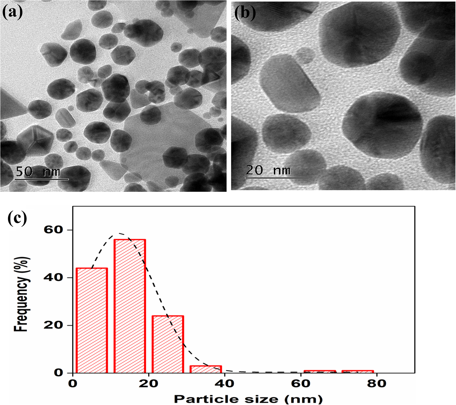
TEM images showing the shape and size of the PgAuNPs. (a) Scale bar—100 nm, (b) scale bar—20 nm, (c) scale bar—10 nm, and (d) TEM image—histogram showing the particle size of PgAuNPs. TEM: transmission electron microscopy; AuNPs: gold nanoparticles; PgAuNPs: Platycodon grandiflorum AuNPs.
Structural analysis
XRD, XPS, and FTIR were performed for the structural characterization of PgAuNPs. Results from XRD analysis confirmed the crystal nature of AuNPs. Figure 4 shows the XRD patterns of AuNPs produced using P. grandiflorum. The 2Θ values of AuNPs at different intense peaks were located at 38.31, 44.46, 64.67, 77.45, and 81.76, which corresponded to 111, 200, 220, 311, and 222 planes of the face-centered cubic (fcc) structure, respectively. The results clearly confirm the formation of fcc crystalline metal ions based on comparison with values reported by the joint committee on powder diffraction standards (JCPDS No. 04-0784). XPS analysis was carried out to identify the elemental composition of the AuNPs, as shown in Figure 5(a)–(f). In the survey scan, the results showed Au4f, C1 s, O1 s, and N1 peaks of the spectra with binding energies of 83.8, 284.7, 532.9, and 399.5 eV, respectively. Oxygen and carbon were the major peaks, and nitrogen and Au were other signals obtained from the AuNPs. The atomic percentages of above elements were shown in Figure 5(f). FTIR was performed to identify the functional groups in P. grandiflorum involved in metal ion reduction during AuNP synthesis. The spectra were determined before and after adding the Au solution to the plant extracts. The major absorption bands in the leaf extract were present at 3400, 1600, 1400, 1300, 1250, 950, and 600 cm−1 (Figure 6(a) and (b)). The broadband at 3300 cm−1 could be attributed to the stretching vibration of the OH group in P. grandiflorum leaf extract. The formation of reduced Au nanoparticle has displayed the occurrence of C–H, C–O, C–C, and C=C bonds, at the appropriate wavenumbers. The major stretching vibrations of the O–H and N–H groups were found to be present between 3200 cm−1 and 3500 cm−1. 48

XRD pattern of synthesized AuNPs from Platycodon grandiflorum at 50°C. XRD: X-ray diffraction; AuNPs: gold nanoparticles.

XPS pattern of PgAuNPs. Data from (a) carbon C1 s, (b) oxygen O1 s, (c) Au4f, (d) nitrogen N1 s, (e) survey scan, and (f) atomic percentage. AuNPs: gold nanoparticles; PgAuNPs: Platycodon grandiflorum AuNPs; XPS: X-ray photoelectron spectroscopy.

FTIR spectrum of (a) Platycodon grandiflorum leaf extract alone and (b) synthesized AuNPs from P. grandiflorum. Dominant IR spectra in the plant extract are at 1600, 1400, 1250, 950, and 600 cm−1. Additional peaks were recorded in AuNP sample. FTIR: Fourier transform infrared; AuNPs: gold nanoparticles; IR: infrared.
Antimicrobial potential
Several researchers have paid great attention to the synthesis of various antibiotics and nanoparticles to effectively control the growth of pathogenic bacteria. However, many bacterial strains are resistant to most of the antibiotics. Therefore, there is an urgent need to synthesize effective Au nanoparticles that can combat the growth of various pathogenic bacterial strains. In this study, the antibacterial activity of the synthesized PgAuNPs against E. coli and B. subtilis was assessed at various concentrations of the NP sample (5, 10, 15, and 20 µg mL−1) using the agar diffusion method. Figure 7(a)–(c) shows the diameter of the inhibitory zone produced by PgAuNPs around the disc. Significant inhibitory activity was observed at a PgAuNP concentration of 20 µg in both bacteria, but a larger inhibitory zone (16 ± 0.7 mm) was noticed in E. coli than that in B. subtilis. The inhibitory activity was greatly increased when the concentration of PgAuNPs increased. In E. coli, lower concentrations of PgAuNPs (10 µg and 15 µg) also significantly inhibited bacterial growth (11 ± 0.7 mm and 13 ± 1.1 mm inhibitory zones, respectively). All the samples with concentration ranging from 5 µg to 20 µg showed inhibitory effect against E. coli (Figure 7(a) and (b)).

(a) Inhibitory zone of AuNPs by Platycodon grandiflorum, (b) antibacterial activity of PgAuNPs against Escherichia coli, and (c) Bacillus subtilis. AuNPs: gold nanoparticles; PgAuNPs: Platycodon grandiflorum AuNPs.
In B. subtilis, the inhibitory activity was detected at 10 µg (8 ± 0.4 mm, inhibitory zone) and its activity increased (9 ± 0 mm and 11 ± 0.7 mm inhibitory zones) at higher concentrations (15 µg and 20 µg, respectively). However, no clear inhibitory zone was observed in the 5 µg sample, possibly because the low sample concentration was not enough to inhibit bacterial growth (Figure 7(a) and (c)). When the concentration of the AuNPs increases, membrane permeability also increases, thus resulting in rapid rupture of the bacterial cell wall. 49 Generally, nanoparticles greatly affect the growth of gram-negative bacteria as they have a thin peptidoglycan layer, which allows the nanoparticles to easily permeate the cytoplasmic membrane and enter the cell, thereby disrupting bacterial cell function and inhibiting growth. 49 However, gram-positive bacteria contain a thick peptidoglycan layer and a linear-polysaccharide chain cross-linked by short peptides, thereby creating a very solid structure that makes it difficult for the nanoparticles to enter the bacterial cell and inhibit the growth. 50,51 Another possible mechanism underlying the antibacterial activity of the nanoparticles is the aggregation of Au ions on the negatively charged cell membrane that can lead to conformational changes in the membrane, loss of permeability control, and ultimately cell death. 36 Many researchers have reported that the small size and spherical shape of nanoparticles allow them to easily permeate the bacterial cell membrane. 52 The antibacterial activity of AuNPs in our study was higher than that reported in other studies, including our previous report on AgNPs. 19,32 The synthesized Au nanoparticles can be used effectively to control bacterial growth during bacterial infections.
Conclusions
This study describes an environmentally safe biological approach to synthesize AuNPs using P. grandiflorum. We confirmed that the temperature and amount of plant extract were both critical for AuNP synthesis. The UV-Vis spectrum confirmed the surface resonance of PgAuNPs at 545 nm. SEM and TEM analyses were used to confirm the morphology (spherical) and average size (15 nm) of the AuNPs. FTIR results confirmed the functional groups involved in reduction of metal ions to AuNPs. Structural analysis revealed the fcc crystalline nature and elemental composition of the AuNPs by XRD and XPS, respectively. The synthesized PgAuNPs significantly inhibited the bacterial growth. However, the antibacterial activity of the PgAuNPs was higher against gram-negative bacteria than that against gram-positive bacteria due to the presence of a thick peptidoglycan cell wall in gram-positive bacteria. Altogether, our results suggest that the AuNPs synthesized from P. grandiflorum have great potential for use in biomedical applications.
Footnotes
Acknowledgments
The author Periasamy Anbu thanks to Inha University for supporting research grant.
Declaration of conflicting interests
The author(s) declared no potential conflicts of interest with respect to the research, authorship, and/or publication of this article.
Funding
The author(s) received no financial support for the research, authorship, and/or publication of this article.