
Abstract
We have developed stable chitosan colloids over a wide pH range without cross-linkers or gelling agents. The colloid was prepared using chitosan nanoparticle obtained from pulverization of bulk chitosan powder, followed by surface treatment using small amount of ascorbic acid (AA) and polyglycerol monostearate (PGMS) in water. Chitosan nanoparticles were well dispersed in a diluted AA solution due to the protonation of the chitosan chain on the surface. And then, the addition of PGMS led them to exhibit highly stable dispersion even in alkali conditions and 50 °C. The hydrodynamic diameter of the colloid was monitored using dynamic light scattering and the real image of the colloid was obtained using cryo-electron microscope measurement. This chitosan colloid will be useful for developing food ingredients or drug carrier templates that should be stable over a wide pH range.
Introduction
Chitosan obtained from deacetylation of chitin is a biodegradable cationic polymer and known as a useful biomaterial for food ingredients 1 –3 and protein delivery, 4 –6 cosmetics, 7 drug 8 –10 and gene delivery, 11 –13 tissue engineering, 14 and so on. In spite of these potential applications, the use of chitosan has been limited because it is soluble only in acidic conditions, where the amine group is protonated to lead electrostatic repulsion between the chitosan chains. In neutral or alkali pH condition, chitosan becomes insoluble by deprotonation. 13 Its usefulness, particularly in pharmaceutical and cosmetic applications, would be extended if the chitosan was soluble at a physiological pH condition. In this regard, much effort has been made, such as blending it with chemical additives 15 and chemical modifications. 16
Nanoparticle form of chitosan has been an attractive prospect as drug and food carriers due to its stability, low toxicity, and versatile routes of administration in various species. 17 Submicron size was suitable for parenteral applications as well as noninvasive mucosal applications, such as oral, nasal, and ocular administrations. 18 There are various methods for preparing chitosan nanoparticles, for example, chemical cross-linking using glutaraldehyde 19 and spray drying, 20 after the chitosan powder was completely dissolved in organic acid solution. For drug applications, cross-linking method was well studied because of the ease of control of time release. However, as some of chemical cross-linking agents may cause cytotoxicity, 21 natural cross-linking agent such as genipin 22 was tried even though it is expensive and in lack of availability.
Instead of chemical cross-linking methods, gelation techniques have been studied, where chitosan was complexed with polyanions by electrostatic interaction. 23 –25 For example, chitosan dissolved in organic acid solution easily aggregated to form nanoparticles by mixing with tripolyphosphate as a gelation agent. 26 Still, the gelation technique could not be applied to the cases that algicidal activity of free amine groups on the chitosan may be necessary 27 and eutrophication caused by polyphosphate might be a concern. 28,29
The main goal of the present study is to prepare a chitosan colloid by not dissolving chitosan in acid solution and not using cross-linking or gelation agent. Instead, chitosan nanoparticle obtained from the pulverization of bulk chitosan powder was mixed with low-concentration L-ascorbic acid (AA) solutions, followed by polyglycerol monostearate (PGMS) treatment. AA, a water-soluble vitamin, which has various biological activity related to free radical scavenging 30 and enhancing immunity 31 was chosen for the study. Chitosan was often dissolved in AA solution, for example, for the antibacterial activity study 32 or the protection of AA from oxidation. 33,34 Chitosan and AA also exhibited synergistic effects, such as wound healing capability on skin 35 and storage stability of fruits. 36,37 The chitosan colloid prepared in AA solution was found to be stable in over a wide pH range from acidic to alkali conditions and exhibits a high thermal stability. This method may offer versatile ways of chitosan nanoparticle to be used as a carrier for foods, cosmetics, drugs, and so on.
Experimental
Materials
Chitosan powder with deacetylation degree of 99 % was purchased from Samsung Chitopia Co. (Seoul, Korea) with a weight-average molecular weight of approximately 100,000 g/mol. The chitosan nanoparticle was prepared by pulverization of the chitosan powder in Apexel Co. (Seoul, Korea) with a dry mill method. AA and PGMS were purchased from Deajung Chemicals Co. (Shiheung, Korea) and Ilshinwells (Seoul, Korea), respectively. Acetic acid, citric acid, and glutamic acid are purchased from Sigma-Aldrich Co. (US). All chemicals were used as obtained.
Preparation of chitosan nanoparticle dispersion
50 mg of chitosan nanoparticle was suspended in 10 ml of organic acid solution with and without PGMS, where PGMS concentration was fixed to 10−5 M. After sonication in a 60 W bath sonicator for 20 min at room temperature, the suspension was centrifuged at 2,500 rpm for 5 min, and then clear supernatant was collected. The precipitate was dried for weighing.
Samples analysis
The size variation of chitosan nanoparticles was observed by scanning electron microscope (SEM), while their morphology in water was viewed using a cryo-electron microscope (cryo-EM) to observe the liquid samples that were dropped and frozen by liquid nitrogen on the carbon grids. Cryo-EM measurements have been performed in the National NanoFab Center, Daejeon, Korea. The average diameters of dispersed chitosan nanoparticles in aqueous solutions were determined using a dynamic light scattering (DLS, Brookhaven Instrument BI-200, US) with a 35 mW He–Ne laser at λ = 632.8 nm and then measured at a fixed angle of 90° to the incident beam at 25 °C. An autocorrelation function of the scattered light was analyzed with a CONTIN algorithm, which is useful for use in polydisperse systems. The hydrodynamic diameter (D h) was then calculated from the diffusion constant obtained from the algorithm.
Results and discussion
Dispersion of chitosan nanoparticle in a dilute organic acid solution
Size of bulk chitosan powder purchased ranged from few to hundred micrometers, as shown in the SEM image in Figure 1(a). After the pulverization, it broke down into smaller and irregular shaped particles with few hundred nanometers in an average size with a wide distribution (Figure 1(b) and inset). This nanoparticle was mixed at various concentrations with four organic acid solutions (acetic acid, citric acid, glutamic acid, and AA) that were used to solubilizing chitosan. 38
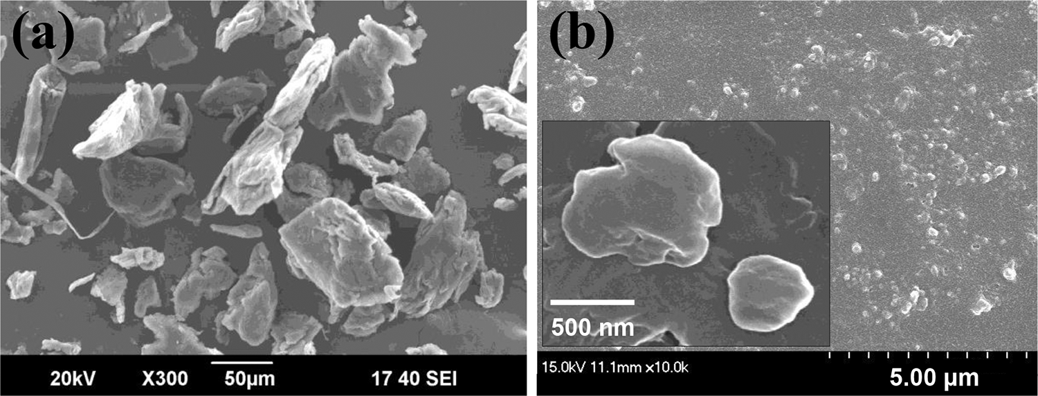
SEM images of chitosan powder (a) before and (b) after the pulverization. SEM: scanning electron microscope.
As shown in Figure 2(a) to (d), at low concentration of organic acids, the solution was turbid and became clear with the increase in concentration, where 50 mg chitosan nanoparticle was mixed with 10 ml organic solution. Organic acid concentration when the solution became clear was approximately 0.12 wt% for acetic acid, approximately 0.7 wt% for citric acid, approximately 0.4 wt% for glutamic acid, and approximately 0.3 wt% for AA, corresponding to 0.20, 0.36, 0.27, and 0.17 mmole, respectively. These values are close to or less than the mole of free amine groups in the chitosan, which is about 0.31 mmol with a molecular weight of chitosan monomer of 161.2 g/mol.

Chitosan nanoparticle mixed with (a) acetic acid, (b) citric acid, (c) glutamic acid, (d) ascorbic acid solution, (e) supernatant collected after centrifugation from (d), and (f) amount of chitosan left in the supernatant as a function of ascorbic acid concentration with and without PGMS. PGMS: polyglycerol monostearate.
As reported previously, 39 –41 chitosan was completely solubilized at an acetic acid concentration such that half of the amine groups in chitosan were protonated. Therefore, acid dissociation behavior (pKa) of the four organic acids listed in Table 1 is important for preparing a chitosan colloid in this study. Here, we chose AA since chitosan solubility behavior in AA solution was not systematically studied yet and acid dissociation behavior of AA is similar to acetic acid compared to the other two organic acids.
MW and pKa of organic acids.

MW: molecular weight; pKa: acid dissociation constant.
After centrifugation of the chitosan-AA solutions, clear supernatants were collected, as shown in Figure 2(e). And the chitosan amount left in the supernatant was calculated by weighing the precipitates after dry, which was plotted as a function of AA concentration in Figure 2(f). The amount gradually increased with AA concentration to reach 50 mg at 0.4 wt% AA, which means all the chitosan stayed in the supernatant. Effects of PGMS in the figure will be discussed in the section “Effects of PGMS.”
We performed DLS measurement on the supernatant. DLS provides D h of a dispersed object in the solution deducted from a correlation function of scattered light intensity based on the CONTIN algorithm. 42 Figure 3(a) shows the correlation function of the clear supernatant collected at 0.1 and 0.2 wt% AA solutions, from which D h distribution profile was obtained. Average D h is also plotted as a function of AA concentration in Figure 3(b). The value was approximately 350 nm at 0.05 wt% and gradually increased to a maximum value of approximately 400 nm at 0.2 wt%. This result is contrary to the expectation that the solubility of chitosan would increase with increasing AA concentration. More reasonable explanation may be that the smaller particle is more easily dispersed than the larger particle because the dispersion is a result of the swelling of the protonated chitosan chains on the surface. Thus, as AA concentration increases, the average size of the dispersed particles would increase because larger particle begins to disperse.

(a) Typical correlation curves from DLS and size distribution from CONTIN algorithm for the supernatant at 0.1 and 0.2 wt% AA and (b) hydrodynamic size (D h) of chitosan colloid as a function of AA concentration with and without PGMS. PGMS: polyglycerol monostearate; DLS: dynamic light scattering; AA: L-ascorbic acid; D h: hydrodynamic diameter.
For more than 0.2 wt% AA, D h decreased with AA concentration and reached approximately 100 nm at 0.35 wt%. At 0.4 wt%, D h was not detected because the correlation curve diminished, indicating that all the chitosan nanoparticles were dissolved. This result confirmed that the chitosan nanoparticle was dispersed by swollen chitosan chains on the surface due to the protonation and then, it gradually lost weight as AA concentration increased. Therefore, for less than 0.4 wt% AA, chitosan nanoparticle in the supernatant was not fully dissolved but dispersed by the swollen chains on the surface, so it can be treated as a “chitosan colloid.”
The chitosan colloid in AA solution was visualized by cryo-EM measurement, as shown in Figure 4. The size of the colloid at 0.2 wt% AA ranged from approximately 150 nm to 400 nm, which appeared to be somewhat smaller compared to the DLS measurement. The larger hydrodynamic size from DLS measurements compared to static measurements from cryo-EM might be reasonable since the electron density contrast between the swollen chitosan chains on the chitosan colloid and aqueous solution is quite small, resulting that the swollen chains might be hardly visible to the electron beam. At 0.35 wt%, the size becomes quite smaller to less than 100 nm, as shown in Figure 4(b). For 0.4 wt% and higher concentration, the particles were not detected because the chitosan colloid might be completely dissolved.
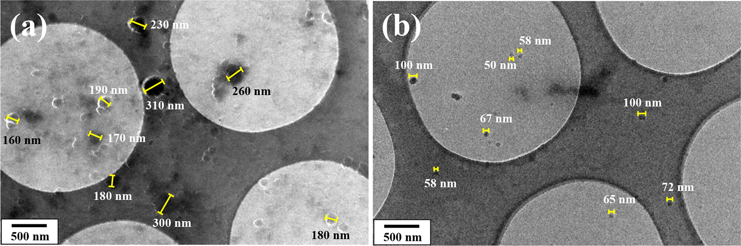
Cryo-EM image of the chitosan colloid in AA with (a) 0.2 wt% and (b) 0.35 wt%. AA: L-ascorbic acid; cryo-EM: cryo-electron microscope.
From DLS and cryo-EM results, one may offer a schematic view of the chitosan colloid, which is composed of swollen chitosan chains around unswollen core, as shown in Figure 5. At low AA concentration, chitosan nanoparticle was swollen to be a stable colloid, where amine groups of chitosan chain on the surface were protonated to form a salt with ascorbate ion providing a repulsive interaction between the particles. As AA concentration increased, it became smaller due to dissolution of chitosan chains from the surface and finally dissolved above 0.4 wt% AA.

Schematic diagram of colloidalization of chitosan nanoparticles in dilute AA solution.
To understand chitosan chain swelling behavior in AA, let f be defined as a mole fraction of AA relative to free amine groups in chitosan, where 50 mg chitosan was dispersed in 10 ml AA solution. We summarized f and the corresponding pH of the solutions in terms of AA concentrations in Table 2. f became unity when the AA concentration reached 0.548 wt%. The fact that all the chitosan were dissolved in 0.4 wt% AA indicates that chitosan can be dissolved even if f is less than unity (f = 0.731). And the solution was still acidic, where pH was 4.95, as presented in Table 2. This means that approximately 27 % of free amine groups was unprotonated in the dissolved chitosan chains, in which phenomenon should also occur in the swollen chains on the surface of the chitosan colloid below 0.4 wt% AA. This behavior is close to the case that chitosan with protonation constant, α = 0.5 on the free amine groups, was fully dissolved in acetic acid solution. 41
Mole fractions of AA relative to amine group in chitosan nanoparticle dependent on the concentrations of AA and pH.
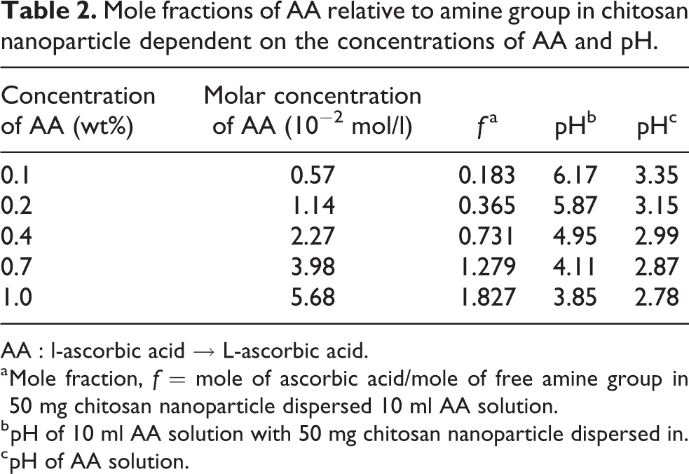
AA : l-ascorbic acid → L-ascorbic acid.
a Mole fraction, f = mole of ascorbic acid/mole of free amine group in 50 mg chitosan nanoparticle dispersed 10 ml AA solution.
b pH of 10 ml AA solution with 50 mg chitosan nanoparticle dispersed in.
c pH of AA solution.
Effect of pH increment
We studied the chitosan colloid stability by increasing pH, where the colloid prepared at 0.2 wt% AA was used. To increase pH, 5 mM NaOH solution was added to the solution drop by drop. D h of the colloid was measured using DLS in terms of pH, as shown in Figure 6. Initial pH was 5.87 (Table 2). D h decreased from approximately 400 nm to approximately 270 nm when pH increases to 7.02, which is because the swollen chain on the surface was collapsed caused by deprotonation of the amine group. 43,44 It is noted that D h of approximately 270 nm at pH 7.02 was close to the colloid size measured using cryo-EM in Figure 4, which confirmed that the size from cryo-EM is smaller than D h from DLS. As pH increased to 7.36, the solution became abruptly cloudy, as shown in the inset photo, and D h was measured to be larger than 1,000 nm, indicating that the deprotonation induces aggregation of the colloids.
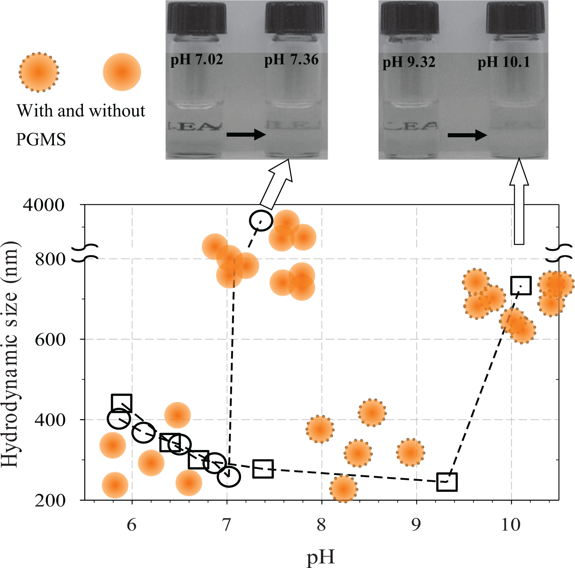
pH-dependent hydrodynamic size of chitosan colloid with and without PGMS, where the colloid was prepared from 0.2 wt% AA solution. PGMS: polyglycerol monostearate; AA: L-ascorbic acid.
Effects of PGMS
PGMS is a commonly used emulsifier in food and cosmetic applications, which is an amphiphilic polymeric material that is composed of a hydrophilic head and a hydrophobic tail. 45,46 The critical micelle concentration of PGMS was measured to be close to 10−5 M using static light scattering with scattering angle of 90° for various solution concentrations. In this experiment, the chitosan nanoparticle was dispersed in 10−5 M PGMS solution.
In the presence of PGMS, the amount of the precipitate during the chitosan colloid preparation in AA solution was a little bit reduced, as shown in Figure 2(c), and D h of the chitosan colloid in the supernatant somewhat increased in Figure 3(b). This result indicated that PGMS helped to disperse chitosan nanoparticle by being adsorbed on the surface. As it is expected that the hydrophobic tail of PGMS must be adsorbed on the surface to allow the hydrophilic head to point toward the water, it is difficult to imagine that the hydrophobic tail could be adsorbed on the protonated (i.e. hydrophilic) swollen chitosan chain on the colloid surface. From the fact that swollen chitosan chains would be partially protonated as the mole fraction analysis in Table 2, we conjectured that unprotonated (i.e. hydrophobic) part of the swollen chain could act as a template for the adsorption of the hydrophobic tail of PGMS. There is other possibility that the chitosan nanoparticles can simply agglomerate with PGMS micelles bridging between them, however, which would be ruled out because of DLS size analysis in Figure 3.
In the presence of PGMS, at pH 7.40, the solution was clear, and D h was equal to approximately 290 nm: This value is larger than the size of chitosan colloid without PGMS measured at pH 7.02, thus further indicating PGMS adsorption. This behavior might be due to the increase in hydrophobicity of the chitosan colloid, which could be a direct consequence of deprotonation, which provides adsorption sites for PGMS and, consequently, repulsive interactions between the colloids. Even at pH 9.32, the solution was still clear, and D h further decreased to approximately 250 nm. For more than pH 10.1, the solution turned to cloudy and D h became larger than 700 nm. Therefore, it is clearly seen that repulsive interaction between the colloids was maintained even at high pH by adsorption of PGMS.
Thermal stability of PGMS-chitosan colloid
Effect of PGMS adsorption on the chitosan colloid could be elucidated by checking thermal stability, where the colloid solution has been kept in a water bath at 50 °C for 3 days. After the treatment, the lower the pH, the smaller the hydrodynamic size, as shown in Figure 7(a), indicating that chitosan chain on the surface was dissolved and disintegrated due to an increase in proton activity at a higher temperature. On the other hand, for the PGMS adsorption case, D h was not reduced at 0.15 wt% AA, as shown in Figure 7(b). For larger than 0.15 wt% AA, D h decreased and was still larger than those without PGMS. As a result, it is concluded that the thermal stability increased due to the surface adsorption of PGMS on the colloid.

Size variation of chitosan colloid (a) without and (b) with PGMS in AA solution before and after heat treatment at 50 °C for 3 days in a water bath. PGMS: polyglycerol monostearate.
Conclusion
We developed a chitosan colloid stable over a wide pH range from acidic to alkali conditions without using a process to dissolve chitosan in AA solution. Here, to fabricate a chitosan colloid, high molecular weight chitosan powder was pulverized to prepare chitosan nanoparticle, which, in turn, was surface-treated by dilute AA solution and further treated using PGMS. The swollen chitosan chains on the nanoparticle surface by protonation of amine group in dilute AA solution were responsible for repulsive interaction between the chitosan colloids. And PGMS adsorption on the swollen chains enhanced dispersion stability of the colloid that is stable even in an alkali solution and also thermal stability upon storage at 50 °C. Size of the colloid was measured in terms of the solution pH using DLS and cryo-EM.
Footnotes
Data availability
The concerning data used to support the findings of this study that are available from the corresponding author upon request.
Declaration of conflicting interests
The author(s) declared no potential conflicts of interest with respect to the research, authorship, and/or publication of this article.
Funding
The author(s) disclosed receipt of the following financial support for the research, authorship, and/or publication of this article: This work was supported by the National Research Foundation of Korea (NRF) grant funded by the Korea government (MSIT) [no. 2019R1F1A1063622] and the Human Resources Development Program of the Korea Institute of Energy Technology Evaluation and Planning (KETEP) grant funded by the Korea Government Ministry of Trade, Industry & Energy (MOTIE) [no. 20184030202260].