
Abstract
Background:
Cyclin-Dependent Kinase 4/6 inhibitors (CDK4/6i) combined with Endocrine Therapy (ET) are the standard treatment for patients with Hormone Receptor-positive/HER2-negative advanced breast cancer (HR+/HER2− aBC).
Objectives:
While CDK4/6i are known to reduce several peripheral blood cells, such as neutrophils, lymphocytes and platelets, the impact of these modulations on clinical outcomes is unknown.
Design:
A multicenter, retrospective-prospective Italian study.
Methods:
We investigated the association between baseline peripheral blood cells, or their early modifications (i.e. 2 weeks after treatment initiation), and the progression-free survival (PFS) of HR+/HER2− aBC patients treated with ETs plus CDK4/6i. Random Forest models were used to select covariates associated with patient PFS among a large list of patient- and tumor-related variables.
Results:
We evaluated 638 HR+/HER2− aBC patients treated with ET plus CDK4/6i at six Italian Institutions between January 2017 and May 2021. High baseline lymphocyte counts were independently associated with longer PFS [median PFS (mPFS) 20.1 versus 13.2 months in high versus low lymphocyte patients, respectively; adjusted Hazard Ratio (aHR): 0.78; 95% confidence interval (CI): 0.66–0.92; p = 0.0144]. Moreover, patients experiencing a lower early reduction of lymphocyte counts had significantly longer PFS when compared to patients undergoing higher lymphocyte decrease (mPFS 18.1 versus 14.5 months; aHR: 0.82; 95% CI: 0.73–0.93; p = 0.0037). Patients with high baseline lymphocytes and undergoing a lower reduction, or even an increase, of lymphocyte counts during CDK4/6i therapy experienced the longest PFS, while patients with lower baseline lymphocytes and undergoing a higher decrease of lymphocytes had the lowest PFS (mPFS 21.4 versus 11 months, respectively).
Conclusion:
Baseline and on-treatment modifications of peripheral blood lymphocytes have independent prognostic value in HR+/HER2− aBC patients. This study supports the implementation of clinical strategies to boost antitumor immunity in patients with HR+/HER2− aBC treated with ETs plus CDK4/6i.
Introduction
The Cyclin-Dependent Kinase 4/6 inhibitors (CDK4/6i) palbociclib, ribociclib and abemaciclib have revolutionized the treatment of Hormone Receptor-positive (HR+), Human Epidermal growth factor Receptor 2 (HER2)-negative advanced (unresectable, locally advanced or metastatic) breast cancer (aBC). Based on results of the randomized phase III trials PALOMA-2, PALOMA-3, MONALEESA-2, MONALEESA-3, MONALEESA-7, MONARCH-2 and MONARCH-3, CDK4/6i have become the standard-of-care treatment for patients with both endocrine-sensitive and endocrine-resistant HR+/HER2− aBC in combination with aromatase inhibitors (AIs) or with the selective estrogen receptor degrader fulvestrant, respectively.1–12 Indeed, when compared to endocrine therapies (ETs) alone, the combination of CDK4/6i and ETs resulted in clinically and statistically significant improvement of patient progression-free survival (PFS)1–4,7 and overall survival (OS).5,6,8–11
The most acknowledged antitumor mechanism of CDK4/6i consists in cancer cell-autonomous (direct) effects, which are mediated by the inhibition of the retinoblastoma protein (RB1) phosphorylation, leading to impaired E2F transcriptional activity and reduced cyclin A and cyclin E expression. 13 These biological modulations result in the inhibition of the G1-to-S transition of the cell cycle, finally causing HR+/HER2− BC cell cycle arrest in G1 phase. 13
However, recent preclinical and clinical evidence indicates that CDK4/6i-induced enhancement of antitumor immunity could play a crucial role in affecting the antitumor effects of this class of drugs. For instance, Goel et al. showed that CDK4/6i can promote cancer cell antigen presentation to antitumor lymphocytes, and that they inhibit the proliferation and immunosuppressive activity of regulatory T cells (Tregs) in murine and human HR+/HER2− BC. 14 In detail, CDK4/6i treatment resulted in E2F inhibition in tumor cells, finally leading to an increased production of type III interferon (IFN) and to enhanced tumor antigen processing and presentation as a result of a global activation of IFN-driven transcriptional programs. Moreover, CDK4/6i promoted tumor infiltration by CD3+ T cells and a concomitant reduction of CD4+FOXP3+ Tregs in murine BC models, paralleled by a reduction of peripheral blood Tregs. 14
One of the most common toxicities associated with CDK4/6i treatment consists of the reduction of total white blood cells (leukopenia) and neutrophil counts (neutropenia), which mainly results from the inhibition of CDK6 in the precursors of these cells in the bone marrow. 15 These hematologic toxicities are more commonly observed with palbociclib and ribociclib than with abemaciclib, which is approximately 13 times more selective against CDK4 as compared to CDK6. 16 Differently from chemotherapy-induced leuko-neutropenia, CDK4/6i-induced leukopenia and neutropenia are not associated with a significantly increased risk of developing severe infections, probably due to the fact that, unlike cytotoxic agents, CDK4/6i do not cause irreversible damage and apoptotic cell death in white blood cell precursors in the bone marrow.1–3,5–8,17 Instead, CDK4/6i-induced leuko-neutropenia could even favor their antitumor activity by relieving neutrophil- and myeloid cell-mediated immunosuppression and by boosting lymphocyte activation and cytotoxicity. 18
Here, we conducted a multicenter, observational study to investigate the potential impact of baseline and on-treatment changes of peripheral blood cells implicated in systemic inflammation and immunity (i.e. total leukocytes, neutrophils, monocytes, platelets and lymphocytes) and their composite scores (i.e. the neutrophil-to-lymphocyte ratio or NLR, the platelet-to-lymphocyte ratio or PLR, the monocyte-to-lymphocyte ratio or MLR, and the Pan-Immune-Inflammation Value or PIV) on the PFS of HR+/HER2− aBC patients treated with CDK4/6i plus ETs.
Methods
Patient population and enrollment criteria
This was an observational, retrospective-prospective, multicenter study conducted in six Italian Cancer Centers [Fondazione IRCCS Istituto Nazionale dei Tumori di Milano (coordinating center); Istituto Oncologico Veneto di Padova; Istituto Europeo di Oncologia, IRCCS – IEO di Milano; Humanitas Clinical and Research Center – IRCCS di Milano; ASST di Cremona; Spedali Civili di Brescia].
Data were collected through an electronic database. Main enrollment criteria were: (i) age ⩾18 years; (ii) histologically/cytologically confirmed diagnosis of HR+/HER2− aBC; (iii) treatment with palbociclib, ribociclib or abemaciclib in combination with ETs (AIs or fulvestrant) as any line of therapy for advanced disease; (iv) pre-, peri- or post-menopausal women [pre- and peri-menopausal patients also received concomitant treatment with a luteinizing hormone releasing hormone (LHRH) analog (i.e. goserelin, triptorelin or leuprolide)]; (v) availability of at least one measurement of blood cell counts at baseline, 2 weeks after treatment initiation, or 12 weeks after treatment initiation; (vi) at least 3 months of follow up at the date of data collection. All patients were followed up until death, loss of contact, or time of data lock (31 May 2021). Written informed consent was obtained from all patients who were alive at the time of study conduction. The study was carried out in accordance with the Good Clinical Practice guidelines and the Declaration of Helsinki.
Study objectives and statistical plan
The objective of this study was to evaluate the association between baseline immune cell crude counts [neutrophils (N_t0), lymphocytes (L_t0), platelets (PLT_t0), monocytes (M_t0)], or their on-treatment modifications (T1: 2 weeks after treatment initiation, and T2: at the time of first radiological disease assessment, that is, approximately 12 weeks after treatment initiation), and the PFS of women with HR+/HER2− aBC treated with CDK4/6i in combination with ET.
To assess the prognostic role of immune cell modulations during CDK4/6i-based therapy, we evaluated the deltas (d1), as defined as absolute cell count difference at T1 as compared to T0 [neutrophil count d1 (N_d1), lymphocyte count d1 (L_d1), platelet count d1 (PLT_d1), monocyte count d1 (M_d1)], or their ratios (r1), here defined as immune cell counts at T1 divided by the same count at T0 [neutrophil count r1 (N_r1), lymphocyte count r1 (L_r1), platelet count r1 (PLT_r1), monocyte count r1 (M_r1)]. We also explored the potential prognostic role of immunological variables in terms of modulation after three treatment cycles (T2) as compared to T0 assessments (here included as delta d2, or ratio, r2).
We also evaluated the association between baseline and on-treatment modulation of NLR (NLR_t0; NLR_d1; NLR_r1; NLR_d2; NLR_r2), PLR (PLR_t0; PLR_d1; PLR_r1; PLR_d2; PLR_r2), MLR (MLR_t0; MLR_d1; MLR_r1; MLR_d2; MLR_r2), or PIV (PIV_t0; PIV_d1; PIV_r1; PIV_d2; PIV_r2), and patient PFS. The NLR was calculated as the ratio between neutrophil and lymphocyte counts; the PLR was calculated as the ratio between platelet and lymphocyte counts; the MLR was calculated as the ratio between monocyte and lymphocyte counts; the PIV was calculated as: (neutrophil count × platelet count × monocyte count)/lymphocyte count.
PFS was defined as the time between the initiation of ET + CDK4/6i therapy and the detection of clinical/radiological disease progression (according to RECIST v1.1 criteria) or patient death from any cause, whichever occurred first. 19 OS was calculated as the time between treatment initiation and patient death from any cause. 19 Patients without an event were censored at the date of last follow up.
Statistical analyses
Standard descriptive statistics were used to describe clinical and biological patients’ characteristics. Spearman’s rank correlation was used to evaluate the correlation between the line of therapy for advanced disease and the line of ET for advanced disease. Unpaired t-test was used to compare peripheral blood immune cell counts, or their combined scores, at baseline, at T1 and T2. Median patient follow-up was calculated with the reverse Kaplan–Meier estimator. 20 Survival curves for PFS or OS were obtained with the Kaplan–Meier method. Optimal cut-points for classifying L_t0 and L_r1 as ‘high’ versus ‘low’ were calculated through the maximally selected rank statistic method. 21 A two-step strategy was used to perform multivariable analysis, as detailed below.
Random forest models
In the first step of multivariable analyses, the impact of several covariates on patient PFS was modeled by using a Random Forest approach. 22 Random Forest method was used with the purpose of (a) detecting and excluding, on the basis of minimal depth statistic, prognostically irrelevant covariates; (b) detecting interactions among covariates and nonlinear effects of continuous predictors; (c) estimating, through error rate estimation, the global predictive performance of the model. The variables selected through Random Forest Models were chosen to be evaluated in multivariable Cox regression models. Depending on the type of analysis, we used either original or imputed data.
Cox regression models
Cox regression modeling was used in the second step of our statistical analyses, with the proportional hazard assumption checked by testing and plotting Schoenfeld residuals. Means of restricted cubic splines were used to handle nonlinear effects for all continuous variables. Results of Cox models were summarized using Hazard Ratios, along with the corresponding 95% confidence intervals (CIs) and Wald p values, while overall model performance was assessed in terms of discrimination with the bootstrap-adjusted Harrell c-index. In Cox models, the HR for continuous variables was reported as the HR related to the inter-quartile range (IQR; interval between the 75th and 25th quantiles). Furthermore, to quantify the effect of high/low values of L_t0 and L_r1 in terms of PFS, we estimated adjusted survival PFS curves. 23 Given the presence of missing data, Cox model analyses were performed both with single and after 10-fold multiple imputations. The main study analyses were repeated in the subset of patients receiving CDK4/6i-based therapy in the first-line treatment setting.
Statistical analyses were performed with SAS (version 9.4, SAS Institute) and R software (version 4.1.2, R Foundation for Statistical Computing). The conventional 5% two-sided threshold was set for statistical significance.
Results
Study cohort
We evaluated a total number of 656 HR+/HER2− aBC patients treated with ET plus CDK4/6i between January 2017 and May 2021. Of these, 13 patients were excluded because they received CDK4/6i monotherapy, 5 patients were excluded because of the lack of at least one measurement of blood cell counts at baseline, at 2 weeks or at 12 weeks after treatment initiation. Finally, 638 patients fulfilling all the enrollment criteria, and who initiated treatment with CDK4/6i plus ET, were enrolled. The study flow diagram is shown in Supplemental Figure 1. In detail, 168 patients were enrolled at Fondazione IRCCS Istituto Nazionale dei Tumori of Milan (coordinating center); 179 patients at Istituto Oncologico Veneto of Padua; 77 patients at Istituto Europeo di Oncologia, IRCCS – IEO of Milan; 53 patients at Humanitas Clinical and Research Center – IRCCS of Milan; 58 patients at ASST of Cremona; 103 patients at Spedali Civili of Brescia.
Baseline patient- and tumor-related characteristics are summarized in Table 1. Median patient age was 61 years (IQR: 52–70 years). The majority of patients were post-menopausal (n = 530; 83.1%), had an Eastern cooperative oncology group performance status (ECOG PS) of 0 (n = 464, 72.8%) and were treated with Palbociclib-based therapy (n = 529; 82.9%). Approximately half of enrolled patients received CDK4/6i-based therapy in the first-line setting (n = 335; 52.5%) and had visceral disease (n = 334; 52.4%).
Patient characteristics.
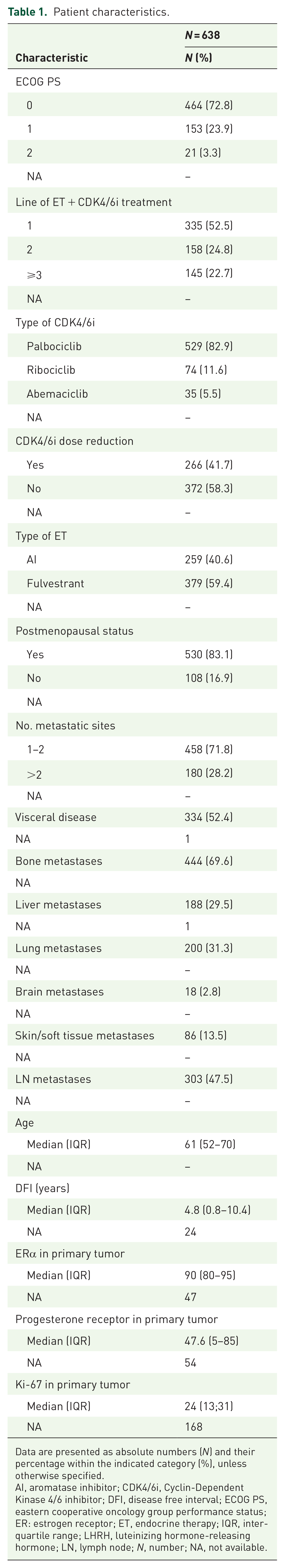
Data are presented as absolute numbers (N) and their percentage within the indicated category (%), unless otherwise specified.
AI, aromatase inhibitor; CDK4/6i, Cyclin-Dependent Kinase 4/6 inhibitor; DFI, disease free interval; ECOG PS, eastern cooperative oncology group performance status; ER: estrogen receptor; ET, endocrine therapy; IQR, inter-quartile range; LHRH, luteinizing hormone-releasing hormone; LN, lymph node; N, number; NA, not available.
At data cut-off and analysis (31 May 2021), 358 patients (56.1%) had experienced disease progression, and 166 patients (26.0%) had died. With a median follow-up time of 25 months (95% CI: 15.2–35.0), median PFS in the whole study cohort was 18.1 months (95% CI: 15.1–22.3). Median PFS was 26.1 months (95% CI: 23.4–30.8) in patients receiving CDK4/6i treatment as a first-line therapy, while median PFS was 11.9 months (95% CI: 9.9–13.9) in patients receiving CDK/4/6i as subsequent lines of therapy (Supplemental Figure 2). These data are consistent with results of phase III clinical trials1–3,5–8,12 and with data from previously published real-world cohorts24–27 of patients treated with ET plus CDK4/6i.
CDK4/6i-induced modulation of peripheral blood immune cells
The modulation of peripheral blood cell counts during CDK4/6i therapy was evaluated by comparing immune cell counts at T0 (baseline), at T1 (2 weeks after treatment initiation) and at T2 (at the time of first radiological disease assessment, that is, approximately 12 weeks after treatment initiation). On-treatment modulation of these parameters is reported in Figure 1. Neutrophil, lymphocyte, platelet and monocyte counts were all significantly and precociously reduced after treatment initiation (T1 versus T0). At T2, there was a partial recovery of monocyte and platelet counts when compared to their values at T1, while neutrophil and lymphocyte counts were not significantly different at T2 as compared to T1. Immune cell scores also underwent significant modifications; in detail, NLR was significantly reduced at T1, and it remained low at T2; conversely, the MLR and the PIV, which were significantly reduced at T1, were partially restored at T2, which reflects the partial restoration of monocyte and platelet counts at this timepoint. Finally, the PLR did not undergo a significant early reduction, while it was minimally increased after 12 weeks of CDK4/6i-based therapy (Supplemental Figure 3).

CDK4/6i-induced modulation of peripheral blood cell counts. Boxplots depicting the kinetics of the indicated peripheral blood cell counts at three different timepoints, namely T0 (baseline, i.e. before CDK4/6i treatment initiation), T1 (~2 weeks after CDK4/6i treatment initiation) and T2 (~12 weeks after CDK4/6i treatment initiation). For each of the indicated comparisons, the p value of the unpaired t-test is reported.
Selection of potentially prognostic clinical and immunological parameters
We used Random Forest models to screen the potential prognostic role of 28 patient- and tumor-related variables. In a first model exploring the role of several clinical variables, and in which missing data were imputed, the following variables emerged as associated with patient PFS: line of therapy for advanced disease, ECOG PS, presence of liver metastases, line of ET for advanced disease, number of metastatic sites, disease-free interval (DFI), percentage of Ki-67-positive cells in primary tumor specimens, patient age, and presence of ERα expression in primary tumor specimens (Supplemental Figure 4). The line of therapy for advanced disease and the line of ET for advanced disease were positively correlated (Spearman’s rank correlation coefficient, ρ = 0.83, p < 0.001), thus indicating that these two variables provide redundant information. Therefore, in subsequent multivariable Cox regression models we only included the line of therapy for advanced disease, while we excluded the line of ET for advanced disease.
Then, we fitted several Random Forest models, with or without imputation of missing data, in which we assessed the potentially prognostic role of previously selected clinical variables, along with peripheral blood immune cells and composite immune scores, evaluated as baseline values (T0), or in terms of their modulation during CDK4/6i-based therapy at T1 or T2 [deltas (d1, d2) and ratios (r1, r2)]. Error rate estimations of these Random Forest Models are summarized in Supplemental Table 1. Among these models, the one with the lowest error rate estimation, that is, the one associated with the best predictive performance in terms of patient PFS, was the model that included the T1–T0 ratio (r1) of immune cells (error rate: 29.2%). Therefore, we selected N_r1, L_r1, PLT_r1 and M_r1 for subsequent evaluation in multivariable Cox regression models. In these models we also included baseline raw immune cell counts (N_t0, L_t0, PLT_t0 and M_t0) in light of their established prognostic role.28,29 Although composite immune scores (NLR, PLR, MLR, PIV) did not improve the predictive performance of Random Forest models when compared to T0 and r1 crude count ratios (Supplemental Table 1), we also evaluated these parameters in multivariable Cox models due to their established prognostic role.30–36
Association between baseline and on-treatment immune parameters with patient PFS
We fitted several multivariable Cox models to investigate the prognostic role of each immune-related variable, as evaluated at T0 and as r1, after adjustment for previously selected clinical covariates (Supplemental Figure 4). In these models, we also explored the impact of nonlinear effects of individual immune cell subsets, as well as of the interaction between t0 and r1 immune cell counts. A summary of results of these models is shown in Supplemental Table 2. Of note, lymphocyte counts and the NLR were the only immune parameters to be independently associated with patient PFS when evaluated both at T0 and as r1.
As for lymphocytes, the higher the baseline lymphocyte counts (L_t0), the lower the risk of disease progression [adjusted HR as a continuous variable (aHR): 0.78; 95% CI: 0.66–0.92; p = 0.0144] (Table 2). Regarding on-treatment lymphocyte modulation, the lower the reduction of blood lymphocytes during the treatment (i.e. the higher L_r1), the lower the risk of disease progression (aHR as a continuous variable: 0.82; 95% CI: 0.73–0.93; p = 0.0037) (Table 2). We did not find an interaction between L_t0 and L_r1 in affecting PFS (p = 0.8465). Similar results were obtained when we fitted the model after multiple data imputation (Supplemental Table 3). Higher baseline L_t0 and L_r1 were also associated with independently better OS (aHR for L_t0: 0.67; 95% CI: 0.52–0.88; p = 0.0052; aHR for L_r1: 0.71; 95% CI: 0.59–0.85; p = 0.0006) (Supplemental Table 4).
Multivariable Cox regression model including L_t0 and L_r1.

The p value is indicated in bold numbers when statistically significant (<0.05).
DFI, disease free interval; ECOG PS, Eastern Cooperative Oncology Group Performance Status; ER, estrogen receptor; L_t0, lymphocyte count at T0; L_r1, lymphocyte T1–T0 ratio.
The continuous relationship between lymphocyte counts at T0 or r1 and the log Relative Hazard is shown in Figure 2, while Supplemental Figure 5 shows the continuous relationship between neutrophils, platelets or monocytes and the log Relative Hazard. We also plotted adjusted PFS curves for L_t0 and L_r1 after dichotomizing L_t0 and L_r1 according to cut-offs calculated through the maximally selected rank statistic method (cut-off for L_t0 = 1376.1; cut-off for L_r1 = 0.711). 37 Patients with higher baseline lymphocytes had better PFS when compared to patients with lower baseline lymphocyte counts (median PFS 20.1 months, 95% CI: 16.8–23.6, versus 13.2 months, 95% CI: 11.7–16.6, for patients with high versus low baseline lymphocyte counts, respectively) [Figure 3(a)]. In addition, patients undergoing a lower decrease, or even an increase of peripheral blood lymphocyte counts during CDK4/6i-based therapy had longer PFS when compared to patients undergoing higher lymphocyte decrease (median PFS 18.1 months, 95% CI: 13.3–23.3, versus 14.5 months, 95% CI: 10.8–19.8) [Figure 3(b)]. When we combined baseline and on-treatment lymphocyte assessments, patients with high L_t0 and high L_r1 had the longest PFS (median PFS: 21.4 months, 95% CI: 17.4–25.8), while patients with low L_t0 and low L_r1 had the worst PFS outcomes (median PFS: 11 months, 95% CI: 7.8–17.3); finally, patients with high L_t0 and low L_r1 and low L_t0 and high L_r1 had intermediate PFS outcomes (median PFS 17.6 months, 95% CI: 11.6–22.8, and median PFS 13.8 months, 11.3–18.4, respectively) [Figure 3(c)]. Therefore, combining baseline and on-treatment modulation of lymphocyte counts helps in discriminating patients with better and worse survival outcomes.

Continuous relationship between peripheral blood lymphocytes and the risk of disease progression. Curves showing the continuous relationship between baseline lymphocyte counts (L_t0), or (a) T1–T0 lymphocyte ratios (L_r1), and (b) log relative Hazard for disease progression.

Impact of baseline lymphocyte counts and their early modulation on patients’ PFS. Adjusted PFS curves according to baseline lymphocyte counts (high versus low). (a) Adjusted PFS curves according to T1–T0 lymphocyte ratio (high versus low). (b) Adjusted PFS curves according to both baseline lymphocyte counts (high versus low) and T1–T0 lymphocyte ratio at T1, or r1 (high versus low). (c) The cut-off values to divide patients with high versus low lymphocyte counts at baseline or high versus low lymphocyte ratios were calculated with maximally rank selected statistics method.
Among composite immune scores, higher NLR was the only variable to be significantly and independently associated with worse patient PFS both at t0 (NLR_t0: HR: 1.21; 95% CI: 1.05–1.40; p = 0.034) and as r1 (NLR_r1: HR: 1.36; 95% CI: 1.16–159; p = 0.0007) (Supplemental Table 2). These data are also reflected by the monotonic relationship between NLR_t0 or NLR_r1 and the risk of disease progression (Supplemental Figure 5). On the other hand, MLR, PLR and PIV did not show a monotonic relationship with the log relative hazard of disease progression (Supplemental Figure 5), in line with the lack of an association between these variables and clinical outcomes (Supplemental Table 2).
Baseline NLR (NLR_t0) has consistently emerged as an independent prognostic variable in several series of patients with different advanced malignancies, including HR+/HER2− aBC.30–32,38,39 For this reason, we fitted another Cox regression model to investigate the independent role of L_r1, which accounts for the modulation of lymphocytes during the treatment, and NLR_t0; in this model, we did not include L_t0 because its prognostic role is already accounted for by NLR_t0 assessment. Of note, higher L_r1 maintained an independent and statistically significant association with better PFS (HR: 0.86; 95% CI: 0.76–0.97, p = 0.0225), while NLR_t0 was not associated with the risk of disease progression (HR: 1.11; 95% CI: 0.97–1.28; p = 0.3110) (Supplemental Table 5). These results indicate that the modulation of lymphocyte counts during ET + CDK4/6i treatment is a better predictor of PFS than baseline NLR in patients with HR+/HER2− aBC.
Association between early lymphocyte modulation and patient PFS in the first-line setting
Since the majority of HR+/HER2− aBC patients nowadays receive CDK4/6i in the first-line treatment setting, we performed a subgroup analysis, in which we evaluated the association between L_t0 and L_r1 and the PFS of patients treated with CDK4/6i in the first-line setting (n = 335; 52.5%). As shown in Supplemental Table 6, multivariable analysis revealed an independent and statistically significant association between higher L_r1 and a lower risk of disease progression; on the other hand, L_t0 was not associated with patient PFS. In this model, a higher DFI was associated with better PFS, while a higher number of metastatic sites, as well as higher ECOG PS, were associated with significantly worse PFS. We also adjusted the prognostic role of L_r1 for NLR_t0 in this patient subset (Supplemental Table 7). Of note, L_r1 was independently associated with patient PFS (HR: 0.83; 95% CI: 0.71–0.99; p = 0.0325), while the NLR_t0 was not [HR: 1.08; 95% CI: 0.86–1.35 (p = 0.5085)]. Together, these data confirm and reinforce data previously observed in the whole patient cohort, that is, that the precocious modulation of peripheral blood lymphocytes has prognostic significance in HR+/HER2− aBC patients treated with first-line CDK4/6i-based therapy.
Discussion
We showed that baseline peripheral blood lymphocyte counts and their early modulation are independently associated with the PFS and OS of HR+/HER2− aBC patients treated with ET plus CDK4/6i as any line of treatment for advanced disease. We also found that precocious modulation of lymphocytes is associated with PFS of HR+/HER2− aBC patients treated with CDK4/6i therapy in the first-line setting.
CDK4/6i-based therapy resulted in a significant reduction of peripheral blood neutrophil, platelet and lymphocyte counts (see Figure 1), thus confirming data previously reported in phase II and phase III clinical trials.1–3,5,6,8 In particular, the reduction of total white blood cell and neutrophil counts is a well acknowledged adverse event (AE) of palbociclib, ribociclib and, less commonly, of abemaciclib-based therapy, and it is frequently associated with drug dose reduction.1–3,5,6,8,15 Regarding other hematological toxicities, any-grade thrombocytopenia was reported in around 9.3–53.2% of patients treated with CDK4/6i in phase III trials, with only around 1% of patients undergoing grade 3 or grade 4 thrombocytopenia.1,2,7–10 Moreover, grade 3/4 lymphopenia was reported in approximately 30% of patients enrolled in the phase II PALOMA-1 trial, 40 but not in the original publications of the phase III trials PALOMA 2 and PALOMA 3.1,2 In a long-term, pooled safety analysis of PALOMA 1, 2 and 3 trials, any-grade 5-year lymphopenia occurred in 2.8% of patients, and 0.9% of patients experienced grade 3/4 lymphopenia. 41 In the MONALEESA-2 trial, the incidence of grade 3 and grade 4 lymphopenia was 7% and 0.9%, respectively. 3 As for abemaciclib, any grade lymphopenia occurred in 52.7–62.9% of patients enrolled in MONARCH-2 and MONARCH-3 trials, with grade 3/4 lymphopenia being reported in 7.3–12% and 0.2–1.3% of patients, respectively.7,8 In the MONARCH-2 trial, 1.4% of abemaciclib-related dose reductions were attributed to the occurrence of lymphopenia. 42 Finally, the impact of CDK4/6i-based therapy on peripheral blood monocyte counts has not been reported in the PALOMA, MONALEESA and MONARCH trials.1–3,5,6,8 Therefore, our analysis is the first study to show that CDK4/6i cause a significant reduction of peripheral blood monocyte counts.
The main aim of our study was to investigate the association between CDK4/6i-induced modulation of peripheral blood immune cell subsets and the PFS of patients with HR+/HER2− aBC. By combining Random Forest and multivariable Cox regression models, we found that baseline and on-treatment lymphocyte counts are consistently associated with clinical outcomes. In particular, patients undergoing a lower decrease, or even an increase of lymphocyte counts during CDK4/6i therapy, had significantly better PFS and OS when compared to patients experiencing a higher decrease of peripheral blood lymphocytes. Other immune parameters explored in our analyses did not show an independent association with patient PFS, with the only exception of high NLR, which was associated with worse PFS when evaluated as NLR_t0 and as NLR_r1. However, the prognostic role of NLR likely reflects the prognostic role of lymphocytes; indeed, when the association between L_r1 and NLR_t0 with PFS was independently evaluated in the same multivariable Cox model, only the early modulation of lymphocytes maintained a significant association with patient PFS.
Since we found that baseline lymphocyte counts and their early on-treatment modulation are stronger predictors of patient PFS in multivariable models, also when adjusted for NLR, results of our study are in part in contrast with a large body of accumulating evidence in other clinical contexts, where composite immune cell parameters, namely NLR, PLR, MLR and PIV, were shown to be better predictors of clinical outcomes than crude immune cell counts in patients with both limited-stage and advanced malignancies, including breast cancer.30–36,43 These results may reflect an especially important role of blood lymphocytes, both at baseline and during CDK4/6i therapy, in affecting the clinical outcomes of HR+/HER2− aBC patients treated with ET + CDK4/6i as any line of therapy.
Consistent with our findings, recent preclinical evidence revealed that CDK4/6i activate specific subsets of cytotoxic and memory T cells that could contribute to the antitumor effects of this class of drugs.18,44,45 For instance, Deng et al. showed that CDK6 but not CDK4 inhibition results in an increased expression of Nuclear Factor of Activated T cells (NFAT) family proteins, which modulate the activation of tumor infiltrating lymphocytes. 18 In addition, intratumor levels of the Th1 chemokines CXCL9 and CXCL10, which modulate the intratumor trafficking of effector T cells, were increased after CDK4/6i-based therapy, while CD11+ myeloid-derived suppressor cells and their products, namely IL-6, IL-10 and IL-23, were reduced. Notably, Tregs were more susceptible to CDK 4/6 inhibition than other T cells, probably as a result of higher expression of CDK6. 18 In the work by Schaer et al. 44 abemaciclib monotherapy was associated with increased intra-tumor immune inflammation in tumor-bearing mice, and the combination of abemaciclib with anti-PD-L1 therapy led to tumor regressions and enhanced T cell immune response and antigen presentation. 44 In another work, CDK4/6 promoted cancer cell senescence through the activation of a senescence-associated secretory phenotype, finally resulting in enhanced tumor immune-cell infiltration. 45 Finally, in a recent biomarker analysis conducted in a subset of patients enrolled in the PALOMA-2 trial, lower expression of the immune checkpoint PD-1 in basal tumor specimens was associated with a relatively higher benefit from palbociclib, thus suggesting that palbociclib might stimulate intratumor immunity in HR+/HER2− aBCs with lower basal lymphocyte infiltration and/or with less efficient antitumor immunity (i.e. those with low PD-1 expression). 46
Based on the immunosuppressive effects of neutrophils and monocytes in cancer patients, 47 we expected that CDK4/6i-induced reduction of peripheral blood neutrophils and monocytes could be associated with better patient PFS. Our data show that this hypothesis was not correct. Indeed, while the precocious reduction of neutrophil counts during CDK4/6i therapy showed a trend toward an association with better patient PFS, CDK4/6i-induced reduction of monocyte and platelet counts was even associated with a trend toward worse PFS (Supplemental Figure 5–7). On the other hand, the finding that the modulation of blood lymphocytes was the only immunological variable to be consistently and independently associated with patient PFS indicates that lymphocytes might play an especially important role in affecting the long-term clinical outcomes in HR+/HER2− aBC patients treated with CDK4/6i-based therapy.
Consistent with recently published works,48,49 we found that CDK4/6i dose reduction was not associated with worse patient PFS (Supplemental Figure 4). However, in our study we made no distinction between early (first 3 months of treatment) and late dose reductions, whereas in the studies published by Kristensen et al. 48 and Roncato et al., 49 a precocious reduction of CDK4/6i dosage was associated with worse clinical outcomes. Therefore, we cannot exclude that we may have failed to capture an effect of dose reduction on patient PFS because of this specific limitation of our study. However, we would like to highlight the fact that lymphopenia is very rarely a cause of CDK4/6i dose reduction. Therefore, the main study results regarding the prognostic significance of early reduction of blood lymphocytes are unlikely to be affected by precocious reduction of CDK4/6i dosage.
Along with preclinical data supporting a role of cytotoxic lymphocytes in mediating the antitumor effects of CDK4/6i, 18 results of our study pave the way for evaluating CDK4/6i in combination with immune checkpoint inhibitors (ICIs) in patients with limited-stage or advanced HR+/HER2− BC. Scirocchi et al. 50 recently found that CDK4/6i + ET treatment downregulates immune suppressive subpopulations, such as Tregs, M-MDSCs, and PMN-MDSCs, in HR+/HER2− aBC patients. Strikingly, patients responding to the treatment showed a higher decrease in Tregs when compared to non-responding patients, and CDK4/6i treatment resulted in an increase in CD4+ T cells and anti-tumor CD137+ CD8+ T cells. 50
In this respect, only few clinical data from prospective trials are available. In the multicenter, non-randomized, open-label, multi-cohort, phase Ib trial NCT02779751, which enrolled 28 endocrine-resistant HR+/HER2− aBC patients treated with abemaciclib plus pembrolizumab after receiving 1 or 2 prior lines of chemotherapy for advanced disease, tumor overall response rate was 29%, and disease control rate was 82%; moreover, median PFS and OS were 8.9 months and 26.3 months, respectively. 51 Another phase I/II trial, namely NCT02778685, enrolled previously untreated HR+/HER2− aBC patients, who received a combination of palbociclib, letrozole and pembrolizumab; of 16 patients, 31% achieved complete response, 25% had partial response (PR) and 31% reported stable disease (SD) as best responses according to RECIST 1.1 criteria. Median PFS was 25.2 months and median OS was 36.9 months. 52 In the recently presented phase II study PACE (NCT03147287), which randomized HR+/HER2− aBC patients progressing to ET plus CDK4/6i to receive fulvestrant alone, fulvestrant plus palbociclib, or fulvestrant plus palbociclib plus avelumab, patients receiving the triple treatment experienced better median PFS (8.1 months) when compared to patients treated with fulvestrant alone (4.8 months), or with fulvestrant plus palbociclib (4.6 months). 53
In the early-stage setting, the ImmunoADAPT (NCT03573648) trial is randomizing stage II–III HR+/HER2− BC to receive ET plus avelumab with or without palbociclib. However, the CheckMate 7A8 phase II study, which evaluated the combination of nivolumab, palbociclib and anastrozole in patients with early HR+/HER2− BC, was closed after the safety run-in due to the increased hepatic toxicities of the experimental triplet treatment. 54 These contradictory findings impose to deeply investigate the safety of combining ET plus CDK4/6i and ICIs in specific clinical contexts.
The following are major strengths of our study: (i) this was the first, relatively large multicenter study to show that baseline lymphocytes and their early modulation during CDK4/6i therapy are associated with PFS and OS in HR+/HER2− aBC patients; (ii) the multicenter nature of the study and the large sample size make our data robust, with PFS data observed in the whole study cohort being consistent with results of phase III trials investigating CDk4/6i and with data from real world cohorts1–12; (iii) the main study findings were confirmed by different multivariable models including clinical and biological variables; (iv) the independent prognostic role of CDK4/6i-induced early lymphocyte modulation was confirmed in the subset of patients treated in the first-line setting.
This study also has several limitations. Firstly, is its retrospective design, with some original data missing at the time of data analysis; however, Random Forest Models fitted with either original or imputed data confirmed the main study findings, thus partially obviating to this limitation (see Supplemental Table 1). In addition, the main Cox models were fitted after both single and multiple imputation to increase the robustness of our results (see Supplemental Table 3). Secondly, the majority of patients enrolled in our study received palbociclib, thus not allowing us to generalize our findings to ribociclib or abemaciclib. In this respect, the ongoing PALMARES-2 study, a large, real-world Italian study involving 23 centers, will clarify if the findings of the current study are also confirmed in larger cohorts of patients treated with ribociclib or abemaciclib. Thirdly, the study results were not validated in an independent study cohort. The ongoing PALMARES-2 study will be instrumental also in this respect. Finally, we did not perform a centralized evaluation of peripheral blood cells; however, blood cell count assessment relies on fully standardized methodologies across laboratories in Italian Cancer Centers. Therefore, we tend to exclude that the study findings may have been influenced by the lack of centralized evaluation of peripheral blood cells.
Conclusion
In conclusion, higher baseline blood lymphocyte counts, or a lower early reduction of lymphocyte counts during CDK4/6i therapy, are independently associated with better PFS and OS in HR+/HER2− aBC patients treated with CDK4/6i plus ET. Among patients receiving CDK4/6i in the first-line treatment setting for advanced disease – that is, the vast majority of HR+/HER2− aBC patients treated with CDK4/6i-based therapy nowadays – a lower early reduction of lymphocyte counts was the only immunological variable to be associated with significantly better PFS. Although results of our study need to be validated in prospective studies, early modifications of lymphocyte counts during CDK4/6i and ET treatment could become a novel and easily assessable prognostic biomarker in the ever-growing population of patients with HR+/HER2− aBC treated with CDK4/6i. Due to the relevance of lymphocytes in affecting the antitumor efficacy of CDK4/6i, experimental combination strategies aimed at boosting antitumor immunity in HR+/HER2− aBC patients, such as ICIs, should be explored to improve the clinical efficacy of CDK4/6i.
Supplemental Material
sj-docx-1-tam-10.1177_17588359231204857 – Supplemental material for Peripheral blood lymphocytes predict clinical outcomes in hormone receptor-positive HER2-negative advanced breast cancer patients treated with CDK4/6 inhibitors
Supplemental material, sj-docx-1-tam-10.1177_17588359231204857 for Peripheral blood lymphocytes predict clinical outcomes in hormone receptor-positive HER2-negative advanced breast cancer patients treated with CDK4/6 inhibitors by Emma Zattarin, Luigi Mariani, Alice Menichetti, Rita Leporati, Leonardo Provenzano, Francesca Ligorio, Giovanni Fucà, Riccardo Lobefaro, Luca Lalli, Andrea Vingiani, Federico Nichetti, Gaia Griguolo, Marianna Sirico, Ottavia Bernocchi, Antonio Marra, Chiara Corti, Paola Zagami, Elisa Agostinetto, Flavia Jacobs, Pierluigi Di Mauro, Daniele Presti, Caterina Sposetti, Carlo Alberto Giorgi, Valentina Guarneri, Rebecca Pedersini, Agnese Losurdo, Daniele Generali, Giuseppe Curigliano, Giancarlo Pruneri, Filippo de Braud, Maria Vittoria Dieci and Claudio Vernieri in Therapeutic Advances in Medical Oncology
Footnotes
Acknowledgements
We would like to thank AIRC, ‘Associazione Italiana per la Ricerca sul Cancro’ (MFAG 2019. Id. 22977. PI: Claudio Vernieri; AIRC IG 2021. PI: Giancarlo Pruneri; 5 per Mille 2019 – ID. 22759 program-G.L. Valentina Guarneri) and the Scientific Directorate of Fondazione IRCCS Istituto Nazionale dei Tumori for funding our research. We would also like to thank Salvatore Lo Vullo and Angela Ficchí for fruitful scientific discussions.
Declarations
Supplemental material
Supplemental material for this article is available online.
References
Supplementary Material
Please find the following supplemental material available below.
For Open Access articles published under a Creative Commons License, all supplemental material carries the same license as the article it is associated with.
For non-Open Access articles published, all supplemental material carries a non-exclusive license, and permission requests for re-use of supplemental material or any part of supplemental material shall be sent directly to the copyright owner as specified in the copyright notice associated with the article.