
Abstract
Background:
Given the low chance of response to neoadjuvant chemotherapy (NACT) in luminal breast cancer (LBC), the identification of predictive factors of pathological complete response (pCR) represents a challenge. A multicenter retrospective analysis was performed to develop and validate a predictive nomogram for pCR, based on pre-treatment clinicopathological features.
Methods:
Clinicopathological data from stage I–III LBC patients undergone NACT and surgery were retrospectively collected. Descriptive statistics was adopted. A multivariate model was used to identify independent predictors of pCR. The obtained log-odds ratios (ORs) were adopted to derive weighting factors for the predictive nomogram. The receiver operating characteristic analysis was applied to determine the nomogram accuracy. The model was internally and externally validated.
Results:
In the training set, data from 539 patients were gathered: pCR rate was 11.3% [95% confidence interval (CI): 8.6–13.9] (luminal A-like: 5.3%, 95% CI: 1.5–9.1, and luminal B-like: 13.1%, 95% CI: 9.8–13.4). The optimal Ki67 cutoff to predict pCR was 44% (area under the curve (AUC): 0.69; p < 0.001). Clinical stage I–II (OR: 3.67, 95% CI: 1.75–7.71, p = 0.001), Ki67 ⩾44% (OR: 3.00, 95% CI: 1.59–5.65, p = 0.001), and progesterone receptor (PR) <1% (OR: 2.49, 95% CI: 1.15–5.38, p = 0.019) were independent predictors of pCR, with high replication rates at internal validation (100%, 98%, and 87%, respectively). According to the nomogram, the probability of pCR ranged from 3.4% for clinical stage III, PR > 1%, and Ki67 <44% to 53.3% for clinical stage I–II, PR < 1%, and Ki67 ⩾44% (accuracy: AUC, 0.73; p < 0.0001). In the validation set (248 patients), the predictive performance of the model was confirmed (AUC: 0.7; p < 0.0001).
Conclusion:
The combination of commonly available clinicopathological pre-NACT factors allows to develop a nomogram which appears to reliably predict pCR in LBC.
Keywords
Introduction
In the 1970s, neoadjuvant chemotherapy (NACT) was developed with the goal of downstage locally advanced breast cancers (BCs) and making them operable. 1 Later, the preoperative strategy was expanded to include early tumors, primarily to enable more conservative surgery. 2 According to guidelines, NACT is advised for patients with inoperable tumors, while it is preferred for those with human epidermal growth factor receptor 2 (HER2)-positive and triple-negative (TN) operable BCs.3,4 Luminal breast cancers (LBCs) are generally less responsive to chemotherapy and may benefit from primary endocrine therapy (ET). 5
Overall, compared to adjuvant therapy, NACT may be more effective in the early eradication of micro-metastatic disease while also providing useful information regarding the sensitivity of the tumor cells to different chemotherapeutics. 6 However, by delaying surgery, NACT may increase the risk of metastasis, especially for chemo-resistant tumors. In addition, an individual patient data meta-analysis demonstrated that NACT was associated with a higher local recurrence rate, although not with a significant increase in distant recurrence or mortality in BC. 6
The pathological complete response (pCR) following NACT for BC represents one of the most powerful independent predictor of excellent long-term prognosis,7–9 although it cannot be considered as a clear surrogate for disease-free survival (DFS) and overall survival.8,10 The association between pCR and long-term outcome is strongest for HER2-positive and TN disease compared to LBCs. However, the lack of statistical significance of the trend toward increased survival on pCR for LBCs applies to low-risk biology tumors8,10,11 and the prognostic effect of pCR may be masked by its limited rate in this subgroup.
The immunophenotype is the strongest predictor of pCR. 11 The largest pCR rates are found in TN and HER2-positive tumors (50% and 30%, respectively). Conversely, pCR dramatically drops for poorly differentiated (16%) and well/moderately differentiated (7.5%) LBCs. 8 Following NACT, the majority of LBC patients will not achieve pCR, with a range of 6–12 patients needed-to-be-treated for one patient to get pCR.
Based on the crucial prognostic role of the pCR at surgery following NACT and on the low pCR rate among LBCs, it is necessary to identify the reliable predictive factors of pCR in this subtype, to develop and validate reproducible tools able to quantify the probability of pCR at diagnosis. To achieve this goal, the current investigation was designed to create and validate a nomogram that took into account pre-surgical clinicopathological characteristics that could be able to forecast pCR for LBC patients who are candidates for NACT.
Materials and methods
A specific methodological protocol was adopted for the development of a nomogram according to Iasonos et al., with respect to the Reporting Recommendations for Tumor Marker Prognostic Studies criteria, 12 for the conduction of a retrospective study in the context of an unselected population. 13
Patients’ population
Luminal breast cancer patients with available clinicopathological features referring to five Italian institutions (Unit of Medical Oncology and Unit of Gynecological Oncology, Fondazione Policlinico Universitario A. Gemelli IRCCS, Università Cattolica del Sacro Cuore, Rome, Medical Oncology ‘A’, Istituto Oncologico Veneto IOV-IRCCS, University of Padova, Medical Oncology, Azienda Ospedaliera Universitaria Integrata, University of Verona, and Unit of Oncology, and University of Udine) were considered eligible [training set (TS)]. Inclusion criteria were as follows: (1) diagnosis of invasive LBC with clinical stage I–II–III; (2) anthracyclines and/or taxane-based NACT, either sequentially or concurrently; (3) patients who had received at least three cycles of chemotherapy; (4) patients who had undergone surgery for primary BC; and (5) diagnosis between 1 January 2000 and 31 December 2018. Data of patients with similar entry criteria, referring to the Unit of Senology, Fondazione Policlinico Universitario A. Gemelli IRCCS, Università Cattolica del Sacro Cuore, Rome, in the same time frame were gathered for the validation set (VS).
The clinicopathological characteristics analyzed at the baseline of NACT were age, menopausal status, primary tumor size (T), clinically defined lymph node status (N), histology, grading (G), estrogen receptor (ER) and progesterone receptor (PR) expression, Ki67 value, and HER2 status. Luminal subtype was defined as ER and/or PR positivity (⩾1%) and HER2 negativity. The samples were considered ER positive and PR positive if the percentage of positive nuclei at immunohistochemical (IHC) method was ⩾1%. The Ki67 proliferative index was evaluated as the percentage of positive nuclei for MIB-1. 14 A sample was defined as HER2 negative if a 0, 1+, or 2+ IHC score with non-amplified FISH was found, according to the ASCO-CAP 2018 guidelines. 15 We classified tumor samples into luminal subtypes A or B based on hormone receptors and Ki67 levels on pre-NACT core biopsy as follows: luminal A: patients with ER- and PR-positive tumors, with PR values ⩾20% and Ki67 <20%; luminal B: patients with ER- and/or PR-positive tumors, with PR values < 20% and/or Ki67 ⩾20%. 16 Pathological response was determined by analysis of surgical specimens. The following data were evaluated on the surgical sample: T size, lymph node status, grading, ER and PR expression, Ki67, and HER2. The institutional ethical review board approved this retrospective analysis of anonymous data (Prot. ID3315 n.0029524/20, 15 July 2020).
Endpoints
The aim of the study was to develop and validate a predictive model of pCR following NACT in a large population of LBC patients, based on well-known pre-treatment clinicopathological features. The primary endpoint was pCR, defined as the absence of invasive cancer in the breast and axillary nodes, irrespective of carcinoma in situ (ypT0/Tis ypN0). 8 The secondary endpoint, the DFS, was assessed as the time from surgery to local and/or disseminated relapse, death for any cause or last follow-up.
Statistical analysis
Descriptive statistics was used to summarize the relevant information on the study. The correlation between the variables was analyzed with the χ2 test. For continuous variables, the receiver operating characteristic (ROC) analysis was used to define the best cutoff that was able of maximizing the endpoint probability difference (pCR). The included variables in the univariate analysis for pCR were age at diagnosis, menopausal status, primary tumor size, lymph node status, clinical stage including T and N, histology, grading, ER and PR expression, Ki67 (with cutoff identified by ROC analysis), HER2 status, luminal subtype, NACT regimen (concomitant or sequential when anthracycline and taxane were associated), the surgery type, and the axillary lymph node dissection. In the univariate analysis for DFS, the pCR, the pathological stage, and the Ki67 change compared to baseline were also considered. The Cox proportional risk model with clinicopathological features was developed using gradual regression (forward selection, enter limit and remove limit, p = 0.10 and p = 0.15, respectively), to identify the independent predictive factors of pCR and DFS. Odds ratios (ORs) and hazard ratios (HRs) with 95% confidence intervals (95% CIs) were estimated for each variable in the context of the univariate and multivariate analyses according to the Cox model for the pCR and DFS, respectively. Only variables significant in univariate were included in the multivariate analysis. The nomogram was developed based on a multivariate analysis exploring the independent impact of these characteristics on the achievement of the pCR. The DFS analysis was performed by the Kaplan–Meier method. The follow-up was analyzed and reported according to Shuster et al. 17 The log-rank test was used to evaluate the differences between curves. The statistical significance was fixed at a value p < 0.05.
Internal validation analysis
To address the multivariate model overfit and to validate the results, a cross-validation technique, which evaluates the replication stability (replication rate) of the final Cox multivariate model in predicting pCR, was investigated, using a resampling procedure considering those variables independent at the multivariate analysis. This technique generates a number of simulation data sets (at least 100, each about 80% of the original size), by randomly selecting patients from the original sample, to establish the consistency of the model across less-powered patient samples.12,18–20
Predictive score assessment
Based on the multivariate analysis of the pCR, independent variables were considered to develop the predictive model. The log-ORs of the resulting independent variables were used to derive weighting factors of a predictive index. Coefficient estimates were extracted and normalized dividing by the smallest one and rounding the resulting ratios to the nearest integer value. Each patient was then assigned a continuous scoring index combining the independent factors at the multivariate analysis (the higher the score, the greater probability of pCR). The ROC analysis was then performed to test the predictive accuracy of the nomogram.19,20
External validation analysis
An external validation of the derived nomogram was explored in the context of the VS. The sample size of the VS was calculated based on a tentative ratio of 2:1 (TS:VS). The ROC analysis allowed to estimate the accuracy of the model. A comparison between the predictive performances at the ROC analyses of the nomogram in the TS and in the VS was carried out.
Results
Clinicopathological information from 787 patients (539 for TS and 248 for VS) were gathered. Patients’ characteristics of the TS are listed in Table 1. The median patients’ age at diagnosis was 49 years (range 28–79). The majority were pre-menopausal (329/539), had luminal B subtype (404/539), and ductal histology (313/539). The pCR was obtained in 11.3% (95% CI: 8.6–13.9) of cases (61 patients).
Patients’ characteristics (training set, N = 539).
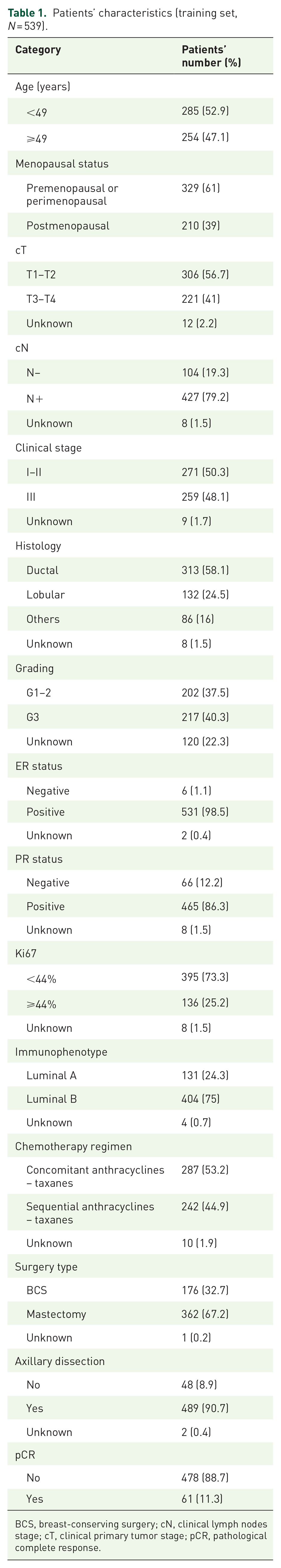
BCS, breast-conserving surgery; cN, clinical lymph nodes stage; cT, clinical primary tumor stage; pCR, pathological complete response.
The pathological features after surgery are shown in Supplemental Table 1. We looked at the distribution of Ki67 values in the pre-NACT biopsy based on the predictive value of Ki67 with regard to the pathological response: the median Ki67 value at baseline in the overall population was 30%, compared to 8% after NACT (p < 0.0001) (Supplemental Figure 1, a). Notably, we found a statistically significant difference in the median pre-NACT Ki67 value between patients who obtained a pCR compared with those who did not (45% versus 28%, respectively; p < 0.0001) (Supplemental Figure 1, b). Then, we determined the optimal Ki67 cutoff on the pre-NACT biopsy to predict pCR: the value obtained from the ROC analysis was 44% [area under the curve (AUC): 0.69, standard error (SE): 0.04; p < 0.001] (Supplemental Figure 2). In the pre-NACT biopsy, most patients (73.3%) showed a Ki67 value < 44% and nearly a quarter (25.2%) presented a Ki67 value ⩾44%.
We assessed the association between pCR rate and markers studied (Table 2). Regarding the association between pathological response and degree of hormone receptors positivity, we observed a statistically significant difference in the pCR rate according to PR expression (9.9% for PR-positive versus 21.2% for PR-negative tumors; p = 0.007). For ER and PR expression, a cutoff of 1% was used. Subsequent sensitivity analyses were performed with cutoffs for ER and PR expression of 50% and 10%, and 50% and 20%, respectively. No statistically significant variation in pCR rate was identified depending on the hormone-positive thresholds examined individually or for combinations of them, although patterns of increasing pCR rate being seen when hormone receptors percentages fell (11.2% for ER > 50% versus 12.2% for ER ⩽50% tumors; p = 0.805; 9.5% for PR > 50% versus 13.4% for PR ⩽50% tumors; p = 0.153; 10.8% for ER > 10% versus 20.0% for ER ⩽10% tumors; p = 0.124; 10.2% for PR > 20% versus 13.4% for PR ⩽20% tumors; p = 0.274). Besides, the pCR rate was significantly associated with the subtype [luminal B-like 13.1% (95% CI: 9.8–13.4) versus luminal A-like 5.3% (95% CI: 1.5–9.1); p = 0.01], while no difference in the pCR rate was obtained in relation to the chemotherapy regimen, although a trend toward increased pCR rate with the sequential anthracycline and taxane-based NACT was seen [sequential versus concomitant with anthracyclines and taxanes: 14.0% (95% CI: 9.6–18.4) versus 9.4% (95% CI: 6.0–12.7), respectively; p = 0.10].
Univariate and multivariate analyses for pCR and replication rate for internal validation.

BCS, breast-conserving surgery; CI, confidence interval; cN, clinical lymph nodes stage; cT, clinical primary tumor stage; ER, estrogen receptor; MT, mastectomy; OR, odds ratio; pCR, pathological complete response; post-m, postmenopausal; PR, progesterone receptor; pre-m, premenopausal.
The statistically significant pre-NACT clinicopathological variables at the univariate analysis for the pCR prediction (clinical stage, tumor size, nodal status, subtype, grading, Ki67, and PR expression) were included in the multivariate analysis (Table 2). The clinical stage I–II (OR: 3.67, 95% CI: 1.75–7.71; p = 0.001), the Ki67 value ⩾44% (OR: 3.00, 95% CI: 1.59–5.65; p = 0.001), and the absence of PR (OR: 2.49, 95% CI: 1.15–5.38; p = 0.019) resulted to be significant independent predictors of pCR, with a high replication rate at the internal cross-validation analysis (replication rate: 100%, 98%, and 87%, respectively) (Table 2).
Based on the ORs of significant pre-treatment variables at multivariate analysis for pCR, each patient was assigned a predictive scoring index combining the normalized scores of the independent variables. Figure 1, a, shows the probability of obtaining a pCR according to the score assigned to each patient by combining the three independent variables, with a proportional relationship between the score and the probability of pCR. By score dichotomizing according to the pCR, we were able to determine that the value 1 was the optimal cutoff point. A statistically significant difference in pCR prediction was found among patients with a continuous score >1 and patients with a continuous score ⩽1. At the ROC analysis, the predictive accuracy of this nomogram was 73% (AUC: 0.73; SE: 0.03) with a high sensitivity and specificity (Figure 1, b).

Clinicopathological nomogram for predicting pCR (a): a predictive score is assigned to each variable and the sum of scores is converted to the probability of pCR; ROC curve for nomogram cutoff in TS (b) and in VS (c).
In the VS, data from 258 early LBC patients were gathered. Patients’ characteristics of the VS are listed in Supplemental Table 2. The predictive nomogram derived in the TS was applied to the VS, providing to be likewise able to discriminate the probability of pCR in the VS. At the ROC analysis, the predictive accuracy was 70% (AUC: 0.7; SE: 0.05) (Figure 1, c). No significant difference between the predictive performance of the model in both patients’ cohorts was found (p = 0.61).
At a median follow-up of 39.4 months (range: 1–187) in the TS, an event occurred in 37.8% of cases (204 patients). With regard to DFS, the surgery type, the pCR, and the grading pre-NACT showed a significant correlation with the risk of disease recurrence at the multivariate analysis (Table 3). A statistically significant difference was found in DFS depending on the grading pre-NACT G1–2 versus G3 (5 years: 58.5% versus 47.4%, p = 0.002; Figure 2, a), on the pCR versus invasive residual disease (5 years: 72.3% versus 52.9%, p = 0.02; Figure 2, b) and on the type of conservative or radical breast surgery (5 years: 64.1% versus 51.6%; p = 0.004; Figure 2, c). A trend toward an improved DFS was observed in the luminal A-like population compared to luminal B-like group, but this increase is not statistically significant (5 years luminal B-like versus luminal A-like tumors: 53.2% versus 58%; p = 0.15; Supplemental Figure 3, a). When taking into account the prognostic significance of pCR and subtype, we performed an exploratory analysis of combined survival by pathologic response (pCR versus invasive residual disease) and subtype (luminal A-like versus luminal B-like) and we found a trend toward increased DFS in the presence of pCR in both luminal sub-populations (Supplemental Figure 3, b and c).
Univariate and multivariate analyses for DFS.

BCS, breast-conserving surgery; CI, confidence interval; cN, clinical lymph nodes stage; cT, clinical primary tumor stage; DFS, disease-free survival; ER, estrogen receptor; HR, hazard ratio; MT, mastectomy; pCR, pathological complete response; pN, pathological lymph nodes; post-m, postmenopausal; PR, progesterone receptor; pre-m, premenopausal; pT, pathological primary tumor.

DFS according to grading pre-NACT (a), pathological response (b), and surgery type (c).
Discussion
According to the predictive performance of the nomogram, the combination of widely accessible clinicopathological criteria allows to reliably evaluate the likelihood of pCR in the context of the luminal subtype. In the proposed model, the probability of pCR ranges from 3.4% to 53.3% with a predictive accuracy of 73%. The nomogram included the variables independently associated with response at multivariate analysis: clinical stage, PR expression, and Ki67 value. At the internal cross-validation analysis, all the independent factors replicate with a high rate. In addition, the validation supported the discrimination performance of the predictive model, highlighting its potential to effectively classify LBC patients according to their unique probabilities of pathological response at diagnosis.
These data are consistent with previous findings.21–24 Tumors with high hormone receptor expression and low proliferative activity benefit less from chemotherapy,21–23 whereas PR negativity and luminal B-like subgroup correlate with better NACT response. 24 However, when examined separately, both PR and Ki67 levels showed only weak predictive ability to predict chemotherapy response. 24 As expected, our study shows that the integration of Ki67 and hormone receptor levels may better predict the pathological response to NACT for LBCs. According to the predictive model herein developed and validated, a LBC patient with clinical stage III, PR expression, and Ki67 <44% has an extremely low likelihood of achieving a pCR following NACT (3.4%). On the other hand, an LBC patient with a clinical stage I or II and Ki67 ⩾44% without PR expression has a high probability of obtaining a pCR (53.3%).
Lower clinical stages have been shown to be related with higher pCR rates than more advanced stages, which is consistent with the literature.25,26 Although there is currently no clear explanation for this, an inverse correlation between the number of tumor infiltrating lymphocytes (TILs), representing a predictor of NACT response27,28 and tumor size and lymph node involvement, has been observed.27,29 This finding is consistent with reduced immune activation in metastatic BC than in early disease stages,30,31 suggesting an improved immune evasion with larger cancer burden. This may support the hypothesis of a higher NACT response for lower disease stage.
The achievement of pCR was found to be a favorable independent prognostic factor, although no statistical significance was proven for either luminal sub-population. Notably, a median follow-up of 39.4 months for LBCs may be insufficient to draw firm conclusions from the survival results.
Grading pre-NACT showed an impact on the DFS as well. Its predictive usefulness is acknowledged, and it has been incorporated into the ClinicoPathological Stage-Estrogen Grade score,32,33 which provides a more refined assessment of prognosis following NACT for LBCs. 32
Consistently with the literature,34–37 our work confirmed the prognostic role of the surgery type. A possible explanation for the improved survival of the BCS group is that patients who undergo mastectomy have a worse prognosis at onset. In addition, not all patients who undergo BCS are downstaged following NACT (some may have suitable for lumpectomy at the onset and have a more favorable prognosis).
Despite the generally better prognosis for LBC patients compared with other BC subtypes due to both the inherent tumor biology and the opportunity for using ET as a therapeutic option, the role of pCR as an independent prognostic factor in BC patients undergoing NACT has been established, although the association’s strength being less evident for LBCs.8,10,38 Therefore, we have developed and validated a nomogram to predict each patient’s likelihood of achieving pCR in a subpopulation such as hormone receptor-positive /HER2-negative, which represents most BC patients but has limited potential to achieve a good pathological response to NACT. Other nomograms have been proposed to predict the chance of pCR in unselected BCs based on hormone receptor expression,22,25,39–41 and previous efforts to combine pre-therapy features to predict the responsiveness of LBCs to NACT have been performed.42–44 Multivariate semi-quantitative models were developed as prognostic scores and then tested as predictive tools for pCR to NACT in LBCs.42–44 However, these approaches have included limited sets of patients and exclusively incorporated pathological variables, such as hormone receptors, Ki67, and HER2 expression, combined with an algorithmic method to derive the predictive score, and have not been implemented in clinical practice. Our nomogram has been developed on a large, multi-institutional dataset, providing a simple, fast, and reliable method for predicting the chemosensitivity and classify LBC patients according to the chance of pCR, integrating the predictive value of each pre-therapy clinicopathological feature. Also, the fact that our tool is based on commonly available clinicopathological features makes it easy to implement in clinical practice. Since the role of NACT for operable LBCs is debated, the nomogram-based predictive tool developed in our study could identify LBC patients who have a greater than 50% chance of achieving a pCR following NACT and consequently a survival advantage due to early exposure to systemic cytotoxic treatment. In contrast, patients with none of the features identified as predictive of response by the nomogram will most likely not obtain a good pathologic response following NACT. Given the prognostic value of residual invasive disease after NACT,45,46 patients with operable LBC at diagnosis and features such as low Ki67, PR expression, and stage III could undergo surgery or neoadjuvant ET to achieve preoperative downstaging. In this scenario, the decision to offer chemotherapy could be reserved for the adjuvant setting, where the use of gene expression panels evaluated on surgical specimens is becoming more widespread and is suggested by international guidelines to guide the choice of adding chemotherapy to ET.47–49
In contrast to well-established prognostic signatures based on pathologic stage following NACT and prognostic and predictive signatures based on gene expression patterns on the surgical specimen that may aid decision-making in the adjuvant setting,46,50–52 we have created and validated a nomogram to predict pathologic response following NACT at diagnosis, informing decision-making at baseline, before any treatment. In this context, evaluable features on pre-NACT biopsies, such as TILs and gene expression risk, can be incorporated with our predictive model, which is based on clinicopathological variables easily accessible in routine clinical practice. Indeed, gene expression panels evaluated on core needle biopsy have demonstrated a predictive role of pathologic response to systemic therapy for LBCs, with a higher likelihood of pCR following NACT for high genomic risk tumors and a higher likelihood of clinicopathological response to neoadjuvant ET for low genomic risk tumors. 53 International guidelines suggest the use of gene expression signatures to consider preoperative ET for LBC patients selected for comorbidities and clinical and genomic features of low risk of recurrence.48,49
We have to acknowledge that the retrospective and non-randomized nature of this analysis represents a limit. Another limitation is that no pathology reports actually mentioned immune cells or infiltrate features; hence, it was not possible to incorporate immune-infiltrate characteristics (last reports refer to late 2018, when TILs were not yet considered essentials for prognosis). Although the great majority received anthracyclines and taxane-based treatment, our results may have been impacted by the NACT regimen and adjuvant ET heterogeneity because of the lengthy inclusion period.
Our data are consistent with a recent report by Sella et al., which found that (1) the 21-gene assay Oncotype DX may predict NACT response of LBCs and (2) in univariate analysis, PR negative, high grade, and a higher recurrence score were associated with the pCR. 54 This suggests that pathological features, such as the absence of PR and the high grade, are related to a better pathological response to NACT, with a potential relationship between these clinicopathological characteristics and the genomic risk as determined by the 21-gene assay.
In conclusion, despite the limitations, this multicenter retrospective analysis suggests that the combination of pre-NACT clinicopathological factors, easily available in clinical practice, in a predictive pCR nomogram is able to powerfully classify LBC patients into NACT responders and non-responders at diagnosis. To the best of our knowledge, the resulting validated model represents the first pCR nomogram exclusively proposed for LBCs, providing predictive information that can support physicians’ decision-making in clinical practice. Indeed, LBCs that have the best chance of achieving a pCR might be exposed early to a systemic cytotoxic treatment. In contrast, LBC patients, who are unlikely to get a pCR following NACT, may undergo surgery or neoadjuvant ET to achieve preoperative downstaging and avoid toxic and potentially harmful NACT. Our prediction tool may also provide the basis for adding new molecular markers to the model. The accuracy of the predictive tool is anticipated to increase with the inclusion of additional pathological characteristics and gene expression signatures.
Supplemental Material
sj-docx-1-tam-10.1177_17588359221138657 – Supplemental material for Development of a nomogram for predicting pathological complete response in luminal breast cancer patients following neoadjuvant chemotherapy
Supplemental material, sj-docx-1-tam-10.1177_17588359221138657 for Development of a nomogram for predicting pathological complete response in luminal breast cancer patients following neoadjuvant chemotherapy by Giovanna Garufi, Luisa Carbognin, Isabella Sperduti, Federica Miglietta, Maria Vittoria Dieci, Roberta Mazzeo, Armando Orlandi, Lorenzo Gerratana, Antonella Palazzo, Alessandra Fabi, Ida Paris, Antonio Franco, Gianluca Franceschini, Elena Fiorio, Sara Pilotto, Valentina Guarneri, Fabio Puglisi, Pierfranco Conte, Michele Milella, Giovanni Scambia, Giampaolo Tortora and Emilio Bria in Therapeutic Advances in Medical Oncology
Supplemental Material
sj-docx-2-tam-10.1177_17588359221138657 – Supplemental material for Development of a nomogram for predicting pathological complete response in luminal breast cancer patients following neoadjuvant chemotherapy
Supplemental material, sj-docx-2-tam-10.1177_17588359221138657 for Development of a nomogram for predicting pathological complete response in luminal breast cancer patients following neoadjuvant chemotherapy by Giovanna Garufi, Luisa Carbognin, Isabella Sperduti, Federica Miglietta, Maria Vittoria Dieci, Roberta Mazzeo, Armando Orlandi, Lorenzo Gerratana, Antonella Palazzo, Alessandra Fabi, Ida Paris, Antonio Franco, Gianluca Franceschini, Elena Fiorio, Sara Pilotto, Valentina Guarneri, Fabio Puglisi, Pierfranco Conte, Michele Milella, Giovanni Scambia, Giampaolo Tortora and Emilio Bria in Therapeutic Advances in Medical Oncology
Supplemental Material
sj-jpeg-1-tam-10.1177_17588359221138657 – Supplemental material for Development of a nomogram for predicting pathological complete response in luminal breast cancer patients following neoadjuvant chemotherapy
Supplemental material, sj-jpeg-1-tam-10.1177_17588359221138657 for Development of a nomogram for predicting pathological complete response in luminal breast cancer patients following neoadjuvant chemotherapy by Giovanna Garufi, Luisa Carbognin, Isabella Sperduti, Federica Miglietta, Maria Vittoria Dieci, Roberta Mazzeo, Armando Orlandi, Lorenzo Gerratana, Antonella Palazzo, Alessandra Fabi, Ida Paris, Antonio Franco, Gianluca Franceschini, Elena Fiorio, Sara Pilotto, Valentina Guarneri, Fabio Puglisi, Pierfranco Conte, Michele Milella, Giovanni Scambia, Giampaolo Tortora and Emilio Bria in Therapeutic Advances in Medical Oncology
Footnotes
References
Supplementary Material
Please find the following supplemental material available below.
For Open Access articles published under a Creative Commons License, all supplemental material carries the same license as the article it is associated with.
For non-Open Access articles published, all supplemental material carries a non-exclusive license, and permission requests for re-use of supplemental material or any part of supplemental material shall be sent directly to the copyright owner as specified in the copyright notice associated with the article.