
Abstract
Background:
Different oncological therapies have been approved for small intestinal neuroendocrine tumors (SI-NETs), but relatively little is known about efficacy and long-term outcome outside of phase III trials.
Methods:
This retrospective analysis assessed patients with well-differentiated, metastatic SI-NETs treated at the Medical University of Vienna, an approved European Neuroendocrine Tumor Society (ENETS) Center of Excellence for neuroendocrine tumors. The primary objective was to assess progression-free survival (PFS) following approved therapies, that is, octreotide, lanreotide, peptide receptor radionuclide therapy (PRRT), and everolimus, in a representative real-world collective.
Results:
A total of 77 patients receiving systemic treatment for advanced SI-NETs between 2010 and 2021 were included, with a median follow-up time of 82.3 months [95% confidence interval (CI), 57.8–106.8 months]. In the entire collective, the estimated median PFS following first-line therapy was 32.0 months (95% CI, 23.5–40.5 months). Peritoneal carcinomatosis was significantly associated with worse PFS (p = 0.016). Regarding therapeutic strategies and outcome, 59 patients received somatostatin analogs first line and no significant difference in PFS was observed between lanreotide and octreotide (29.3 versus 35.5 months, p = 0.768). Across all treatment lines, 42 patients underwent PRRT (estimated median PFS: 32.0 months; 95% CI, 25.6–38.3 months) and a small subgroup of 7 patients received everolimus (estimated median PFS: 9.2 months; 95% CI, 1.6–17.0 months). For the total cohort, the estimated median OS following first-line therapy was 100.6 months (95% CI, 82.3–118.8 months), but the high proportion of deaths attributed to NET (77.8%) underlines the lethal nature of the disease. No unexpected toxicities were observed.
Conclusions:
While peritoneal carcinomatosis emerged as an adverse prognostic factor for PFS in this collective, the long-term outcome of patients treated at a specialized NET center using approved therapies appeared comparable to pivotal studies in SI-NET.
Keywords
Introduction
Neuroendocrine neoplasms (NENs) are rare, solid tumors originating from the diffuse neuroendocrine cell system distributed throughout the body.1,2 Small intestinal (SI) NENs are among the most common NENs in the gastroenteropancreatic (GEP) tract and comprise neoplasms arising in the jejunum and ileum.3–5 Based on mitotic count/Ki-67 index and histomorphological presentation, these neoplasms are dichotomized into well-differentiated neuroendocrine tumors (NETs) and poorly differentiated neuroendocrine carcinomas, 6 with the former progressing more slowly and being prognostically more favorable. 7 Furthermore, expression of somatostatin receptors (SSTR) is a characteristic feature of NETs and is of specific biological, diagnostic, and therapeutic relevance.8,9
For Austria, a 1-year prospective study reported a GEP-NET incidence of 2.39 per 100.000 inhabitants per year and 15.4% of GEP-NETs were of SI origin. 4 In total, 48.4% of patients were metastatic at the time of initial diagnosis, 4 highlighting that these patients are frequently diagnosed with advanced disease. SI-NETs generally have a good prognosis, but it depends heavily on staging and grading. 10 Apart from histology, major predictors of survival are age, sex, presence of liver metastases, elevated tumor markers, and carcinoid syndrome caused by active production of serotonin by tumor cells.11–13 Overall, according to Surveillance, Epidemiology, and End Results registry data, one-third (32.4%) of all SI-NET patients and half (55.5%) of patients with metastatic disease have carcinoid syndrome, 14 which can result in characteristic symptoms such as diarrhea or flushing, but eventually also in lethal right-sided heart failure.15,16
In terms of therapy, surgery is the only potentially curative treatment option for both localized and advanced SI-NETs.5,17 For advanced or metastatic SSTR-positive SI-NETs grade (G) 1 or G2, the standard antiproliferative treatment, regardless of functional activity, consists of the somatostatin analogs (SSAs) octreotide and lanreotide, commonly followed by peptide receptor radionuclide therapy (PRRT) with 177lutetium-DOTATATE (or with 90yttrium-DOTATOC) in case of progression. 17 Furthermore, the mammalian target of rapamycin (mTOR) inhibitor everolimus is an option for patients with progressive disease irrespective of SSTR status and pretreatment. 17 All of these compounds have been evaluated in randomized phase III studies with the primary endpoint progression-free survival (PFS) and were compared to placebo (SSA18,19 or everolimus 20 ) or high-dose SSA in case of PRRT 21 (second line after SSA and progression). Importantly, none of these studies was designed to show improved overall survival (OS), and crossover impedes clear interpretation of survival outcomes.22,23 Thus, quality of life and toxicity results should also be taken into consideration when applying these therapies. 24
Owing to the strict eligibility criteria of randomized controlled trials (RCTs), study populations may differ considerably (e.g. with regard to performance score or comedication) from the patient population seen in day-to-day clinical practice and hence also long-term data might not be comparable. 25 Furthermore, SI-NENs are relatively rare neoplasms and some of the trials mentioned above did not exclusively study well-differentiated neuroendocrine tumors of SI origin, thus additional evidence regarding this specific subgroup could be insightful.
The Division of Oncology at the Medical University of Vienna is a tertiary referral center for patients with a diagnosis of NEN and a certified European Neuroendocrine Tumor Society (ENETS) Center of Excellence, thus overseeing a considerable number of patients with this diagnosis. As systematically analyzed real-world data (RWD) can complement RCT data, we present RWD regarding oncological therapy, treatment patterns, clinical characteristics as well as short- and long-term outcomes of patients with advanced SI-NETs receiving systemic therapy at our tertiary referral center.
Methods
This study is a noninterventional, retrospective data analysis, which collected RWD on advanced SI-NETs receiving systemic therapy at the Medical University of Vienna. The study population was defined by screening of electronic health records and by selecting patients with histologically confirmed SI (jejunoileal) NETs of grade G1 or G2 treated at the Division of Oncology, Department of Medicine I, Medical University of Vienna between January 2010 and January 2021. In addition, patients with cancer of unknown primary (NET CUPs) with serotonin- and CDX2-positive immunohistochemistry (thus most likely to originate from the small intestine) were included. A total of 135 patients with SI-NET or CUP were identified from this period, but 18 were excluded as they presented with grade 3 NET. Furthermore, patients with insufficient (histological or radiological) information (n = 12) or patients without systemic therapy (n = 19) or follow-up imaging (n = 9) were excluded. All histological reports were (re)assessed by a reference pathologist at our center according to the most recent WHO classification. Response and progression were determined based on radiological assessment following RECIST principles and tumor board review. Response evaluation intervals were at the discretion of the attending physician and in line with current guidelines, that is, at least 3 or 6 months apart depending on tumor biology. The disease control rate (DCR) was the percentage of patients with complete response, partial response, or stable disease for at least 6 months. Data on the sites of metastasis were collected from surgery/histology and radiological imaging (primarily positron emission tomography/computed tomography). All patients were routinely discussed by a multidisciplinary tumor board. The analysis had received approval (EK No: 2488/2020) from the local Ethics Committee of the Medical University of Vienna.
Patient data were extracted from electronic health records of the Medical University of Vienna and stored in a database built up with FileMaker (Claris International Inc.). Apart from collection of basic patient characteristics (age, sex, performance status, and symptoms) and NET-specific characteristics [tumor stage, primary localization, absence/presence and localization of metastases, date of diagnosis, histology, functional imaging, endocrine activity, biochemical markers chromogranin (CgA) and urinary 5-hydroxyindoleacetic acid (5-HIAA)], the chronological sequence of various lines of systemic therapy was reconstructed for each patient, including information on the type of treatment (drugs and dosages), adverse events, and outcome (objective response, PFS, OS, and possibly cause of death).
IBM SPSS Statistics 27 (IBM Corp., Armonk, NY, USA) was used to calculate statistical measures. For categorical variables, frequencies and percentages were calculated, and quantitative variables were presented as ranges, means, medians (or quartiles), and with standard deviations. A Sankey diagram of treatment sequencing was developed with Google Charts. PFS and OS curves were calculated with the Kaplan–Meier method, and subgroups were compared using the log-rank test. If univariate analysis found these differences to be statistically significant, regression analyses with Cox proportional hazards model were performed. A two-tailed p value below the level α = 0.05 was regarded as statistically significant. Dates of death were only obtainable if they were available in the in-house electronic health records (e.g. autopsy reports) or if they were listed in the cause-of-death statistics from the IT4Science service of the Medical University of Vienna/Statistics Austria. Median follow-up was determined using the reverse Kaplan–Meier estimator. 26
Results
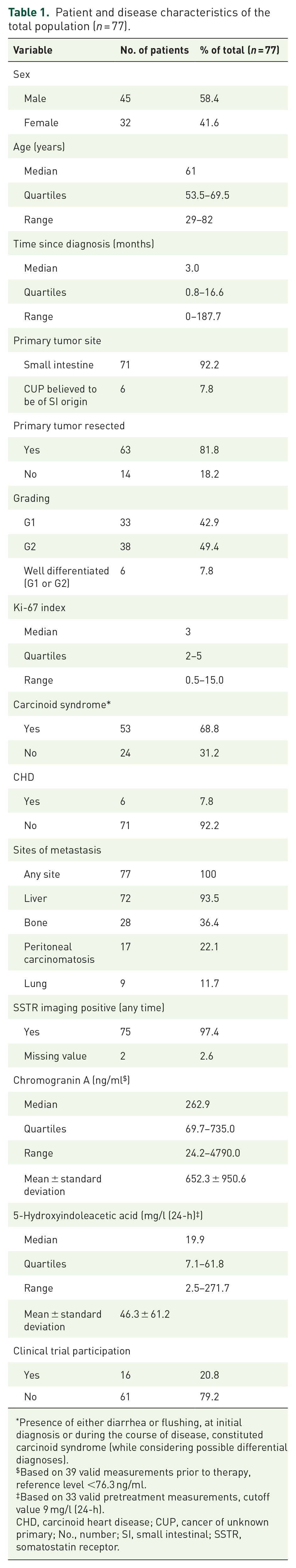
A total of 77 patients were included in this analysis; 32 (41.6%) were female and 45 (58.4%) were male. The median age at diagnosis was 61 years (range, 29–82 years) and the median follow-up time calculated from diagnosis was 82.3 months [95% confidence interval (CI), 57.8–106.8 months]. Functional symptoms were present at the initial diagnosis in 26 patients (33.8%) and additionally developed in 27 patients (35.1%) during the course of the disease. In total, 19 patients (24.7%) had diarrhea and flushing, 26 (33.8%) reported only diarrhea, seven (9.1%) experienced only flushing, and one patient suffered from other hormonal symptoms. Hence, overall, 24 (31.2%) had non-functioning tumors. Corresponding to the high percentage of symptomatic patients, the proportion of patients with elevated 5-HIAA levels was high (69.7%). However, valid pretreatment measurements were only available for 42.9% (33/77) of the study population and missing information on non-hormonal causes of diarrhea, for example, short bowel syndrome, cannot be ruled. Ultimately, six patients (7.8%) were diagnosed with carcinoid heart disease (CHD). While screening for CHD by transthoracic echocardiography is performed annually at our center in patients with increased 5-HIAA levels, and NT-proBNP is determined in routine laboratory tests depending on clinical presentation, we cannot exclude that a few cases may have remained undetected.
Regarding primary localization of the tumor, six patients (7.8%) had no known primary and were classified as CUP, while the remaining patients had a primary tumor of SI origin. The primary tumor was resected in 63 patients (81.8%), and 6 (9.5%) of those did not have metastatic disease documented at the time of diagnosis (stage III disease) but developed metastases metachronously. In the 14 patients (18.2%) without primary tumor resection, histological information was obtained from metastases only. Concerning histological grading, 33 patients (42.9%) had G1 tumors, 38 (49.4%) had NETs G2, and in 6 individuals (7.8%) the grading information was limited to characterization as well-differentiated NET, with the Ki-67 index being unavailable. SSTR imaging was positive in all patients, except two where this information was missing. For detailed information regarding metastatic spread, biochemical markers (i.e. CgA and 5-HIAA), and further clinical features see Table 1.
Patient and disease characteristics of the total population (n = 77).
Presence of either diarrhea or flushing, at initial diagnosis or during the course of disease, constituted carcinoid syndrome (while considering possible differential diagnoses).
Based on 39 valid measurements prior to therapy, reference level
Based on 33 valid pretreatment measurements, cutoff value 9 mg/l (24-h).
CHD, carcinoid heart disease; CUP, cancer of unknown primary; No., number; SI, small intestinal; SSTR, somatostatin receptor.
SSAs were the first line treatment for 59 (76.6%) of 77 patients, PRRT was the initial therapy in 7 (9.1%), everolimus in 1 patient (1.3%), and other treatment strategies were applied in 10 patients (13.0%), which included compounds evaluated in clinical trials at that time, liver-directed therapies and radiotherapy. Altogether, 167 therapeutic interventions were documented, and the median number of treatment lines per patient was 2. See Figure 1 for an overview of first-line and sequentially applied therapies.

Sankey diagram of treatment sequencing in the study population of 77 patients (diagram created with Google Charts).
Outcome of the patient cohort overall
For the entire study population (n = 77), the estimated median PFS following first-line therapy was 32.0 months (95% CI, 23.5–40.5 months), and the estimated median OS from initiation of first-line therapy was 100.6 months (95% CI, 82.3–118.8 months), see Figure 2. The 5- and 10-year survival rate from treatment start were 81.9% (95% CI, 71.4–92.2%) and 39.3% (95% CI, 20.4–58.2%), respectively. At the data cutoff date, 27 patients (35.1%) had died and 50 (64.9%) were alive with disease. In 21 patients (77.8%), the cause of death was NEN-related and four (14.8%) died due to non-cancer or non-index-cancer causes (one case each of atherosclerotic heart disease, acute myeloblastic leukemia (AML), esophageal cancer, and pancreatic cancer). Furthermore, in two patients (7.4%), this information was not available. The patient with AML died 5 years after receiving four cycles of PRRT, followed by salvage PRRT with two further doses 2 years later, and 2 years after radiation therapy for spinal metastases, which additionally resulted in an exposure of 30 Gy. Looking at survival measured from the date of diagnosis, the estimated median OS in the entire collective was 137.4 months (95% CI, 87.6–187.2 months).

Kaplan–Meier analysis of (a) PFS and (b) OS (from treatment initiation) of the entire study population.
Subgroup analyses for the entire collective were performed to compare PFS and OS of patients based on predefined clinical characteristics (sex, grading, presence of carcinoid syndrome, bone metastases, peritoneal carcinomatosis, and CgA elevation). Interestingly, only the presence of peritoneal carcinomatosis (n = 17, 22.1%) had a significant negative impact on PFS (p = 0.016) with a corresponding hazard ratio (HR) of 2.07 (95% CI, 1.13–3.78; p = 0.018), indicating that peritoneal carcinomatosis doubles the risk of progression. The negative prognostic effect of peritoneal carcinomatosis was maintained in a multivariate analysis considering sex, age at diagnosis, and grade (HR: 3.53; 95% CI, 1.77–7.07; p = 0.0004). As can be extracted from the respective Kaplan–Meier plot for PFS in Figure 5, all but one patient (16/17) progressed if peritoneal carcinomatosis was present, while 36 events were reported and 24 were censored in the subgroup without peritoneal carcinomatosis.
Somatostatin analogs
As mentioned above, SSAs were the first-line treatment for 59 patients (76.6%) and 26 (33.8%) received octreotide versus 33 (42.9%) lanreotide. A total of 51 patients (86.4%) were administered the recommended dose of 120 mg lanreotide autogel every 4 weeks or 30 mg octreotide LAR every 4 weeks. For the remaining eight individuals, either dosage information was not available (n = 3), or doses/intervals deviated from the approved drug regimen (n = 5). The estimated median PFS following SSA therapy was 33.6 months (95% CI, 23.8–43.4 months) in the patient cohort overall. In terms of differences between the two compounds, the estimated median PFS was 29.3 months (95% CI, 18.6–40.1 months) in the 33 lanreotide recipients, whereas it was slightly longer at 35.5 months (95% CI, 24.9–46.0 months) in the octreotide subgroup (n = 26). However, there was no statistically significant difference in the PFS between the two approved drugs (p = 0.768), and the curves were nearly identical (see Figure 3(a)). In line with the entire collective, presence of peritoneal carcinomatosis was associated with a significant decrease in PFS (p = 0.034), while no further negative prognostic markers were identified. No patient on SSA alone experienced a complete or partial response. The DCR was 86.4% (95% CI, 77.7–95.2%), as 51 patients had radiologically documented stable disease following SSA therapy. The estimated median OS from initiation of first-line SSA treatment was 124.2 months (95% CI not calculable) in the lanreotide subpopulation, 90.3 months (95% CI, 82.5–98.1 months) in the octreotide subgroup (p = 0.098), and 100.6 months (95% CI, 85.8–115.3 months) overall, see Figure 3(b).

Kaplan–Meier analysis of (a) PFS and (b) OS (from treatment initiation) of SSA-treated patients.
PRRT
Administration of PRRT was documented in a total of 42 patients (54.5%) during the course of the disease. PRRT was the first-line treatment for seven patients (e.g. in view of tumor inoperability and symptom burden), and one patient had received everolimus prior to PRRT. The remaining 34 patients (44.2%) had disease progression during SSA therapy before starting PRRT. All patients received 177lutetium-based PRRT, except for one who received 90yttrium-based PRRT and for another one who received both radionuclides. Most patients (n = 30, 71.4%) received four doses of the radionuclide, five individuals (11.9%) 3 infusions, five patients (11.9%) 2 cycles, and two (4.8%) a single PRRT administration. In total, 28 patients (68.3%) were treated with SSA concomitantly and subsequently had also SSA maintenance therapy. The estimated median PFS was 32.0 months (95% CI, 25.6–38.3 months; Figure 4(a)). Although exact measurements of predefined lesions were not documented according to RECIST, a relevant tumor regression was described in the radiology reports of 11 patients [objective response rate (ORR) of 29.7%; 95% CI, 15.0–44.5%; excluding 5 patients with pending assessment]. In addition, disease stabilization was experienced by 20 patients, resulting in a DCR of 83.8% (95% CI, 71.9–95.7%). The estimated median OS from the start of therapy was 65.8 months (95% CI, 41.8–89.9 months; Figure 4(b)) and the median follow-up time was 32.2 months (95% CI, 22.9–41.6 months). Apart from more general adverse events (e.g. fatigue, nausea, diarrhea, and peripheral edema), specific hematological effects of PRRT were documented (information invalid or missing in four patients). Following PRRT cycles, 23 patients (60.5%) had anemia, 17 (44.7%) thrombocytopenia, 19 (50.0%) leukopenia, 31 (81.6%) lymphopenia, and 1 (2.6%) neutropenia which were mostly low grade according to the CTCAE version 5.0, but 8 patients (21.1%) had grade 3 lymphopenia, one grade 4 lymphopenia, and one patient grade 3 thrombocytopenia. One case of AML was observed during prolonged follow-up, as described above in the section regarding long-term outcome in the patient cohort overall. In terms of nephrotoxicity, four patients (10.5%) presented with grade 1 elevation in creatinine.

Kaplan–Meier analysis of (a) PFS and (b) OS (from treatment initiation) of patients after PRRT.
Everolimus
Everolimus was administered to a total of seven patients (9.1%), including one patient who received the treatment upfront and one who had previously received more than four treatment lines. Three patients were treated with 10 mg/day (as evaluated in RADIANT-4), two patients were started with 5 mg/day due to frailty/reduced performance status and dosages were then escalated to 10 mg/day, and two individuals remained at 5 mg/day. The estimated median PFS was 9.2 months (95% CI, 1.6–17.0 months), and the estimated median OS calculated from treatment initiation was 16.9 months (95% CI, 6.8–27.1 months), with patients followed for a median time of 18.9 months (95% CI, 10.9–26.9 months). With regard to safety and tolerability, every single patient experienced adverse events that resulted in either temporary treatment interruptions with subsequent dose reductions (n = 4) or discontinuation of therapy (n = 2) or dose modifications alone (n = 1). The most frequent toxicity was rash (n = 5; two patients had grade 2–3 rash), followed by diarrhea (n = 3). Stomatitis, peripheral edema, and pruritus occurred in two patients each. Furthermore, one instance of cytopenia (grade 2–3) and one case of hyperglycemia was documented in patient records.
Discussion
This real-world assessment includes a homogeneous population of 77 patients with metastatic SI-NETs G1 or G2 receiving approved systemic therapies at a tertiary referral center, capturing the course of treatment for almost every patient in its entirety. The long time until tumor progression (estimated median PFS following first-line therapy of 32.0 months) as well as the encouraging OS (estimated median OS of 100.6 months) highlight the chronic but still ultimately lethal nature of this disease, with 77.8% of recorded deaths being NET related. Naturally, there is inherent variability in survival time that is also determined by various factors.11,12,27,28 Some of those influencing clinical factors were analyzed in this collective, but exclusively the presence of peritoneal carcinomatosis had a significant effect on median PFS in the entire collective (p = 0.016) and the SSA-treated collective (p = 0.034), respectively. No differences in OS were detected (see Figure 5), but this analysis was probably underpowered for detecting an impact of other factors on survival. Interestingly, grading was not a significant predictive marker for OS, despite a (non-significant) trend for a better PFS in G1 versus G2 tumors. Possible explanations include lack of power but also changes in grading during the course of the disease. In our patients, 11 re-biopsies were performed in case of suspected changes of tumor biology (e.g. based on imaging/SSTR uptake), but no treatment relevant changes, that is, switch to G3, were documented in this collective. Finally, most data on the prognostic effect of G1 versus G2 derive from an earlier era, where not as many approved and effective treatments were available. Thus, one could speculate that modern treatments partly counterbalance the differences in outcome.

Kaplan–Meier analyses and log-rank tests comparing PFS and OS (from treatment initiation) of the entire study population according to various subgroups.
In our cohort, the percentage of patients with CHD is low at 7.8% (or 11.3% of patients with carcinoid syndrome), considering the results of a 2007 study in which 150 midgut tumor patients with carcinoid syndrome were screened by transthoracic echocardiography and 30 patients (20%) were diagnosed with CHD. 29 While we cannot exclude that a few cases may have remained undetected until the cutoff date for data collection, it also seems plausible that improved treatment might have further lowered the prevalence of CHD, as it did earlier (56% of carcinoid syndrome patients had CHD in a study from 1993). 30 With regard to survival, there was no statistically significant difference in PFS or OS based on the presence of carcinoid syndrome (p = 0.458 and p = 0.757, respectively, see Figure 5), and the same applies to consecutively performed survival analyses based on 5-HIAA elevation and CHD (PFS: p = 0.167 and p = 0.400, respectively, and OS: p = 0.504 and p = 0.651, respectively). Again, carcinoid syndrome is a known predictor of survival, 12 but the number of cases in this study was probably insufficient to detect a significant effect of this and other factors on survival.
Several findings regarding specific treatment strategies may be discussed. First, the present data further support the available evidence for the use of first-line SSA in an all-comer cohort of metastatic SI-NET patients. As previously shown by the two randomized trials PROMID 18 and CLARINET, 31 the antitumor effect of SSA therapy translates into durable disease stabilization rather than reduction in tumor size. 32 Thus, not surprisingly, no complete or partial response was seen in this collective, while the disease stabilization rate for all patients treated with SSA was high at 86.4%. Interestingly, the present collective differs considerably from the two clinical trials, both in terms of baseline characteristics and in terms of outcome. For instance, the PROMID 18 trial almost exclusively studied NET G1 tumors (95.3% versus 42.3% in our octreotide collective), while the CLARINET 19 trial only enrolled asymptomatic patients (versus 63.6% symptomatic in the present subgroup). Such dissimilarities and a presumably different tumor biology 19 may explain the pronounced PFS difference (by a factor of 4) between the midgut subpopulation of the CLARINET open-label extension study (median PFS of 61.5 months) 23 and the active drug group of PROMID (median TTP of 14.3 months), 18 and they may put the present PFS data (estimated median PFS of 33.6 months) into perspective – which, however, still underline that durable disease control can be achieved in a majority of patients. In the current dataset, there was no survival difference between the patient collectives treated with either compound. For further comparison, the presented survival results following SSA treatment at our ENETS Center are similar to the findings from a study of an institutional database including 224 SI-NET patients treated with SSA (median PFS of 2.2 years and a median OS from therapy initiation of 8.1 years). 33 Beyond that, several studies on more heterogeneous populations are in line with our outcome data on SSA treatment.34–36
In addition, also PRRT as a meanwhile approved therapy for midgut NET was assessed in this analysis. The antitumor efficacy as well as limited toxicity of PRRT was demonstrated in the NETTER-1 trial, 21 where 177lutetium-DOTATATE (four intravenous infusions every 8 weeks) plus octreotide was compared to high-dose octreotide alone in 221 patients with progressive disease after SSA treatment. Importantly, the ORR and DCR estimates reported in the current analysis are consistent with results of the PRRT group of the NETTER-1 trial 21 (ORR of 17.8%) and of a meta-analysis 37 of six studies with 473 GEP-NEN patients in total. Furthermore, also the present PFS and OS figures recapitulate the data from the clinical trial 21 and from numerous other studies.38–40 While the retrospective approach of the current study is inadequate to determine the frequency of general adverse events (like gastrointestinal disorders, fatigue, or peripheral edema), analysis of laboratory findings revealed hematotoxicity data showing blood abnormalities twice to five times more frequent than in the NETTER-1 trial. 21 Whereas most hematological adverse events were mild, higher toxicity (grade 3 or 4) occurred primarily as (asymptomatic) lymphopenia. 41 Furthermore, delayed hematological effects and malignancies such as the reported case of AML are rare events that should not be attributed to PRRT as the sole or immediate cause right away but nevertheless require attention by the treating physician.42,43 Regarding renal toxicities and in line with previously published results, 44 no grade 3 or 4 nephrotoxicity (indicated by an elevation in serum creatinine) was observed in this analysis but a grade 1 (or 2) increase in creatinine was common (10.5%), although less frequent than previously reported (25.5%) in a larger collective treated with 177lutetium-DOTATATE. 42
Finally, also treatment with the mTOR inhibitor everolimus was assessed, though in a rather limited number of patients. SI-NET patients receiving everolimus are generally in a late line of therapy and therefore have a rather unfavorable outcome. This is illustrated by the present findings and notably by the two major trials investigating everolimus in this setting (RADIANT-4 trial 20 and RADIANT-2 study45,46), both of which, however, provided more optimistic PFS and OS figures than the estimates reported here. Interestingly, in the present collective, 5/7 patients had concomitant SSA therapy for functional symptoms. In the pivotal RADIANT-4 trial only non-functional midgut NET were allowed. In contrast, the phase III RADIANT-2 trial compared everolimus plus octreotide with placebo plus octreotide in NETs associated with carcinoid syndrome and showed that everolimus improved median PFS, but this difference was not statistically significant. While there are two phase II trials showing no survival benefit of concomitant SSA treatment in pancreatic, lung, and thymus NET,47,48 there is extensive evidence of its clinical benefit in terms of symptomatic relief, and this is again supported by our data. 17 Similar to the present population, in which all patients underwent a dose alteration or drug discontinuation, 67% of everolimus patients in the RADIANT-4 trial 20 had treatment interruptions or dose reductions, and 12% terminated therapy due to grade 3–4 adverse events. Likewise, the common toxicities described in these studies were also found in our small collective.
While the long follow-up time (median, 82.3 months) and the single center approach allow extensive clinical analyses, this analysis is subject to limitations. First, this is an experience from a tertiary referral center; thus, a selection bias with inclusion of predominantly late-stage and pre-treated patients may have occurred. This bias could be possibly counterbalanced by a population-based study, which, however, would provide less detailed clinical experiences. Second, the retrospective nature of the analysis and hence the lack of standardized data collection during the treatment of patients might result in a potential data bias. Finally, we cannot completely exclude that deaths may possibly be underreported in the dataset, as no additional survival follow-ups regarding patients not regularly seen at the center were performed and dates of death were only obtainable if available in the inhouse electronic health records or reported by Statistics Austria.
An interesting finding in this collective is the fact that peritoneal carcinomatosis was a prognostic factor in patients with SI-NET. It had a significant negative impact on PFS (p = 0.016) which was maintained in a multivariate analysis considering sex, age at diagnosis, and grade (HR, 3.53; 95% CI, 1.77–7.07; p = 0.0004) and which was even seen in the smaller subset of patients treated with first-line SSA.
To conclude, the long-term outcome and safety of the four approved therapies for metastatic SI-NETs (octreotide, lanreotide, PRRT, and everolimus) are in line with pivotal studies if applied at an experienced center. This, however, also emphasizes the importance of treating those patients at specialized institutions with multidisciplinary expertise and availability of the full therapeutic spectrum for NET patients.