
Abstract
Though distinct in terms of pathology, natural history and therapeutic approach, irritable bowel syndrome (IBS) and inflammatory bowel disease (IBD) have some features in common. These include shared symptomatology and largely similar demographics. However, in most instances, clinical presentation, together with laboratory, imaging and endoscopic findings will readily permit the differentiation of active IBD from IBS. More problematic is the situation where a subject with IBD, in apparent remission, continues to complain of symptoms which, in aggregate, satisfy commonly employed criteria for the diagnosis of IBS. Access to methodologies, such the assay for levels of calprotectin in feces, now allows identification of ongoing inflammation in some such individuals and prompts appropriate therapy. More challenging is the IBD patient with persisting symptoms and no detectable evidence of inflammation; is this coincident IBS, IBS triggered by IBD or an even more subtle level of IBD activity unrecognized by available laboratory or imaging methods? Arguments can be advanced for each of these proposals; lacking definitive data, this issue remains unresolved. The occurrence of IBS-type symptoms in the IBD patient, together with some data suggesting a very subtle level of ‘inflammation‘ or ‘immune activation‘ in IBS, raises other questions: is IBS a prodromal form of IBD; and are IBS and IBD part of the spectrum of the same disease? All of the available evidence indicates that the answer to both these questions should be a resounding ‘no’. Indeed, the whole issue of overlap between IBS and IBD should be declared moot given their differing pathophysiologies, contrasting natural histories and divergent treatment paths. The limited symptom repertoire of the gastrointestinal tract may well be fundamental to the apparent confusion that has, of late, bedeviled this area.
Keywords
Introduction
At first sight, irritable bowel syndrome (IBS) and inflammatory bowel disease (IBD) appear to be quite separate, well-defined entities. Yet they do share some similarities [Barbara et al. 2014b]. Both run chronic relapsing courses and can incur significant impacts on quality of life and social functioning [Tang et al. 2008; Naliboff et al. 2012]. Symptom onset and clinical relapse in both IBS and IBD involve multifactorial, yet incompletely understood, triggers that likely include variable combinations of environmental, psychological and genetic components and, possibly, feature complex relationships with the gut microbiome [Grover et al. 2009; Vermeire et al. 2011; Camilleri and Katzka, 2012; Pellissier et al. 2014; Kuo et al. 2015; Bharadwaj et al. 2015]. Beyond these generalities, similarities wither and fade.
Though evidence has been advanced to implicate immune activation [Öhman and Simrén, 2010; Rodríguez-Fandiño et al. 2010; Barbara et al. 2011; Del Valle-Pinero et al. 2011; Camilleri and Katzka, 2012; Vivinus-Nébot et al. 2012; Bashashati et al. 2012, 2014; Chang et al. 2012; Theoharides, 2014; Pike et al. 2015] and defects in the gut barrier [Camilleri, 2012; Camilleri et al. 2012a, b; Matricon et al. 2012; Pastorelli et al. 2013; Odenwald and Turner, 2013; Quigley, 2013; Hyland et al. 2014; Lopetuso et al. 2015; Barmeyer et al. 2015], in IBS, it is quite clear that ‘inflammation‘ in IBS never even approximates the extent of the inflammatory state that characterizes IBD [van Limbergen et al. 2007; Schoepfer et al. 2008; Danese, 2011]. A reduction in bacterial diversity in the gut microbiome has been described in both IBS and IBD [Codling et al. 2010; Dalal and Chang, 2014; Casén et al. 2015] and limited observations on the microbiome in these conditions suggest that they are quite different in terms of the detailed microbial composition of their microbiotas [Kassinen et al. 2007; Krogius-Kurikka et al. 2009; Noor et al. 2010; Rajilić-Stojanović et al. 2011, 2015; Saulnier et al. 2011; Ghoshal et al. 2012; Carroll et al. 2010, 2012; Parkes et al. 2012; Chaussard et al. 2012; Jeffery et al. 2012a; Durbán et al. 2013; Ringel and Maharshak, 2013; Simrén et al. 2013; Lopez-Siles et al. 2014; Jalanka-Tuovinen et al. 2014; Pozuelo et al. 2015; Sundin et al. 2015]. It must be emphasized that the study of the microbiome in IBS and IBD is still in its infancy and many issues, such as the site of sampling, correlation with disease activity and stability over time complicate the interpretation of available data [Ghoshal et al. 2012; Jeffery et al. 2012b; Simrén et al. 2013; De Palma et al. 2014; Collins, 2014; Dupont, 2014; Mayer et al. 2014, 2015; Dalal and Chang, 2014; Major and Spiller, 2014; Bennet et al. 2015; Öhman et al. 2015].
Epidemiological and genetic data also suggest fundamental differences between IBS and IBD. IBS is much more common, affecting 10–15% of the adult population in Europe and North America; like IBD, IBS most commonly affects individuals under the age of 50 while IBS, in sharp contrast to IBD, demonstrates a marked predilection for females [Choung and Locke, 2011; Camilleri and Katzka, 2012; Quigley et al. 2012]. Unlike IBD, whose occurrence seems to closely mirror Westernization and urbanization [Shanahan and Bernstein, 2009; Ananthakrishnan, 2015], IBS, despite some variations in prevalence, seems to be common worldwide among both urban and rural populations and does not demonstrate the strong ethnic associations that IBD so clearly does. Though familial occurrence is common in both disorders, in IBD this is firmly based on genetic transmission of predilection genes [de Lange and Barrett, 2015]; in contrast, a clear genetic pattern has not been delineated for IBS [Camilleri and Katzka, 2012; D’Amato, 2013; Cheung and Wu, 2014] and multigene analysis has been employed to differentiate between the two disorders [von Stein et al. 2008]
For the most part, risk factors also separate IBS and IBD. For example, such risk factors as cigarette smoking, living in an urban environment, use of oral contraceptives, perinatal or childhood exposure to infection and/or antibiotics and atypical mycobacterial infections have all, at one time or another, been implicated in the pathogenesis of IBD [Shanahan and Bernstein, 2009; Ananthakrishnan, 2015]. Far less is known of causative factors in IBS; associated factors have included a history of functional pain in childhood, anxiety and stress [Choung and Locke, 2011; Camilleri and Katzka, 2012; Quigley et al. 2012]. One environmental factor that they do share is bacterial infection: enteric pathogens may initiate IBS and IBD or lead to symptom relapse in those already affected [Shanahan and Bernstein, 2009; Beatty et al. 2014; Hutfless et al 2015].
The management of IBD involves highly structured, hierarchical top–down or bottom–up approaches based on a range of anti-inflammatory strategies from 5-aminosalicylate derivatives, through corticosteroids to immunomodulators and biologicals [Bernstein, 2015]. None of these approaches, though little studied, have proven of value in IBS whose management remains largely symptomatic and, reflecting our fundamental ignorance of its pathophysiology(ies), somewhat empirical. No truly disease-modifying approaches are available for the IBS sufferer [Barbara et al. 2014a].
Why then even consider the possibility of overlap between IBD and IBS? Several observations and no little speculation have muddied the formerly clear blue waters that so clearly separated IBS and IBD (Table 1). Each of these is now considered in turn.
Irritable bowel syndrome (IBS) and inflammatory bowel disease (IBD) overlap: issues to be considered.
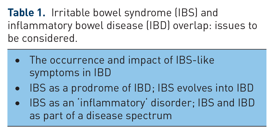
The occurrence and impact of IBS-like symptoms in IBD
Although surveys have recorded a high prevalence of IBS-type symptoms among patients with active IBD, drawing any inferences from such data, is in my mind, pure nonsense. The nitpicker will immediately insist that, as ‘Rome’ hath decreed, a diagnosis of IBS cannot even be made in an individual who has another ‘organic‘ explanation for their symptoms [Longstreth et al. 2006]. In this instance I unashamedly side with the erstwhile pedant; given the very limited symptomatic repertoire of the intestines (pain, bloating, distension, diarrhea, constipation), it is to be expected that a patient with active Crohn’s disease (CD) or ulcerative colitis (UC) will satisfy criteria for IBS! More problematic is the interpretation of such symptoms when the patient appears to be in remission.
Ongoing, undetected levels of inflammation/disease activity
Much of the impetus behind the current interest in a possible overlap between IBS and IBD comes from studies suggesting a high prevalence of IBS-type symptoms in IBD patients [Kruis, 2013; Halpin and Ford, 2012] (up to 59.7% in CD and 38.6% in UC) who are in apparent clinical remission [Simrén et al. 2002; Keohane et al. 2010; Jonefjäll et al. 2013; Jelsness-Jørgensen et al. 2013; Fukura et al. 2014; Gracie and Ford, 2015]. This phenomenon was first reported by Isgar and colleagues who documented IBS-type symptoms in 33% of their patients with UC in remission [Isgar et al. 1983]. In their meta-analysis of 13 studies incorporating 1703 patients, Halpin and Ford calculated a pooled prevalence for IBS symptoms among IBD subjects in remission of 35% [Halpin and Ford, 2012]. IBS symptoms were more likely among those with CD than UC. The occurrence of such symptoms in the IBD patient represents a source of considerable stress, incurs considerable morbidity and impairs quality of life [Simrén et al. 2002; Jelsness-Jørgensen et al. 2014]. Of concern, such symptoms are a significant risk factor for narcotic use in IBD [Long et al. 2012]. How is the clinician to interpret these symptoms?
In my opinion, one’s first obligation in this context is to determine whether, indeed, the patient truly is in remission or whether inflammation, undetected by conventional means, persists. This may not be as easy as it sounds. Thus, the various clinical indices that have been traditionally used to define activity in both CD and UC bear a far from perfect correlation with more direct measures of inflammatory activity [Vilela et al. 2012; Levesque et al. 2015]. In other words, relying, for example, on the Crohn’s disease activity index (CDAI) alone will overestimate rates of true remission in CD. Recently developed and now widely available methodologies that detect low levels of inflammatory activity have proven of great value in addressing this dilemma [Sands, 2015; Alibrahim et al. 2015; Dhaliwal et al. 2015]. Of these, the fecal level of calprotectin has proven to be a very sensitive measure of disease activity in IBD and its use in the IBD patient with IBS symptoms has revealed that many have active, if subclinical, activity of their IBD [Keohane et al. 2010; Jelsness-Jørgensen et al. 2013]. Other markers of inflammation such as levels of highly sensitive C-reactive protein in the circulation [Hod et al. 2015], of lactoferrin in feces [Zhou et al. 2014] or levels of the pro-inflammatory cytokine tumour necrosis factor-α (TNF-α), as well as numbers of intra-epithelial lymphocytes (IELs) in cecal mucosal biopsies [Danese, 2011; Vivinus-Nébot et al., 2014], levels of nitric oxide in rectal biopsies [Reinders et al. 2005] and the response of cultured mucosal biopsies to lipopolysaccharide [Danese, 2011; Vivinus-Nébot et al. 2014] have also helped to define the IBD patient with ongoing activity. Unfortunately, though they assessed their patients carefully using conventional methods, none of the aforementioned studies simultaneously measured fecal levels of calprotectin. We cannot, therefore, predict whether any or all of these other markers could detect inflammatory activity undetected by calprotectin. Though slightly elevated levels of some pro-inflammatory cytokines have been detected in uncomplicated IBS [Bashashati et al. 2014], these have never been of the magnitude observed in IBD. Furthermore, calprotectin levels are normal in IBS [Keohane et al. 2010; David et al. 2015; Caviglia et al. 2014; Moayyedi, 2014; Kalantri et al. 2015; Agilli et al. 2015].
The delineation of this patient population with ongoing activity is critical in directing a management strategy; equally important is the avoidance of corticosteroids, immunomodulators and biologics when there is no active IBD. For now, calprotectin plays a critical role in this decision point. What should be the cutoff value for determining whether or not there is ongoing activity?
Looking, firstly, at IBD, in general, D’Haens and colleagues found that a cutoff value of ⩽250 μg/g predicted endoscopic remission [Crohn’s Disease index of Severity (CDEIS) ⩽3] with 94.1% sensitivity and 62.2% specificity [positive predictive value (PPV) 48.5%, negative predictive value (NPV) 96.6%] in CD [D’Haens et al. 2012]. Similarly, in UC, a fecal calprotectin >250 μg/g gave a sensitivity of 71.0% and a specificity of 100.0% (PPV 100.0%, NPV 47.1%) for active mucosal disease (Mayo score >0) [D’Haens et al. 2012]. When combined with the Harvey Bradshaw Index, a cutoff of <100 μg/g proved superior to other levels in the prediction of endoscopic healing in CD in another study [Björkesten et al. 2012].
Turning to differentiating between IBS and IBD, Jelsness-Jørgensen and colleagues suggested that a cutoff of <100 μg/g provided optimal differentiation [Jelsness-Jørgensen et al. 2013]. However, based on a systematic review of 28 studies, Waugh and colleagues calculated a pooled sensitivity and specificity for fecal calprotectin at a cutoff level of 50 µg/ml of 93% and 94%, respectively, in the detection of active IBD and in differentiating IBS from IBD [Waugh et al. 2013]. Similarly, Menees and colleagues [Menees et al. 2015] and based on another meta-analysis, estimated that the probability of an individual with IBS with a C-reactive protein level of ⩽0.5 mg/dl and a fecal calprotectin level of ⩽40 µg/g harboring IBD was ⩽1%. In deciding on a cutoff value for calprotectin there will always be a tradeoff between sensitivity and specificity. Furthermore, it is evident that there remains a gray area between that level of calprotectin (40–50 µg/g) which is reliably indicative of absolutely no inflammatory activity and a higher level (100–250 µg/g) which indicates active IBD.
It is also interesting to note that cigarette smoking, a known risk factor for CD, was also predictive of IBS symptoms associated with low-grade activity, as detected by calprotectin, in a group of patients with CD [Keohane et al. 2010].
It must be conceded that, while calprotectin has served as a reliable predictor of low-grade inflammatory activity in IBD populations with IBS-type symptoms and in apparent remission, we do not know how the detection of such activity predicts therapeutic response to anti-inflammatory therapies, as this has not been directly studied. Furthermore, while an elevated calprotectin level will ‘rule in’ IBD we do not, at this time have an equally effective strategy to ‘rule in’ IBS; clearly our goal should be to make positive diagnoses of both IBS and IBD.
Coincident IBS or undetectable levels of IBD activity?
Not surprisingly, the story does not end here. Not all individuals with IBD and IBS-type symptoms apparently in remission have elevated levels of fecal calprotectin in the feces [Jonefjäll et al. 2013; Berrill et al. 2013], suggesting that not all IBS-like symptoms in IBD can be explained by ongoing inflammation, at least, as detected by fecal calprotectin assays. Is this simply a case of our being unable to detect even the most subtle levels of ongoing IBD activity or are we dealing with coincident IBS or, even, IBS triggered by IBD?
Whether these symptoms, in the face of normal levels of calprotectin in the stool, are harbingers of IBD activity can only be addressed by either large-scale studies at the ultrastructural and/or molecular level which examine for even more subtle levels of inflammation, or by trials of anti-inflammatory activity. Pending their completion let us ponder upon an alternative explanation, that these symptoms reflect IBS or IBS triggered by IBD. Given the high prevalence of IBS in the general adult population and the relatively high prevalence of IBD in Western nations, it should come as no surprise that IBS and IBD could coexist, purely on the basis of chance [Quigley et al. 2012; Shanahan and Bernstein, 2009]. Furthermore, it is statistically possible, given the high background prevalence of IBS, that some IBD patients would develop IBS de novo while in remission from IBD. Though this seems unlikely based on the prevalence rates of 40–60% for IBS in IBD [Keohane et al. 2010; Barratt et al. 2011a, 2011b; Halpin and Ford, 2012; Jelsness-Jørgensen et al. 2012, 2013; Berrill et al. 2013; Jonefjäll et al. 2013], rates that are 4–5 fold higher than those in the general population, this is a possibility that must be considered. Other observations, indeed, speak to these occurrences as being reflective of IBS as it is seen in the general, non-IBD, population. Thus, at least two studies [Berrill et al. 2013; Jonefjäll et al. 2013] have reported a higher prevalence of IBS-like symptoms in female than in male patients with IBD, a feature of ‘true’ IBS, and others have shown a positive correlation with anxiety [Jelsness-Jørgensen et al. 2012, 2013; Jonefjäll et al. 2013], a known trigger of IBS symptoms in otherwise healthy populations.
Furthermore, neither the extent nor the severity of inflammation at initial presentation predicted the likelihood of IBS symptoms developing once remission was induced in UC [Jonefjäll et al. 2015]. Similarly, Berrill and colleagues showed that IBS symptoms in IBD patients occur regardless of IBD disease type, the nature and intensity of IBD treatment regimens and the duration of remission [Berrill et al. 2013]. Taken together, these two studies suggest a poor correlation between IBD disease activity and IBS-like symptoms. In contrast to the above described lack of correlation with objective evidence of disease activity at initial presentation, symptom severity at this same time has proven predictive of the later emergence of IBS-type symptoms following remission [Jonefjäll et al. 2015], further supporting the suggestion that these patients have an underlying IBS-type phenotype.
Reports of familial aggregation of IBS and IBD are also consistent with this concept [Aguas et al. 2011]. Indeed, at the cellular and molecular level, patients with IBS-like symptoms in IBD have been shown to exhibit findings in keeping with commonly observed phenomena in IBS, in general: increased intestinal permeability; a profusion of transient receptor potential vanilloid receptor 1 (TRPV1) pain receptor fibers; and decreased expression of the tight junction proteins zonulin (ZO) 1 and α-cathenin [Akbar et al. 2010; Keszthelyi et al. 2013]. Other features of the immunopathology or biology of IBS in IBD and IBS, per se, may not be so convergent. In the future, studies of other relevant biomarkers, such as neutrophil gelatinase-associated lipocalin (NGAL) [Oikonomou et al. 2012] which has been linked to IBD or of vinculin, recently associated with diarrhea-predominant IBS [Pimentel et al. 2015], may provide further insights into relationships between these entities.
The availability of the fecal calprotectin assay has fulfilled our first goal in the assessment of this patient population: to detect those with ongoing subclinical activity of IBD (Figure 1a). In this way, anti-inflammatory regimens should be reserved for those IBD patients with ongoing, active IBD. But how do we approach the IBD patient who manifests IBS-like symptoms in the face of a normal level of calprotectin in the feces [Berrill et al. 2013; Jonefjäll et al. 2013] (Figure 1b)? Available evidence, summarized above, suggests that this subgroup bears more resemblance to IBS as we know it outwith the context of IBD than to IBD and that it should be managed according to those strategies that we employ, admittedly with limited success, in IBS, in general. Data supporting this approach are very limited though one very pilot study suggested that instituting a low Fermentable Oligo-, Di-Monosaccharides and Polyols (FODMAP) diet may be of some benefit here as in IBS, in general [Gearry et al. 2009; Schwender and Floch, 2014]. Other, uncontrolled studies suggest benefits for other dietary strategies [McDermott, 2007], a tricyclic antidepressant [Iskander et al. 2014] and mindfulness therapy [Berrill et al. 2014].

Approach to the patient with IBD in apparent remission based on fecal calprotectin level: (a)Calprotectin elevated – search for and treat active IBD. (b) Calprotectin normal – for now, pending better data, manage as IBS.
It is possible that the characteristics of IBS (or more accurately an IBS-like syndrome) occurring in the IBD patient who is in remission are so influenced by the underlying IBD that they no longer completely resemble IBS as seen in an otherwise healthy population. It is not implausible to propose that IBS, in the context of IBD, would involve an expanded spectrum of pathophysiologic features. Understanding IBS symptoms in the IBD patient as a form of IBS that is transformed because of the underlying IBD would help explain the occurrence of pro-inflammatory features, such as the TNF-α alpha response to lipopolysaccharide (LPS), seen in this phenotype [Vivinus-Nébot et al. 2014]) but not otherwise in IBS. It is possible that, in these particular patients, different components of the immune system interact with the host’s neuromuscular apparatus and brain–gut axis to produce a new syndrome distinct from ‘true’ IBS, full-blown IBD or subclinically active IBD. In this way, we can simultaneously accept that IBS symptoms in IBD are not ‘simply’ superimposed or coincidental IBS, as reflected by the presence of biological features that are not typical of ‘pure’ IBS, nor do they inevitably represent ongoing activity of IBD defined by every means that is currently at our disposal.
Elsewhere, we have proposed the term ‘irritable inflammatory bowel syndrome’ (IIBS) to refer to this clinical scenario [Stanisic and Quigley, 2014]. Various components of the brain–gut axis could be ‘upregulated’, or ‘primed’ in the IBD patient by the inflammation that is fundamental to this disorder. This results in disordered motility, altered visceral sensation and dysregulated brain–gut signaling; abnormalities that may affect noninflamed intestine or persist following resolution of active inflammation. For example, various disturbances in gut motor function have been documented in IBD [Bassotti et al. 2014] and visceral pain can be a very significant issue for some IBD patients [Neri, 2013]. It stands to reason that these and other motor and sensory phenomena could be accentuated in an individual with pre-existing IBS or who possesses psychosocial, genetic or physiological characteristics that predispose one to IBS. The term IIBS indicates the nature of the symptoms: namely, IBS-like, the context; occurring in an IBD patient; and signifying that it is a syndrome rather than a clearly delineated disease entity.
IBS as a prodrome of IBD
Retrospective and population-based studies indicating the presence of IBS-type symptoms for many years (up to 20) before a diagnosis of IBD and CD, in particular, is made raise the possibility that IBS may be a prodrome of IBD [García Rodriguez et al. 2000; Minderhoud et al. 2004; Burgmann et al. 2006; Olbe, 2007; Barratt et al. 2011a; Porter et al. 2012; Card et al. 2014; Canavan et al. 2014]. For example, Porter and colleagues found that IBS was a risk factor for the development of IBD [Porter et al. 2012], finding that the incidence of IBD was 8.6 fold higher among those diagnosed with IBS compared with those who did not carry this diagnosis. Card and colleagues [Card et al. 2014] made similar observations based on their analysis of the General Practice Research Database in the UK; IBD patents were three times more likely to have a prior diagnosis of IBS. They also found that a prior diagnosis of IBS was more likely among those with a final diagnosis of CD in the young and in females. It is important to note that most, but certainly not all, of these IBS diagnoses occurred in the year before the diagnosis of IBD. Other data [Burgmann et al. 2006] also indicate that, despite increased awareness and advances in diagnostics, the diagnosis of IBD and of CD, in particular, is still long delayed.
These sobering observations, together with the recognition that symptoms of IBS and IBD (CD more so than UC) overlap, suggest that misdiagnosis rather than an evolution from one disorder into another largely explains the apparent predisposition of IBS sufferers to ‘develop’ IBD. Though scarcely meeting any standard of scientific credibility, it can also be said that it has been the experience of those who have cared for decades for those with well-established and even severe IBS that an evolution to IBD is never seen. One could also speculate that, in patients with a documented history of IBS symptoms prior to the diagnosis of IBD, the appearance of IBS following the induction of remission of IBD could then represent the re-emergence of pre-existing IBS.
IBS as an ‘inflammatory’ disorder: IBS and IBD as part of a disease spectrum
The aforementioned contemplation of a possible evolution from IBS into IBD should lead, logically, to a reflection on the possibility that IBS and IBD represent a part of the spectrum of the same disease process. This notion has emerged from a large volume of data indicating contributions from both the microbiome [Kassinen et al. 2007; Krogius-Kurikka et al. 2009; Noor et al. 2010; Rajilić-Stojanović et al. 2011, 2015; Saulnier et al. 2011; Carroll et al. 2012; Parkes et al. 2012; Chaussard et al. 2012; Jeffery et al. 2012a; Durbán et al. 2013; Ringel and Maharshak, 2013; Simrén et al. 2013; Lopez-Siles et al. 2014; Jalanka-Tuovinen et al. 2014; Pozuelo et al. 2015; Sundin et al. 2015] and host immune response [Öhman and Simrén,. 2010; Rodríguez-Fandiño et al. 2010; Barbara et al. 2011; Del Valle-Pinero et al. 2011; Camilleri and Katzka, 2012; Vivinus-Nébot et al. 2012; Bashashati et al. 2012; Chang et al. 2012; Theoharides, 2014; Bashashati et al. 2014; Pike et al. 2015] to the pathophysiology of IBS-type symptoms. These studies have been extensively reviewed elsewhere [Ghoshal et al. 2012; Jeffery et al. 2012b; Simrén et al. 2013; Dupont, 2014; Major and Spiller, 2014; Bennet et al. 2015; Öhman et al. 2015].
For the purposes of this discussion, it is important to conclude that findings, in IBS, in relation to both the microbiome and the immune response have been far from consistent and highly variable. While the intrinsic vagaries of the IBS phenotype inevitably confound such studies, it is clear, that unlike IBD, a consistent and reproducible pattern of immune response has not emerged from these IBS studies. Furthermore, immunological findings in IBS are, at best, subtle and are qualitatively and quantitatively very different from those that characterize IBD, in any form. Though some familial clustering of IBS and IBD has been reported [Aguas et al. 2011], none of the many genes that have been associated with IBD (many of whom relate to the immune response) have been linked to IBS. One exception in this regard relates to postinfectious IBS (PI-IBS) where polymorphisms in genes related to the immune response and intestinal barrier function have been associated with susceptibility to this rather rare phenotype of IBS [Villani et al. 2010]. The example of PI-IBS should warn us of the danger of absolutism when referring to any aspect of IBS; it is certain, not just likely, that a disorder so poorly and loosely defined will incorporate pathological entities that we are currently incapable of detecting. The recent description of primary bile acid diarrhea [Camilleri, 2015] as a cause of some instances of what were formerly classified as diarrhea-predominant IBS serves as a timely reminder of the porous nature of the very concept of IBS. The possibility that, under that very broad and inclusive umbrella that is IBS, lurks a group of individuals whose symptoms are driven by an immune response and/or the microbiome should not, therefore, be discounted. This subgroup, if it exists, may share some pathogenetic features with IBD or perhaps with another ‘inflammatory’ bowel disorder that in times gone by also masqueraded as IBS: microscopic colitis. Only prospective studies of the immune response, the microbiome and the host genome in carefully phenotyped (in terms of symptoms, demographics and psychological makeup) and well-defined patient populations will answer these questions.
One pathophysiologic process that could link the two disorders and contribute to the excess of IBS symptoms in IBD relates to intestinal permeability. Alterations in barrier function seem to be a very sensitive indicator of disease activity in IBD [D’Inca et al. 1999; Arnott et al. 2000]. It is possible that the epithelium in IBD patients who are in remission is already ‘primed’ by a low level of inflammation related to IBD, per se. IBS-type triggers, such as stress or anxiety, then lead to the release of histamine and other chemokines from mast cells [Kiank et al. 2009; Overman et al. 2012; Vivinus-Nébot et al. 2012; Matricon et al. 2012; Vivinus-Nébot, 2014] and, thereby, foster further recruitment of lymphocytes and the production of pro-inflammatory cytokines. These, in turn, result in a further increase in intestinal permeability, as well as histamine release and upregulation of TRPV1 pain receptors, generating IBS-like symptoms [Akbar et al. 2010; Keszthelyi et al. 2013; Neri, 2013]. Meanwhile their effects on an IBD-primed molecular milieu would lead to activation of inflammatory pathways and production of cytokines, such as TNF-α, and the release of calprotectin. This proposal is supported by the findings of Vivinus-Nébot and colleagues who documented features characteristic of both IBS and IBD in biopsies taken from their patient population, as well as the shared feature of increased intestinal permeability [Vivinus-Nébot et al. 2014]. Specifically, they found that the expression of the tight junction proteins ZO-1 and α-cathenin were decreased both among those with IBS without IBD and those with IBS-like symptoms in IBD. However, they also found that patients with IBS-like symptoms in IBD had increased expression of TNF-α mRNA and increased numbers of intra-epithelial leukocytes, features of IBD [Vivinus-Nébot et al. 2014]. It should be noted that TNF-α protein expression, though numerically higher, was not significantly elevated in IBS in IBD patients though the TNF-α response to lipopolysaccharide was significantly accentuated in this patient population compared with those IBD patients who did not have IBS-like symptoms, further supporting the presence of an exaggerated inflammatory response in these patients [Vivinus-Nébot et al. 2014; Gracie and Ford, 2014].
Conclusion
IBD patients in apparent clinical remission who present with IBS-like symptoms currently pose a diagnostic and therapeutic dilemma for their treating physicians. Though not as yet supported by therapeutic trials, it seems reasonable to initiate the evaluation of these patients by measuring the level of calprotectin in a fecal sample. If this lies clearly in a range associated with active inflammation, then further assessments of IBD activity and appropriate anti-inflammatory therapy should be initiated. Randomized controlled trials supporting this approach are urgently needed. If the level of calprotectin lies within the normal range or, more difficult still, in a ‘gray’ zone, the clinician may initiate symptomatic therapy based on dominant symptomatology and current best practice for IBS. Again, clinical trials are needed to support this, or any other, approach to these patients. For now the precise nature of this latter patient population; overlapping IBS, undetectable IBD activity or some intermediate entity (IIBS), is unknown but clearly, given its prevalence, deserves a concerted effort on many fronts.
Footnotes
Funding
This research received no specific grant from any funding agency in the public, commercial, or not-for-profit sectors.
Conflict of interest statement
The author declares no conflict of interest in preparing this article.