
Abstract
Objective:
Our study aimed to evaluate the effect of circular RNA ABCB10 (circ-ABCB10) on proliferation and apoptosis of clear cell renal cell carcinoma (ccRCC) cells, and its prognostic value in patients with ccRCC.
Methods:
Circ-ABCB10 expression in five ccRCC cell lines and normal kidney epithelial cell line was measured by quantitative polymerase chain reaction (qPCR). Empty overexpression, circ-ABCB10 overexpression, empty shRNA, and circ-ABCB10 shRNA plasmids were transfected into A498 cells as negative control for circ-ABCB10 over expression {NC (+)}, Circ-ABCB10(+), negative control (−){NC(−)}, and Circ-ABCB10(−) groups, then cell proliferation and apoptosis were evaluated by Cell Counting Kit-8 and annexin V/propidium iodide. Meanwhile, apoptotic markers were measured by western blot. Subsequently, circ-ABCB10 expression in tumor tissues and paired adjacent tissues from 120 ccRCC patients was measured by qPCR.
Results:
Circ-ABCB10 expression was elevated in all the ccRCC cell lines compared with the normal kidney cells line. A498 cell proliferation was enhanced in the Circ-ABCB10(+) group compared with the NC(+) group, while it was inhibited in the Circ-ABCB10(−) group compared with the NC (−) group; and A498 cell apoptosis was repressed in the Circ-ABCB10(+) group than the NC(+) group, but was promoted in the Circ-ABCB10(−) group compared with the NC(−) group. In addition, circ-ABCB10 was up-regulated in tumor tissues compared with paired adjacent tissues, and its high expression correlated with the advanced pathological grade and the tumor node metastasis stage as well as independently predicting worse overall survival in ccRCC patients.
Conclusion:
Circ-ABCB10 promotes tumor progression and correlates with pejorative prognosis in ccRCC.
Introduction
Kidney cancer is the eleventh most common cancer in male, and the fifteenth most frequent cancer in female, which results in 403,262 new cases and 175,098 related deaths in 2018. 1 Renal cell carcinoma (RCC) occupies almost 90% of all kidney cancer cases, most of which were clear cell RCC (ccRCC). 2 ccRCC patients are at high risk of recurrent disease and fatality, even in patients with localized ccRCC who receive radical nephrectomy.3,4 To improve the survival of ccRCC patients, increasing efforts have been made, including exploring new target therapies, developing a multidisciplinary team, and finding novel prognostic biomarkers.5,6
Circular ribonucleic acid (circRNA)s, an innovative type of non-coding RNAs, are transcripts with deficiency of 5′-3′ ends and poly (A) tails that form a covalently closed circle.7,8 Recently, there has been increasing concern regarding the role of circRNAs in oncology; for example, emerging studies report that multiple circRNAs can regulate cancer cell functions and potentially serve as prognostic factors in cancer patients.9–12 Circ-ABCB10 is a novel circRNA that displays promising oncological effect, which was identified by a recent study that discloses that it enhances cell proliferation of breast cancer cells by sponging microRNA (miR)-1271. 13 It also expresses in the kidney according to the Tissue-Specific CircRNA Database (http://gb.whu.edu.cn/TSCD/). Moreover, our preliminary experiment revealed that circ-ABCB10 was up-regulated in various RCC cell lines; therefore, we speculated that circ-ABCB10 might participate in ccRCC etiology and have the potential to be a prognostic marker in ccRCC patients.
Thus, the aim of our study was to evaluate the effect of circ-ABCB10 on the proliferation and apoptosis of ccRCC cells and its prognostic value in patients with ccRCC.
Methods
Cell sources
ccRCC cell lines A498, Caki-2, and Cal-54 were purchased from Deutsche Sammlung von Mikroorganismen und Zellkulturen (DSMZ) (Braunschweig, German). ccRCC cell lines ACHN and Hs891.T and the normal kidney epithelial cells line HREC were purchased from the American Type Culture Collection (ATCC) (Rockefeller, USA).
Cell cultures
A498, Cal-54, and ACHN cells were cultured in 90% minimum eagle’s medium (MEM) (Sigma-Aldrich, USA) supplemented with 10% fetal bovine serum (FBS) (Gibco, USA) in a humidified incubator under 5% CO2 at 37°C; Caki-2 cells were cultured in 90% McCoy’s 5A medium (Sigma-Aldrich, USA) supplemented with 10% FBS (Gibco, USA) in a humidified incubator under 5% CO2 at 37°C; Hs891.T cells were cultured in 90% dulbecco’s modified eagle medium (DMEM) supplemented with 10% FBS (Gibco, USA) in a humidified incubator under 5% CO2 at 37°C; and HREC cells were cultured in 90% renal epithelial cell basal medium (RECBM) (ATCC, USA) supplemented with 10% FBS (Gibco, USA) in a humidified incubator under 5% CO2 at 37°C.
Transfection of circ-ABCB10 overexpression and shRNA
Circ-ABCB10 expression was measured by quantitative polymerase chain reaction (qPCR) in A498, Caki-2, Cal-54, ACHN, Hs891.T, and HREC cell lines. A498 was then chosen for followed functional experiments. Empty overexpression plasmid, circ-ABCB10 overexpression plasmid, empty shRNA plasmid and circ-ABCB10 shRNA plasmid were transfected into A498 cells as negative control for circ-ABCB10 over epxression {NC(+)}, Circ-ABCB10(+), NC(−) and Circ-ABCB10(−) groups for evaluation of the effect of Circ-ABCB10 on A498 cell proliferation and apoptosis. The corresponding plasmids used in this study were constructed by Shanghai GenePharma Bio-Tech Company (Shanghai, China).
Measurement of proliferation and apoptosis of A498 cells
Cell proliferation of A498 cells was measured at 0h, 24h, 48h, and 72h after transfection using Cell Counting Kit-8 (CCK-8) (Thermo, USA) according to the instructions of the manufacturer. As to cell apoptosis of A498 cells, it was measured at 48h after transfection using Annexin V (AV) apoptosis detection kit with propidium iodide (PI) (Invitrogen, USA) according to the instructions of the manufacturer. Meanwhile apoptotic marker C-Caspase 3 and anti-apoptotic marker Bcl-2 expressions were measured using western blot.
Patients
A total of 120 ccRCC patients who underwent nephrectomy in The Central Hospital of Wuhan, Tongji Medical College, Huazhong University of Science and Technology, China, from January 2014 to June 2015 were consecutively recruited in this study. The inclusion criteria were as follows: (a) diagnosed as primary ccRCC confirmed by clinical, radiographic, and histopathological examination; (b) without metastasis; (c) age over 18 years old; (d) scheduled to receive nephrectomy; and (e) able to be followed up regularly. The exclusion criteria included: (a) non-clear cell RCC or secondary ccRCC; (b) underwent neoadjuvant therapy; (c) with a history of other malignant tumors; (d) life expectancy less than 12 months; and (e) pregnant or lactating women. Approval of this study was granted by Ethics Committee of The Central Hospital of Wuhan, Tongji Medical College, Huazhong University of Science and Technology, and the written informed consents were signed and collected from all patients or their guardians before enrollment.
Baseline data collection
Demographic and clinicopathologic characteristics of patients were collected at baseline, including age, gender, tumor location, pathological grade, tumor size, T stage, N stage, and tumor node metastasis (TNM) stage.
Specimens collection and follow up
All patients underwent nephrectomy, and the tumor tissue and paired adjacent tissue specimens resected from patients were immediately frozen in liquid nitrogen post-nephrectomy and stored in a freezer for the detection of circ-ABCB10. After surgery, all patients were continuously followed-up for 3 years until 30 June 2018, and overall survival (OS) was calculated from surgery to patients’ death.
Western blot
After extraction of total protein, the bicinchoninic acid (BCA) disodium kit (Pierce Biotechnology, USA) was used for assessing total protein concentration, which was calculated by making a standard curve. Afterward, the total protein was separated by sodium dodecyl sulfate-polyacrylamide gel electrophoresis (SDS-PAGE) electrophoresis and was transformed on the polyvinylidene fluoride (PVDF) membranes (Millipore, Bedford, USA). Subsequently, the PVDF membranes were blocked and primary antibody was added and incubated in 37℃ for 1–3 h or under 4℃ overnight. Then the secondary antibody was added, which was also incubated in 37℃ for 1h. Finally, enhanced chemiluminescence (ECL) kit (Millipore, USA) was used for visualizing the bands under x-ray. Antibodies used in western blot were listed in Supplementary Table 1.
qPCR
Tissues and cells were digested using 0.25% Trypsin (Gibco, USA) and collected, followed by extraction of total RNA using TRIzol Reagent (Invitrogen, USA). Subsequently, the linear RNA in the 1ug total RNA was digested by RNase R (Epicentre, USA), and then the 1ug total RNA was reverse transcribed by PrimeScript™ RT reagent Kit (TAKARA, Japan) into complementary deoxyribonucleic acid (cDNA). Afterward, qPCR was performed using the QuantiNova SYBR Green PCR Kit (Qiagen, German) to assess the expressions of circ-ABCB10 and glyceraldehyde-phosphate dehydrogenase (GAPDH). The primers used in qPCR were listed in Supplementary Table 2, and the result was calculated using 2−△△Ct with GAPDH as an internal reference.
Statistical analysis
Statistic Package for Social Science (SPSS) 22.0 software (SPSS Inc., USA) and GraphPad Prism 7.00 (GraphPad Software, USA) were used for statistical analyses and figure creation. Measurement data were presented as mean ± SD if normally distributed, or median (25th–75th quantiles) if not normally distributed; and count data were expressed as count (percentages). Comparisons of measurement data were determined by t test, Wilcoxon rank sum test or Kruskal–Wallis H rank sum test; paired data comparison was determined by paired t test. Survival analysis was performed using Kaplan–Meier (K–M) curve, and the difference of survival was determined by the Log-rank test. Factors affecting OS were determined by univariate and multivariate Cox’s proportional hazards regression analyses with Forward Stepwise (Conditional) method. P value <0.05 was considered as significant.
Results
Circ-ABCB10 expression in ccRCC cell lines and normal kidney epithelial cell line
The circ-ABCB10 expression was elevated in ccRCC cell lines A498 (P < 0.001), Caki-2 (P < 0.001), ACHN (P < 0.05), Hs891.T (P < 0.01) and Cal-54 (P < 0.05) compared with normal kidney epithelial cell line HREC (Figure 1), which indicated that the circ-ABCB10 expression was upregulated in ccRCC cells than that in normal kidney cells. Subsequently, further experiments were conducted in A498 cells.
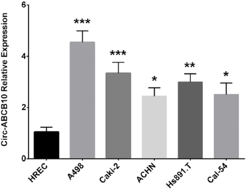
circ-ABCB10 expression in ccRCC cell lines and normal renal cell lines.
Effect of circ-ABCB10 on cell proliferation and cell apoptosis in A498 cells
After transfections of empty overexpression plasmid, circ-ABCB10 overexpression plasmid, empty shRNA plasmid, and circ-ABCB10 shRNA plasmid in A498 cells, the circ-ABCB10 expression was up-regulated in the ABCB(+) group compared to the NC(+) group (P < 0.001). However, it was decreased in the ABCB(−) group compared with the NC(−) group (P < 0.001) (Figure 2), which indicates that the transfections were successful. Afterward, CCK-8 was conducted for cell proliferation assessment, which showed that the cell proliferation of A498 cells was promoted in the ABCB(+) group compared with the NC(+) group at 48h (P < 0.05) and 72h (P < 0.01) after transfection. However, it was repressed in the ABCB(−) group compared with the NC(−) group at 48h (P < 0.05) and 72h (P < 0.01) (Figure 3). Apoptosis was decreased at 48h after transfections in the ABCB(+) group compared with the NC(+) group (P < 0.01), while was elevated in ABCB(−) group compared to in NC(−) group (P < 0.01) (Figure 4(a and c)). Moreover, the pro-apoptotic protein C-Caspase 3 expression was down-regulated in the ABCB(+) group compared with the NC(+) group; however, it was up-regulated in the ABCB(−) group compared with the NC(−) group (Figure 4(b)). The expression of the anti-apoptosis protein Bcl-2 was elevated in the ABCB10(+) group compared with the NC(+) group, while it was decreased in the ABCB(−) group compared to the NC(−) group. These data indicated that circ-ABCB10 promoted the proliferation and inhibited the apoptosis of ccRCC cells.
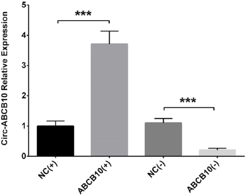
circ-ABCB10 expression in A498 cells after transfections.
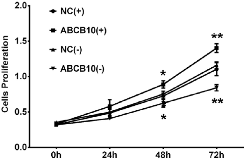
Cell proliferation in A498 cells after transfections.
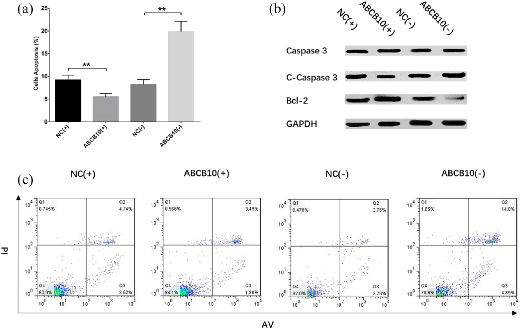
Cell apoptosis in A498 cells after transfections.
Baseline characteristics of ccRCC patients
In 120 ccRCC patients, the mean age was 58.9±12.1 years, and there were 50 (41.7%) females and 70 (58.3%) males (Supplementary Table 3). The number of patients with a tumor located in the right kidney and the left kidney was 62 (51.7%) and 58 (48.3%), respectively. In addition, the number of patients with a pathological grade of 1, 2, and 3 was 58 (48.3%), 45 (37.5%), and 17 (14.2%), respectively. The number of patients at TNM stage I, II, and III was 78 (65.0%), 28 (23.3%), and 14 (11.7%), respectively. Other baseline clinical characteristics of ccRCC patients are listed in Supplementary Table 3.
Circ-ABCB10 expression in tumor tissue and paired adjacent tissue in ccRCC patients
The circ-ABCB10 expression was evaluated by qPCR in tumor tissue and paired adjacent tissue, which disclosed that it was up-regulated in the former compared with the latter (P < 0.001) (Figure 5).
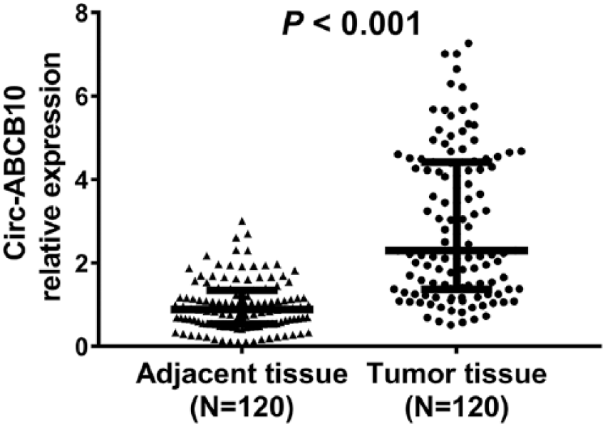
circ-ABCB10 expression in tumor tissue and paired adjacent tissue.
Correlation of circ-ABCB10 with characteristics in ccRCC patients
Tumor tissue circ-ABCB10 expression was positively correlated with pathological grade (P = 0.034) (Supplementary Figure 1(d)) and TNM stage (P = 0.002) (Supplementary Figure 1(f)), while it was not associated with age (P = 0.635) (Supplementary Figure 1(a)), gender (P = 0.095) (Supplementary Figure 1(b)), tumor location (P = 0.796) (Supplementary Figure 1(c)), or tumor size (P = 0.334) (Supplementary Figure 1(e)).
Correlation of circ-ABCB10 with OS in ccRCC patients
The K–M curve and log-rank test were conducted to evaluate the OS, which showed that the OS was worse in ccRCC patients with high tumor tissue circ-ABCB10 expression compared with ccRCC patients with low tumor tissue circ-ABCB10 expression (P = 0.004) (Supplementary Figure 2).
Factors affecting OS in ccRCC patients
Univariate and multivariate Cox’s proportional hazards regression analysis were performed to determine the factors affecting OS in ccRCC patients. The univariate Cox’s proportional hazards regression analysis disclosed that high tumor tissue circ-ABCB10 expression (P = 0.010), pathological grade 2/3 (P = 0.013), and TNM stage II/III (P = 0.023) were correlated with worse OS in ccRCC patients (Supplementary Table 4). Subsequently, multivariate Cox’s proportional hazards regression analysis was conducted using the forward stepwise (conditional) method, which elucidated that high tumor tissue circ-ABCB10 expression (P = 0.009) and pathological grade 2/3 (P = 0.012) were independent factors for predicting shorter OS in ccRCC patients.
Discussion
In this study, we discovered that: (a) circ-ABCB10 promoted cell proliferation and inhibited cell apoptosis of ccRCC cells; (b) circ-ABCB10 was up-regulated in tumor tissue compared with paired adjacent tissue, and was positively correlated with pathological grade and TNM stage; and (c) the OS was worse in patients with high tumor tissue circ-ABCB10 expression, and high circ-ABCB10 expression in tumor tissue was an independent predictive factor for shorter OS.
As the most common histological type of kidney cancer, ccRCC has shown some improvements in its prognosis in recent years with the introduction of novel drugs, such as vascular endothelial-growth-factor-targeted drugs and immunotherapeutic agents; however, there is still a large number of patients not benefiting from this progress and presenting with an unsatisfactory prognosis.14–16 Fortunately, the research on non-coding RNAs as biomarkers and treatment targets predominantly provides new insight in improving the prognosis of ccRCC patients. An increasing number of non-coding RNAs have been identified with the potential of being biomarkers or treatment targets in ccRCC, such as miR-32-5p, miR-224, and long non-coding RNA colorectal neoplasia differentially expressed.17–19 Moreover, circRNA, as the novel class of non-coding RNA, is also becoming increasingly popular in ccRCC research. 20 For example, an experiment reveals that Hsa_circ_0001451 is down-regulated in ccRCC tissues compared with paired normal tissues, and is associated with the clinicopathological characteristics of ccRCC patients. 20
A growing number of noteworthy studies that investigate the effect of circRNAs in cancers have been reported, which disclose that many circRNAs might act as oncogenic or anti-oncology genes in various carcinomas.10,21,22 With respect to the oncogenic function of circRNA, a previous experiment revealed that circRNA-homeodomain-interacting protein kinase (HIPK3) increases gallbladder cancer cell proliferation by sponging miRNA-124. 23 Circ-zinc finger RNA (ZFR) binding protein advocates cell proliferation by sponging miR-1261 and promoting the expression of C8orf4 in papillary thyroid cancer. 24 Another experiment reveals that overexpressed circ-myosin light chain kinase (MYLK) enhances cell proliferation, invasion, and migration of prostate cancer cells via regulating miR-29a. 25 In addition, circ-SMARCA5 promotes prostate cancer progression through advancing the cell cycle and repressing cell apoptosis of cancer cells. 26 For circ-ABCB10, only one study has revealed that circ-ABCB10 is overexpressed in breast cancer tissue compared with paired adjacent non-cancerous tissue, and the knockdown of circ-ABCB10 represses proliferation while it enhances apoptosis of breast cancer cells. 13 Subsequently, a complement sequence between circ-ABCB10 and miR-1271 was found in their study, and their subsequent experiments discovered that circ-ABCB10 might regulate breast cancer cell function through sponging miR-1271. 13 In the Tissue-Specific CircRNA Database (http://gb.whu.edu.cn/TSCD/), we found that circ-ABCB10 is specifically expressed in kidney tissue, and our preliminary experiment disclosed that circ-ABCB10 was up-regulated in ccRCC cell lines compared with normal kidney cells. Subsequently, we further performed functional experiments and found that circ-ABCB10 promoted cell proliferation while it inhibited cell apoptosis of ccRCC cells, which could be explained by the following: (a) circ-ABCB10 might regulate ccRCC cell function through sponging anti-oncogene miRNAs, such as miR-331-3p and miR-1228-5p, which are derived from the Tissue-Specific CircRNA Database (http://gb.whu.edu.cn/TSCD/); and (b) circ-ABCB10 might promote ccRCC progression via regulating the expression of its located gene ABCB10.
Although increasing reports have disclosed that some dysregulated circRNAs that correlate with tumor progression and patient prognosis in many cancers (e.g. lung and breast cancer, and hepatocellular carcinoma), the investigations of circRNAs in ccRCC development and progression is seldom reported.27–29 A previous study disclosed that the estrogen receptor beta enhances invasion of ccRCC by mediating circ-ATPase plasma membrane Ca2+ transporting 1 (ATP2B1) and miR-204-3p/ fibronectin 1 axis. 30 Another study reveals that androgen recep-tor represses circ-Hippocampus Abundant Transcript 1 (HIAT1), which subsequently results in the promotion of ccRCC cell migration and invasion via regulating the expressions of miR-195-5p, miR-29a-3p, miR-29c-3p, and cell division cycle 42 (CDC42). 31 Furthermore, a previous study explains that circ_0001451 expression is down-regulated in tumor tissue compared with paired normal tissue of ccRCC, and its expression is negatively correlated with the pathological grade in ccRCC patients. 20 However, no study has been shown to evaluate the application of circ-ABCB10 as a biomarker for ccRCC progression and prognosis. In this present study, we first discovered that circ-ABCB10 was up-regulated in tumor tissue and correlated with higher pathological grade and TNM stage in ccRCC patients. In addition, high circ-ABCB10 expression in tumor tissue was an independent predictive factor of worse OS in ccRCC patients. The possible explanations might be (a) circ-ABCB10 promoted the progression of ccRCC through increasing cancer cell proliferation and inhibiting cancer cell apoptosis, as discovered in the cells experiment in our study; (b) circ-ABCB10 might sponge several anti-tumor miRNAs, such as miR-331-3p and miR-1228-5p (http://gb.whu.edu.cn/TSCD/), and subsequently promote the progression of ccRCC; and (c) the pejorative role of circ-ABCB10 in ccRCC progression is likely related to the function of ABCB10 gene.
In conclusion, circ-ABCB10 promotes tumor progression and correlates with pejorative prognosis in ccRCC.
Supplemental Material
Supplementary_Figure_1_1 – Supplemental material for Circular RNA ABCB10 promotes tumor progression and correlates with pejorative prognosis in clear cell renal cell carcinoma
Supplemental material, Supplementary_Figure_1_1 for Circular RNA ABCB10 promotes tumor progression and correlates with pejorative prognosis in clear cell renal cell carcinoma by Yunfang Huang, Yun Zhang, Lin Jia, Changxuan Liu and Fang Xu in The International Journal of Biological Markers
Supplemental Material
Supplementary_Figure_2 – Supplemental material for Circular RNA ABCB10 promotes tumor progression and correlates with pejorative prognosis in clear cell renal cell carcinoma
Supplemental material, Supplementary_Figure_2 for Circular RNA ABCB10 promotes tumor progression and correlates with pejorative prognosis in clear cell renal cell carcinoma by Yunfang Huang, Yun Zhang, Lin Jia, Changxuan Liu and Fang Xu in The International Journal of Biological Markers
Supplemental Material
Supplementary_Table_1 – Supplemental material for Circular RNA ABCB10 promotes tumor progression and correlates with pejorative prognosis in clear cell renal cell carcinoma
Supplemental material, Supplementary_Table_1 for Circular RNA ABCB10 promotes tumor progression and correlates with pejorative prognosis in clear cell renal cell carcinoma by Yunfang Huang, Yun Zhang, Lin Jia, Changxuan Liu and Fang Xu in The International Journal of Biological Markers
Supplemental Material
Supplementary_Table_2 – Supplemental material for Circular RNA ABCB10 promotes tumor progression and correlates with pejorative prognosis in clear cell renal cell carcinoma
Supplemental material, Supplementary_Table_2 for Circular RNA ABCB10 promotes tumor progression and correlates with pejorative prognosis in clear cell renal cell carcinoma by Yunfang Huang, Yun Zhang, Lin Jia, Changxuan Liu and Fang Xu in The International Journal of Biological Markers
Supplemental Material
Supplementary_Table_3 – Supplemental material for Circular RNA ABCB10 promotes tumor progression and correlates with pejorative prognosis in clear cell renal cell carcinoma
Supplemental material, Supplementary_Table_3 for Circular RNA ABCB10 promotes tumor progression and correlates with pejorative prognosis in clear cell renal cell carcinoma by Yunfang Huang, Yun Zhang, Lin Jia, Changxuan Liu and Fang Xu in The International Journal of Biological Markers
Supplemental Material
Supplementary_Table_4 – Supplemental material for Circular RNA ABCB10 promotes tumor progression and correlates with pejorative prognosis in clear cell renal cell carcinoma
Supplemental material, Supplementary_Table_4 for Circular RNA ABCB10 promotes tumor progression and correlates with pejorative prognosis in clear cell renal cell carcinoma by Yunfang Huang, Yun Zhang, Lin Jia, Changxuan Liu and Fang Xu in The International Journal of Biological Markers
Footnotes
Author contributions
YH and YZ contributed equally to this work and should be considered as co-first authors.
Declaration of conflicting interests
The author(s) declared no potential conflicts of interest with respect to the research, authorship, and/or publication of this article.
Funding
The author(s) received no financial support for the research, authorship, and/or publication of this article.
Supplemental material
Supplemental material for this article is available online.
References
Supplementary Material
Please find the following supplemental material available below.
For Open Access articles published under a Creative Commons License, all supplemental material carries the same license as the article it is associated with.
For non-Open Access articles published, all supplemental material carries a non-exclusive license, and permission requests for re-use of supplemental material or any part of supplemental material shall be sent directly to the copyright owner as specified in the copyright notice associated with the article.