
Abstract
Objective
To explore the relationship between the serum concentration of IL-17 and the pathogenesis of psoriasis.
Methods
ampere database was established through literature search, and meta-analysis was used to evaluate the effect indicator of the included literatures.
Results
5 articles were included in the study, and the quality evaluation score was 6–7 points by the Newcastle-Ottawa Scale (NOS) criteria. There was heterogeneity in the standardized mean difference (SMD) between the two groups (I2 = 98.63%). The random effect model analysis showed that the combined SMD = 2.11, 95%CI (0.79, 3.43), the effect size between studies was statistically significant (z = 3.13, p = 0.00).
Conclusion
There was a statistical difference in serum IL-17 concentration between the case group and the control group. The serum concentration of IL-17 in the case group was higher than that in the control group.
Introduction
Psoriasis is a chronic, recurrent and inflammatory skin disease, which is a common and frequently occurring disease in dermatology, accounting for 2–3% of the world’s population. 1 Psoriasis is a T-cell-mediated inflammatory systemic disease that mainly affects the skin 2 and is characterized by the proliferation of keratinocytes and the appearance of erythematous patches on the skin covered by white silver scales.3–5
Interleukin 17(IL-17), can be secreted not only by Th17 cells, but by innnate immune cells like ILC3, gammmadelta T cells, MAIT cells, is believed to play a crucial role in the pathogenesis of psoriasis, atherosclerosis and other inflammatory diseases. 6 The main mechanism of its involvement in the pathogenesis of psoriasis is activating MAP kinase, NF-kB and other inflammatory signaling pathways, inducing the secretion of a variety of proinflammatory factors and chemokines, and further synergizing with IL-6, TNF-α and other proinflammatory factors and enhancing their inflammatory response, through binding to the corresponding receptors. 7
With the deepening of the research, there are six members of IL-17 cytokine family (IL-17A to IL-17F), 8 among which IL-17 A/IL-17F has the highest biological activity. 9 The main effects of IL-17 A/IL-17F are activation of T lymphocytes, recruitment of neutrophils, chemotactic monocytes, expansion of myeloid cells, induction and aggravation of local inflammatory reaction.10,11 Psoriasis is associated with the involvement of IL17 A/IL-17F in the recruitment of neutrophils to form microabscesses.
Previous studies have shown that serum IL-17 levels in psoriasis patients are higher than those in healthy subjects, while other studies have shown that there is no significant difference in serum IL-17 levels between psoriasis patients and healthy subjects. In this study, in order to explore the association between serum IL-17 level and the pathogenesis of psoriasis, relevant literature was retrieved and meta-analysis was performed on the included studies.
Materials and methods
Outcomes
The outcomes of interest were: 1) occurrence of psoriasis (all cases were in accordance with the diagnostic criteria of Clinical Dermatology); 2) Serum IL-17 concentration.
Study eligibility
Studies were deemed eligible if they: 1)were full-length publications in peer-reviewed journals; 2) evaluated occurrence of psoriasis; 3) ampere case-control study was conducted; 4) Detection of serum IL-17 concentrations.
Literature search
We searched PubMed/Web of Science/The Cochrane Library databases dating back 10 years, using the search terms ((Psoriasis) AND ((IL-17) OR (Interleukin-17)) AND ((Case control study)OR (Case-control study)OR (case-control trials))), limiting to human studies using search tools available in the database. The search scope is the article title, abstract and keyword of each database. Final search with verification for inclusion was completed in June 2022.
Extraction of data
Two researchers checked and screened the retrieved literature independently, and jointly determined the included literature. The data extraction of the included literature was also carried out by two researchers independently, and those with different opinions were decided through discussion.
Quality evaluation
Case-control studies usually have selection bias, confounding bias and measurement bias. The Newcastle-ottawa Scale (NOS) was selected to evaluate the selected literature. 12
Statistical analysis
Meta-analysis was performed by STATA software (V.16.0, www.stata.com). Standardized mean difference (SMD) was used as the effect index to analyze the relevant statistics. The clinical heterogeneity and methodological heterogeneity of the included literatures were analyzed first. Subgroup analysis was conducted according to clinical homogeneity and methodological homogeneity of the included studies, and then the statistical heterogeneity within the subgroup was analyzed. Chi-square test was used for statistical heterogeneity analysis, with the test level α = 0.01. At the same time, I2 was used to quantitatively analyze the heterogeneity, and the test level was set as 50%. If p < 0.01 and I2 > 50%, the heterogeneity among the results of multiple studies was considered to be significant, and the random effect model was used to analyze. When p > 0.01 and I2 < 50%, there was no statistical heterogeneity among the results of each study, and the fixed effect model was used for combined analysis.
Results
Characteristics of the included studies
A total of 57 literatures were retrieved from the three databases, and 34 were left after removing duplicate literatures. After screening, 5 studies13–17 were finally included. The literature screening process is shown in Figure 1. There were 548 patients and 732 normal controls. The basic characteristics of the included literature are shown in Table 1. Only one of the five studies did not describe the age distribution and sex composition of healthy controls. In the remaining four studies, there were no statistically significant differences in age distribution and gender composition between the case and control groups. One of the five studies tested serum IL-17A, and serum IL-17/IL-17A levels were detected by ELISA. Flowchart showing the literature search and database establishment process for case-control studies of psoriasis and interleukin-17. Basic information of the included literatures

Quality evaluation of included studies
The quality assessment of the included literatureusing the NOS criteria.

Result of meta-analysis for all 5 articles
Heterogeneity analysis for the included studies, I2 = 98.63% > 50% between the two groups, it can be considered that the heterogeneity between the study results is significant, so it is appropriate to use the random effect model for meta-analysis. Serum IL-17/IL-17A level in psoriasis patients was significantly higher than that in control group, with the SMD = 2.11, 95%CI(0.79, 3.43), and the effect size between studies was statistically significant (Z = 3.13, P = 0.00), as shown in Figure 2.This indicated that there was a statistical difference in serum IL-17/IL-17A concentration between the case group and the control group. The serum concentration of IL-17/IL-17A in the case group was higher than that in the control group.

Meta-analysis of serum IL-17 and IL-17A concentrations in case and control groups included in all 5 literature.
Subgroup results of meta-analysis
Among the five articles, four discussed the change of IL-17 concentration and one discussed the change of IL-17A concentration. For these four papers of IL-17 concentration, serum IL-17 level in psoriasis patients was significantly higher than that in control group, with SMD = 2.47, 95%CI(1.00, 3.94), and the effect size between studies was statistically significant (Z = 3.30, p = 0.00), as shown in Figure 3. There was one paper discussing the IL-17A concentration, and the result showed that the serum IL-17A level in psoriasis patients was significantly higher than that in control group, SMD = 0.70, 95%CI(0.52, 0.88), as shown in Figure 2. Meta-analysis of serum IL-17 concentrations in case and control groups included in the literature. (1) Ethics approval, Not applicable, (2) Informed consent Not applicable.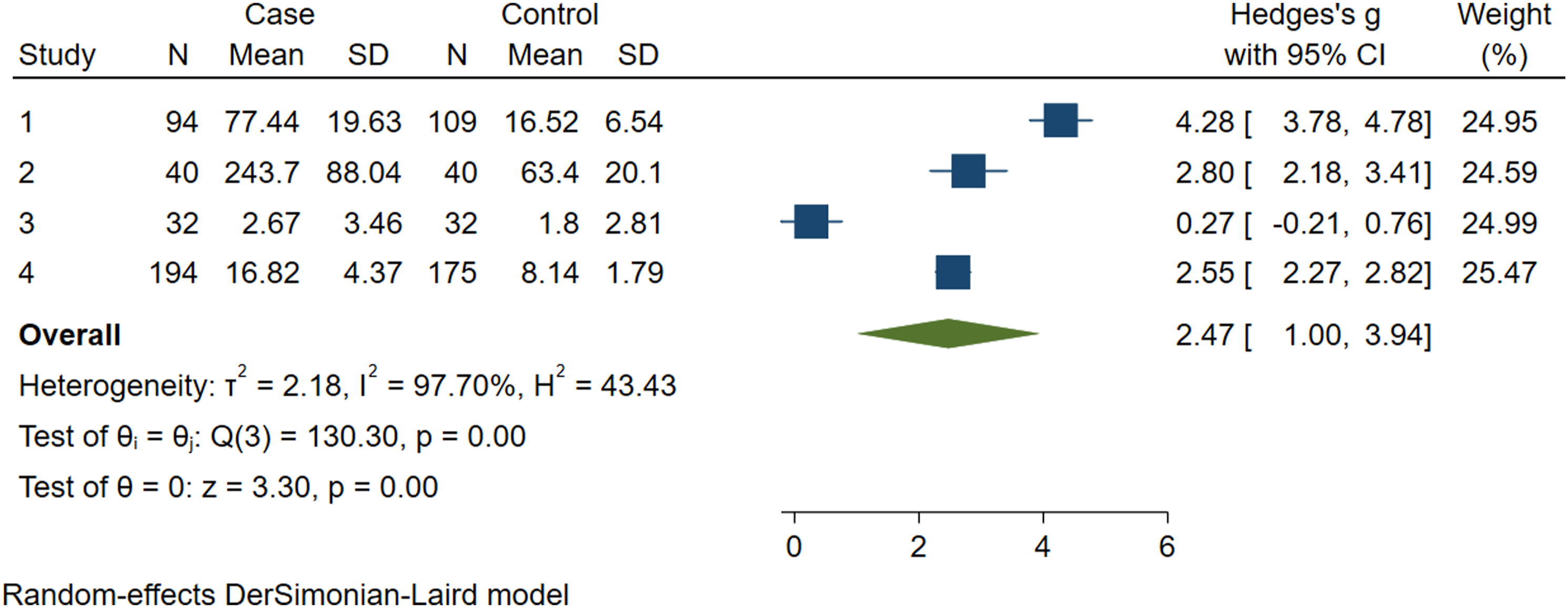
Discussion
Current studies on the pathogenesis of psoriasis have found that the IL-23/Th17 immune axis is the key driving mechanism of the pathogenesis of psoriasis. IL-23 activates Th17 cells to secrete IL-17, and major targets of IL-17 in psoriasis include keratinocytes, endothelial cells, and innate immune cells. In keratinocytes, 18 IL-17A, as the most important effector cytokine in psoriasis in IL-17 cytokine family, can induce various inflammatory mediators by synergizing with TNF-α, and can facilitate the abnormal proliferation of keratinocytes, leading to epidermal hyperplasia. In endothelial cells, IL-17 interacts to promote tissue inflammation and procoagulant activity through upregulation of IL-6, IL-8, and intracellular adhesion molecule-1, which may contribute to development of cardiovascular comorbidities in psoriasis. Lastly, IL-17A has proinflammatory effects on antigen-presenting cells, including macrophages
Witowski et al. 19 first found the expression of IL-17 in the serum of patients with psoriasis, and its expression level was higher than that of healthy controls. Subsequently, many scholars found that the serum levels of IL-17 were highly expressed in patients with psoriasis, but some studies showed that there was no significant difference in the serum levels of IL-17 between psoriasis patients and healthy people. The studies included in this systematic review had large sample size, high quality evaluation, and high reliability of the results. For the five articles in this study, the serum concentration of IL-17/IL-17A in the case group was higher than that in the control group. The subgroup results of Meta-analysis showed that serum IL-17 and serum IL-17A concentration of patients with psoriasis were significantly higher than that of healthy controls, respectively. The results showed that the serum IL-17/IL-17A concentration of psoriasis patients was significantly higher than that of normal controls.
Tang Juan, et al. 20 found that in the skin lesions of psoriasis patients, the expression levels of IL-17 mRNA and protein were higher than those of normal controls. Studies have shown when the activity of IL-17A is specifically inhibited, the skin lesions of psoriasis are significantly improved. 21 In addition, clinical trials have shown that anti-IL-17 biologic agents such as secukinum-Ab, ixekizumab, and brodalumab have achieved good efficacy in the treatment of psoriasis. 22 This confirms that IL-17, as a proinflammatory factor, is involved in the pathogenesis of psoriasis and plays a crucial role in it.
This study showed that the serum IL-17/IL-17A concentration of patients with psoriasis was significantly higher than that of healthy controls. This is consistent with the results of studies on the expression levels of IL-17 mRNA and protein in the skin lesions. It may be related to the secretion of IL-17 from skin lesions with the exocytosis of Th17 cells, and part of IL-17 factors entered the blood system due to the circulation of body fluids. It was found that the expression level of IL-17 in serum of patients with psoriasis vulgaris was positively correlated with PASI score.23–25 In conclusion, serum IL-17 concentration can be used as a response index of psoriasis severity. Compared with the detection of IL-17 in skin lesions, which requires tissue samples by surgery and immunohistochemical method, the detection of serum IL-17 concentration is relatively easy to operate with ELISA method, and more convenient for the observation in the process of treatment.
The limitation of this paper is that there are few included articles, and the research indicators of included articles are not consistent, including IL-17 and IL-17A. Therefore, subgroup analysis was performed, and the results of IL-17 and IL-17A subgroup analysis were consistent with the results of IL-17/IL-17A combination analysis.
Conclusion
Serum IL-17 concentration can be used as a response index of psoriasis severity. Compared with the detection of IL-17 in skin lesions, which requires tissue samples by surgery and detection with immunohistochemical method, the detection of serum IL-17 concentration is relatively easy to operate with ELISA method, and more convenient for the observation in the process of treatment.
Footnotes
Acknowledgements
The authors thank Professor Na Wang from the Fourth Hospital of Hebei Medical University for her guidance.
Author Contributions
Conception and design: JD Liu, JF Zhang and GQ Zhang; administrative support: JF Zhang and GQ Zhang; participate in the meta analysis: all authors; collection and assembly of data: all authors; manuscript writing: JD Liu, JF Zhang and GQ Zhang; and final approval of manuscript: all authors.
Declaration of Conflicting Interests
The author(s) declared no potential conflicts of interest with respect to the research, authorship, and/or publication of this article.
Funding
The author(s) disclosed receipt of the following financial support for the research, authorship, and/or publication of this article: This work was supported by The Hebei Provincial government funded clinical medical talents under Grant number 2020-4 and Open project of Key Laboratory of Ministry of Education under Grant number 201702029.
Data Availability
The data are from published articles, which have been annotated as references, and readers can refer to them by themselves.