
Abstract
The breast cancer is most frequently diagnosed cancer in the women worldwide. Our study investigated the anticancer effect of alternariol, a secondary metabolite originated from endophytic fungi, against DMBA induced breast carcinoma on Wistar rats. The toxicity study investigated the LD50 and the subsequent doses of alternariol for the carcinogenic study. The breast cancer was developed in rats via induction of DMBA (5 mg/kg, i.v.) and the carcinogenic study was continued for 24 weeks. The induction of breast cancer and the chemotherapeutic effect of alternariol were assessed through histopathological analysis of rat mammary tissue, followed by immunohistochemical analysis, cell proliferation assay and apoptosis assay by TUNEL method. The result showed that alternariol therapy decreased the hyperplastic lesions of mammary tissue and restored the normal histopathological characteristics of breast tissue. Furthermore, alternariol treatment downregulated the expression of carcinogenic markers such as PI3K and Akt increased the expression of apoptotic markers including p53, caspase-3 and Bax. Alternariol therapy also decreased the cellular proliferation and enhanced the apoptotic events. In conclusion, the breast cancer progression was significantly reduced via induction of apoptotic events and inhibition of cell propagation which allowed constructing of suitable mechanism for alternariol mediated chemotherapeutic approach.
Introduction
Breast cancer is the most frequent oncological threat and a substantial cause of death for women worldwide, according to the World Health Organization. It has been reported that breast cancer is the most often diagnosed malignancy with an estimated 4,820,000 new cases and 3,210,000 deaths in China. 1 A further alarming trend in China is the sharp decline in breast cancer survival rates. 2 The alteration of cellular physiology and signaling pathways of the healthy cells leads to cancer initiation via uncontrolled expression of various regulatory proteins and triggers cellular proliferation.
The inactivation of tumor suppressor proteins and oncogenic mutations of PI3K (phosphoinositide 3-kinase)/Akt/mTOR (mammalian target of rapamycin) signaling pathway due to PIK3CA mutations is one of the key reasons for breast cancer development in estrogen receptor (ER) positive breast cancer. 3 The frequent activation of PI3K/Akt/mTOR signaling pathway is associated with cell survival, cellular growth and proliferation of cancer cells by cellular metabolism and reorganization of cytoskeleton. 4 Besides that activation this signaling pathway also causes inhibition of apoptosis in the cancer cells 5 which leads to cancer cell survival, increase invasiveness, angiogenesis. 6 Additionally, the activation of this signaling pathway is also associated with the resistance to endocrine therapy, human epidermal growth factor receptor 2 (HER2)-directed therapy and cytotoxic therapy in breast cancer. 7 The reactive oxygen species (ROS) plays a crucial role in the process of cellular oxygen metabolism which accounts for numerous cellular activities responsible for cellular growth and proliferation at physiological concentrations. However, increased production of ROS leads to disruption of redox homeostasis and causes the ROS mediated destruction of several important macromolecules, which includes DNA, proteins, and lipids. 8 Thus, eventually promotes the carcinogenesis. 9 Furthermore, p53, A tumor suppressor protein, plays an important role in intrinsic apoptotic pathway through the regulation of proapoptotic markers such as Bax and caspase-3 expression. Thus any alterations of p53 regulation due to epigenetic modification hamper the cell growth and survival machinery and leads to cancer. Thus, the current therapeutic approaches are focusing on developing alternative chemotherapeutic compounds with distinctive anticancer mechanisms that can alter resistance and sensitize malignant cells to chemotherapies through the inhibition of associated signaling pathways, prevention of ROS generation by antioxidants, and induction of apoptosis.
Recently, the utilization of mycotoxins, biologically active secondary fungal metabolites, has gain interest among researchers due to its excellent cytotoxicity towards the mammalian cells. 10 The mycotoxin alternariol (AOH) is generally produced by Alternaria alternate, which can be found as natural food contaminant in small grains, wheat, fruits, vegetables and as well as in the processed fruit products like juices and wines. 11 The derivatives of AOH possess a wide range of biological activities especially anticancer activity, as it comprises two free hydroxyl groups and one cyclic ester group. 12 There are mainly two mechanisms of actions which have been identified for AOH, causing the coordinated induction of toxicant induced cytotoxicity including apoptosis, and necrosis which is characterized by inflammation mediated rupture of plasma membrane. 13 AOH also has been reported to reduce the cellular proliferation, leads to cell cycle arrest, and corresponds to its DNA damaging activity. 14 Additionally, it also stimulates the generation of reactive oxygen species (ROS), causing oxidative DNA damage.14,15 AOH has an inhibitory activity against topoisomerase II, which stabilizes the cleavable complex of DNA and causes DNA double strand breaks (DSBs). This inhibitory effect of AOH results in increased level of phosphorylated histone H2AX (g-H2AX), a marker of DSBs, and triggers G2 cell cycle arrest. 12 Furthermore, the induction of autophagy and senescence has been reported for AOH. It is also associated with the stimulation of AhR/ARNT pathway, which regulates CYP1A1 activity and apoptosis. 16 and modulates human endocrine systems by activating human estrogen receptor (hERα) and androgen receptor (hAR) signaling. 17 Alternariol also affect the viability and motility of breast epithelial cells through the induction of oxidative stress, DNA damage, and cell cycle arrest in the G2/M cell cycle phase. Moreover, it also activates nuclear factor erythroid 2-related factor 2 (NRF2) signaling pathway and inhibits Akt/ERK pathway to regulate viability the mammary tissue on molecular level. 18 However, another study revealed that Alternaria toxin mixtures caused activation of aryl hydrocarbon receptor which induced Cytochrome P450 (CYP) 1A1/1A2/1B1 activity in phase I metabolism and activates the breast cancer cells. 19
Till now there is lack of documented evidences demonstrating the cytotoxic effect of AOH through the induction of apoptosis on the breast cancer model. Additionally, the agonistic activity of AOH against human estrogen receptor (hERα) and androgen receptor (hAR) showcasing its affinity towards the endocrine system which might be a key chemotherapeutic target in breast cancer. In this study the breast cancer was induced by 7,12-dimethylbenz[α]anthracene (DMBA), a polycyclic aromatic hydrocarbon, that is widely used as immunosuppressor and a powerful organ-specific laboratory carcinogen which serves as a tumor initiator. Thus the present study is focusing upon the AOH mediated chemotherapeutic evaluation on DMBA-induced breast cancer through in vivo study on rats.
Materials and methods
Chemicals and reagents
Alternariol (#35758), biotinylated goat anti-rabbit IgG (#21537), streptavidin peroxidase (#S5512), 3,3ʹ-diaminobenzidine (DAB) (#D12384) and proteinase K (#1.07393) were procured from Sigma Aldrich Chemical Co. (St Louis, MO). Anti-mouse p53 (#GTX70214), anti-rabbit PI3K (#GTX100462), Akt (#GTX121937), Bax (#GTX109683), caspase-3 (#GTX110543), PCNA (#GTX100539) were purchased from GeneTex International Corporation (Global). The apoptosis detection kit (#MK500) was procured from Takara Bio Inc. (Kusatsu, Japan). All the other reagents were purchased from the local suppliers at its purest grade.
Animal husbandry and maintenance
All the animal experimentations were performed according to the Institutional Animal Ethical Committee of Taizhou First People’s Hospital (Approval no. 2021KYB420) and all the procedures performed throughout the research have complied with ethical guidelines and were included in institutional ethical policies.
The animals were obtained from Wenzhou Medical University Animal Center. For toxicity study, Balb/c mice (20–25 g), 6–8 weeks old, of both sexes were used and for the chemotherapeutic study female Wistar rats (70–120 g) of 6 weeks old were utilized. The animals were kept in the polypropylene cages with free access to food and tap water, at a temperature of 22 ± 3oC, with relative humidity 50–58% and 12 h light/dark cycle. The animals were acclimatized for 1 week before the initiation of the experiment.
Toxicity study
Acute toxicity study
The toxicological evaluation of alternariol was assessed by acute toxicity study in order to determine the LD50 value, as per the guidelines of Organization for Economic Cooperation and Development (OECD) for testing of chemicals, TG 420 (adopted December 2001) with slight alterations. The animals were arbitrarily distributed into five experimental groups, each of which contains 6 animals, three of each sex. The groups were followed as one normal control group receives only vehicle and the other four groups were selected as per the different doses selected for alternariol which includes 2000, 1000, 800 and 600 mg/kg body weight. After the drug introduction the animals were supplied with food and water immediately and observed for 3 days for any mortality.
Sub-acute toxicity study
Following the acute toxicity study the sub-acute toxicity study was carried out in order to select the doses for chemotherapeutic study. The 28-days repeated oral toxicity study was performed according to the guidelines of Organization for Economic Cooperation and Development (OECD) for testing of chemicals, TG 420 (adopted December 2001) with slight alterations. The animals were arbitrarily divided into five experimental groups and each group consists of 6 animals, 3 of each sex. The groups were followed as one normal control group receives only vehicle and the other four groups were selected as per the different doses selected for alternariol which includes 400, 200, 100 and 50 mg/kg body weight. During the experimental period the animals were observed for general clinical appearance and mortality. At the end of the repeated oral toxicity study, all the animals were euthanized and the vital organs (liver, kidney, stomach and testis) were excised for histopathological evaluation. Furthermore, the hematological and serum biochemical parameters were also assessed.
Hematological and serum biochemical analysis
The hematological parameters were assessed by using Medonic CA-620 cell analyzer systems (Boule Medical, Stock-Holm, Sweden). The blood samples were obtained from the retro-orbital plexus of the animals and it was centrifuged for 10 min at 3000 r/min in order to separate the serum form the blood. The serum samples obtained was then assessed through automated biochemistry analyzer/Pentra C400 (Horiba Medical, Kyoto, Japan).
Histopathological evaluation
At the end of sub-acute toxicity study the animals were euthanized and the vital organs were excised from them which include liver, stomach, kidney and testis. The organs were then kept into 10% formalin solution overnight for fixation. The organs were then treated with graded alcohol for dehydration and mounted on low melting paraffin wax upon the glass slides of 5 micron. It was then deparaffinized and rehydrated using xylene and graded alcohol respectively. The breast tissue samples were then stained by heamatoxylin and eosin stain (H&E) and evaluated under light microscope.
Experimental protocol
At the end of the acclimatization period the animals were distributed randomly into five experimental groups in which each group was consisting of 6 animals. The rats of 7–8 weeks old (except group I) were administered with a single i.v. injection of DMBA (0.5 mg/100 g body weight) in corn oil through the tail vein and the breast cancer was induced. Following the introduction of carcinogen the rats were treated with alternariol through oral gavage which continued for 24 weeks. The experimental groups were followed as:
Group I- vehicle control animals, Group II- treated with DMBA and allotted as carcinogen control, Group III- treated with DMBA + received 50 mg/kg alternariol, Group IV- treated with DMBA + received 100 mg/kg alternariol, Group V- treated with DMBA + received 200 mg/kg alternariol.
After completion of chemotherapeutic study, the breast tissues were excised from the euthanized animals and stored in formalin for fixation. The tissues were assessed for appearance of any tumor structure. Breast tissues were evaluated through histopathological analysis, immunohistochemistry and cell proliferation assay.
Histopathological evaluation of breast tissue
The animals were ether anaesthetized and the thoracic and abdominal inguinal mammary tissue was collected. Fixation of the breast tissues were done in 10% formalin solution, followed by embedding in paraffin wax. The tissues were cut into 5 μm thickness and placed on slides. The staining was done using H&E and evaluated under light microscope.
Antioxidant activity of breast tissue
The breast tissues were homogenized (10% w/v) in 0.1 M of phosphate buffer (pH 7.0) for the assessment of antioxidant activity. The homogenized mixture was then centrifuged for 15 min, the supernatant was decanted and used for enzymatic analysis. 20
Catalase activity
As per the method of Sinha et al. (1972) the catalse activity of homogenized breast tissue was carried out. The absorbance was noted at 620 nm and the catalase activity was expressed in terms of μmol of H2O2 consumed/min/mg protein. 21
Superoxide dismutase activity
The SOD activity was determined as per the method described by Awasthi et al. (1984). The SOD activity was assessed as units/min/mg protein. 22
Glutathione peroxidase activity
The activity of GPx was determined according to the method described by Rotruck et al. (1973). The GPx activity was denoted as μmol of GSH consumed/min/mg protein. 23
Immunohistochemistry
The excised breast tissue were embedded in paraffin, cut into 5 μm thickness and mounted on poly-L-lysine coated slides. The tissues were then deparaffinized and immersed into the solution of H2O2. The sections were then treated with protein blocking solution for 1 h and then incubated with anti‐mouse p53 (1:1000), anti-rabbit PI3K (1:500), Akt (1:500), caspase-3 (1:500) and Bax (1:500) antibodies overnight at a temperature of 4oC. It was then followed by PBS washing and treated with HRP conjugated secondary antibody for 30 min. The development of the sections was done using DAB and H&E staining was carried out for counterstaining. The slides were placed under light microscope (OLYMPUS CX 21i TR) and images were captured followed by quantification of the immune-positive cells by ImageJ software (version 1.8.0).
Cell proliferation assay
The tissue section of 5 μm thickness was mounted on poly‐L‐lysine coated glass slides. The slides were then depraffinized, rehydrated and treated with H2O2. After the PBS wash, the slides were treated with anti‐rabbit PCNA antibody (1:500) overnight at 4oC. The slides were then again treated with HRP conjugated secondary antibody at room temperature for 30 min. The development of the sections was done using DAB and H&E staining was carried out for counterstaining followed by evaluation under light microscope.
Apoptosis assay by TUNEL method
The breast tissues mounted on poly‐L‐lysine coated glass slides were treated with proteinase K (20 μg/ml in PBS) at room temperature for 10 min, in order to digest the nonspecific proteins. The peroxide quenching of the tissues were done by H2O2 (3% in PBS) treatment for 5 min at room temperature. The tissue were then treated with terminal deoxynucleotidyl transferase (TdT) buffer (30 mM of Trizma base, pH 7.2, 140 mM sodium cacodylate, 1 mM of cobalt chloride) and further treated with TdT reaction solution containing TdT and dUTP for 90 min at 37°C. The reaction was stopped by treating the sections with 2% standard saline citrate for 10 min at room temperature. The slides were then incubated with anti‐digoxigenin peroxidase for 30 min at room temperature. The development of the sections was done using DAB and H&E staining was carried out for counterstaining followed by evaluation under light microscope.
Assessment of labeling index and apoptotic index
The labeling index (LI) was determined as the % PCNA‐positive cells/total number of cells. The apoptotic index (AI) was expressed as the % TUNEL‐positive cells/total number of cells.
Statistical analysis
The results were expressed as mean ± standard error mean (SEM). The one‐way analysis of variance (ANOVA) followed by post‐hoc test (Tukey’s test) was carried out for the evaluation of statistical significance by using Graph pad prism software (Version 5). The variations were supposed to be statistically significant, with p < 0.05.
Results
Toxicity study
Acute and sub-acute toxicity
After performing the acute toxicity study the LD50 dose of alternariol has been found to be 600 mg/kg body weight. According to the LD50 value of AOH the doses for the sub-acute toxicity study was selected which was followed as 400, 200, 100, 50 mg/kg body weight. During the repeated oral toxicity study treatment related mortality was observed for the dose level of 400 mg/kg. Additionally, the food and water consumption was decreased for the animals treated with 400 mg/kg of AOH. The body weight of the animals was also reduced for the same treatment group.
Hematological and serum biochemical analysis
Hematological finding in male Balb/c mice treated with alternariol for 28 days repeated-dose oral sub-acute toxicity study.

Standard error of mean = standard deviation (SD)/√ Total subject. Result are analysed by one-way ANOVA and confirmed by Tukey’s multiple comparison post-hoc analysis. MCV: mean corpuscular volume; MCH: mean corpuscular haemoglobin; MCHC: mean corpuscular haemoglobin concentration; RBC: red blood cell; WBC: white blood cell.
*Significant difference as compared to the normal control (p < 0.05).
#Significant difference as compared to the alternariol 50 mg/kg (p < 0.05).
$Significant difference as compared to the alternariol 100 mg/kg (p < 0.05).
αSignificant difference as compared to the alternariol 100 mg/kg (p < 0.05).
Hematological finding in female Balb/c mice treated with alternariol for 28 days repeated-dose oral sub-acute toxicity study.
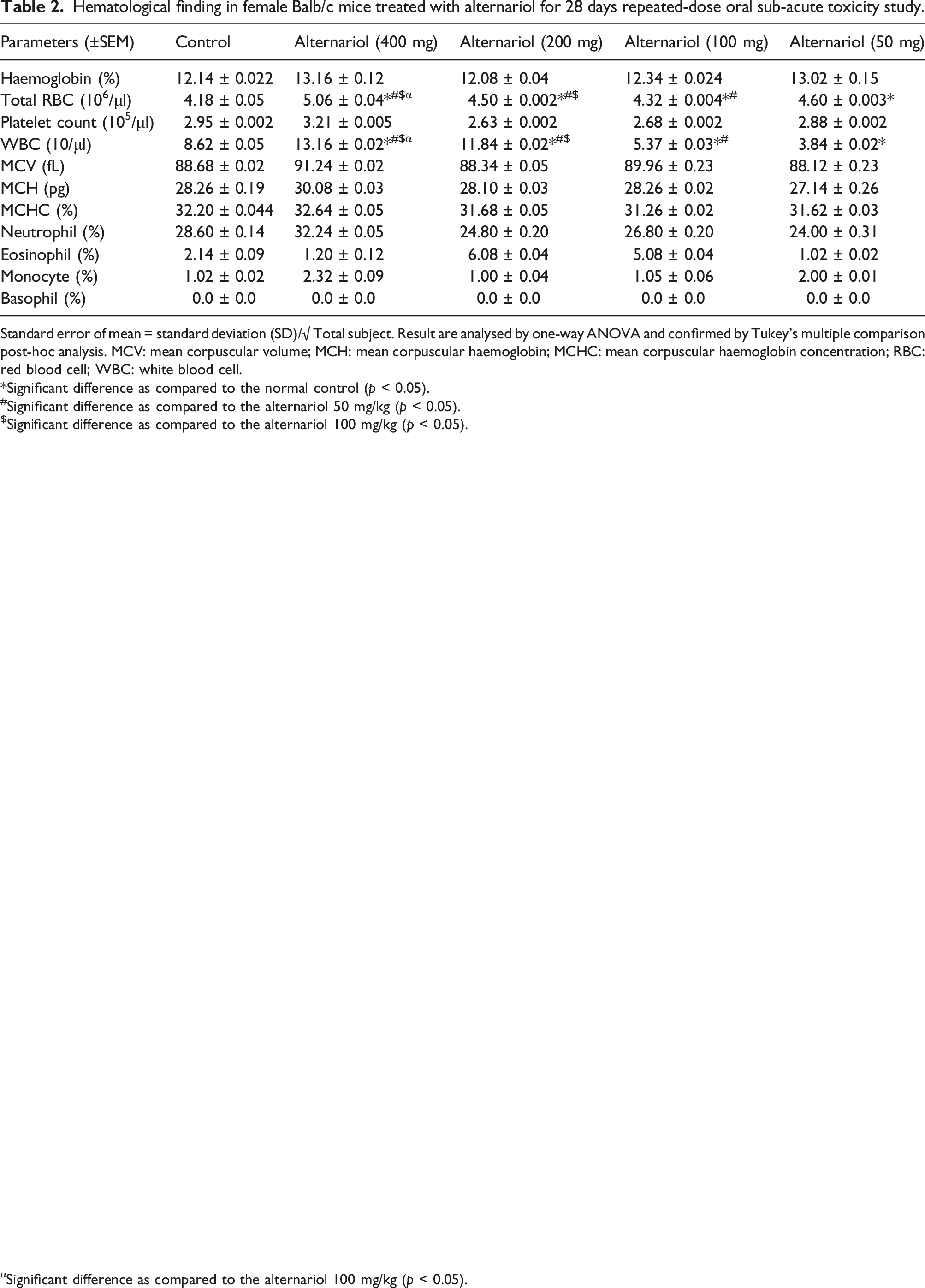
Standard error of mean = standard deviation (SD)/√ Total subject. Result are analysed by one-way ANOVA and confirmed by Tukey’s multiple comparison post-hoc analysis. MCV: mean corpuscular volume; MCH: mean corpuscular haemoglobin; MCHC: mean corpuscular haemoglobin concentration; RBC: red blood cell; WBC: white blood cell.
*Significant difference as compared to the normal control (p < 0.05).
#Significant difference as compared to the alternariol 50 mg/kg (p < 0.05).
$Significant difference as compared to the alternariol 100 mg/kg (p < 0.05).
αSignificant difference as compared to the alternariol 100 mg/kg (p < 0.05).
Serum biochemical finding in male Balb/c mice treated with alternariol for 28 days repeated-dose oral sub-acute toxicity study.

Standard error of mean = standard deviation (SD)/√ Total subject. Result are analysed by one-way ANOVA and confirmed by Tukey’s multiple comparison post-hoc analysis.
*Significant difference as compared to the normal control (p < 0.05).
#Significant difference as compared to the alternariol 50 mg/kg (p < 0.05).
$Significant difference as compared to the alternariol 100 mg/kg (p < 0.05).
αSignificant difference as compared to the alternariol 100 mg/kg (p < 0.05).
Serum biochemical finding in female Balb/c mice treated with alternariol for 28 days repeated-dose oral sub-acute toxicity study.

Standard error of mean = standard deviation (SD)/√ Total subject. Result are analysed by one-way ANOVA and confirmed by Tukey’s multiple comparison post-hoc analysis.
*Significant difference as compared to the normal control (p < 0.05).
#Significant difference as compared to the alternariol 50 mg/kg (p < 0.05).
$Significant difference as compared to the alternariol 100 mg/kg (p < 0.05).
αSignificant difference as compared to the alternariol 100 mg/kg (p < 0.05).
Histopathological analysis of organs
Followed by sub-acute toxicity study of AOH, the histopathological analysis of the vital organs like kidney, liver, stomach, and testis were performed. In the normal control group the morphological architecture of kidney demonstrated glomerulus, bowman’s capsule, proximal tubule, and distal tubule (Figure 1[i] (a)). The 400 mg/kg group depicted thickening of capsular membrane, ruptures, desquamated nuclei, vacuolization (Figure 1[i] (e)). The 50, 100, 200 mg/kg treated group did not show any significant alterations in kidney tissue (Figure 1[i] (b)–(d)). In normal control group, the histopathology of liver showed portal vein, bile duct, hepatic artery, hepatocytes, sinusoids and the kupffer cells (Figure 1[ii] (a)). The 400 mg/kg treated group depicted periportal mononuclear infiltrates, degeneration of hepatocytes, focal inflammation of hepatocytes (Figure 1[ii] (e)). Whereas, 50, 100, and 200 mg/kg treated group did not caused any histological alterations of liver tissues (Figure 1[ii] (b)–(d)). In normal control group the histology of the stomach tissues showed sub-mucosa, muscularis mucosa, and mucosal layers with distinct gastric pits, chief cells, parietal cells, mucosal cells, and surface epithelial cells (Figure 1[iii] (a)). The 50, 100, and 200 mg/kg treated group (Figure 1[iii] (b)–(d)) denoted no histopathological alterations in the stomach tissue. On the other hand, 400 mg/kg treated group was associated with hemorrhages between villus, hyperplasia, and leukocyte infiltration (Figure 1[iii] (e)). In normal control group and AOH treated group (50, 100, and 200 mg/kg) normal morphological architecture of testis were observed (Figure 1[iv] (a)–(d)) while, 400 mg/kg AOH treated group indicated edema in interstitial tissue, degeneration of seminiferous tubule and hyperplasia (Figure 1[iv] (e)). [i] Histopathological representation of Kidney of Balb/c mice. (a) Normal tissue showing (g) glomerulus, Bowman’s capsule (bc), Macula densa (md), Proximal convoluted tubule (pct), Distal convoluted tubule (dct) (b) kidney tissue exposed to 50 mg/kg alternariol (c) kidney tissue exposed to 100 mg/kg alternariol (d) kidney tissue exposed to 200 mg/kg alternariol (e) exposed to 400 mg/kg alternariol showing thickening of capsular membrane (tbm), ruptures (r), desquamated nuclei (dn), vacuolization [ii] Histopathological representation of Liver of Balb/c mice (a) Normal control showing the Central vein (cv), Bile duct (bd), Sinusoidal dilation (sd), Kupffer cell (kc), Lymph vessel (lv) (b), (c) and (d) liver tissue exposed to 50 mg/kg, 100 mg/kg and 200 mg/kg of alternariol. (e) Liver tissue exposed 400 mg/kg alternariol showing periportal mononuclear infiltrates (pmi), degeneration of hepatocytes (dh), focal inflammation (fi). [iii] Histopathological representation of stomach of Balb/c mice (a) Normal control showing muscularis externa (me), Submucosa (sm), Muscularis mucosa (mm), Lamina propia (lp), Gastric pit (gp), epithelial lining (epl) (b), (c) and (d) Stomach tissue exposed to 50 mg/kg, 100 mg/kg and 200 mg/kg of alternariol. (e) Stomach tissue exposed 400 mg/kg alternariol showing Hemorrhages (h) between villus, Hyperplasia (hyp), leukocyte infiltration (Li). [iv] Histopathological representation of testis of Balb/c mice (a) Normal control showing Sertoli cell (sc), Spermatogonia (Sp), Seminiferous tubule (St), Interstitial tissues (It) is seen within the tubular lumen (b), (c) and (d) testis exposed to 50 mg/kg, 100 mg/kg and 200 mg/kg of alternariol. (e) Testis exposed to 400 mg/kg alternariol showing edema in interstitial tissue (e), Degeneration of seminiferous tubule (d) and hyperplasia (Hyp). (H&E) 10× magnification [inset 40×].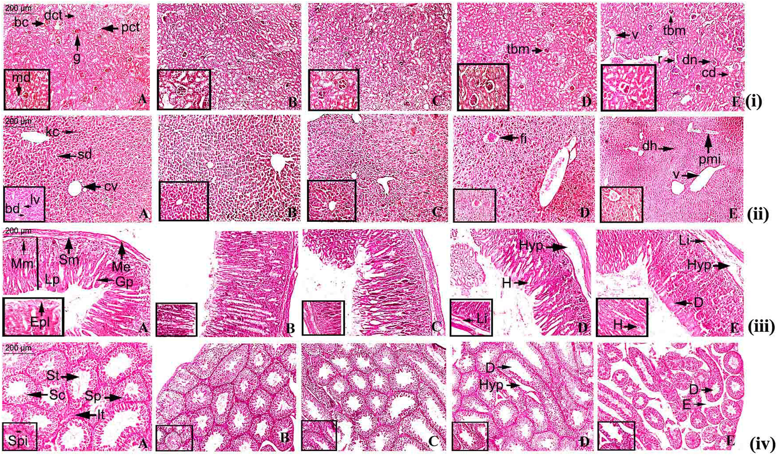
Antioxidant status
In the carcinogen control group, a significant reduction of SOD, CAT and GST levels were observed as compared to normal control group (p < 0.05). AOH treatment increased the levels of SOD, CAT and GST significantly in breast tissue in which 400 mg/kg group showed significant upregulation of antioxidants in breast tissue as compared to the normal control group and other treatment groups (p < 0.05) (Figure 2). Effect of alternariol on in vivo antioxidant enzymes SOD (superoxide dismutase) and CAT (catalase), (glutathione) GST. *Significant difference as compared to normal control group (p < 0.05). #Significant difference as compared to carcinogen control group (p < 0.05). $Significant difference as compared to 50 mg/kg group (p < 0.05). αSignificant difference as compared to 100 mg/kg group (p < 0.05).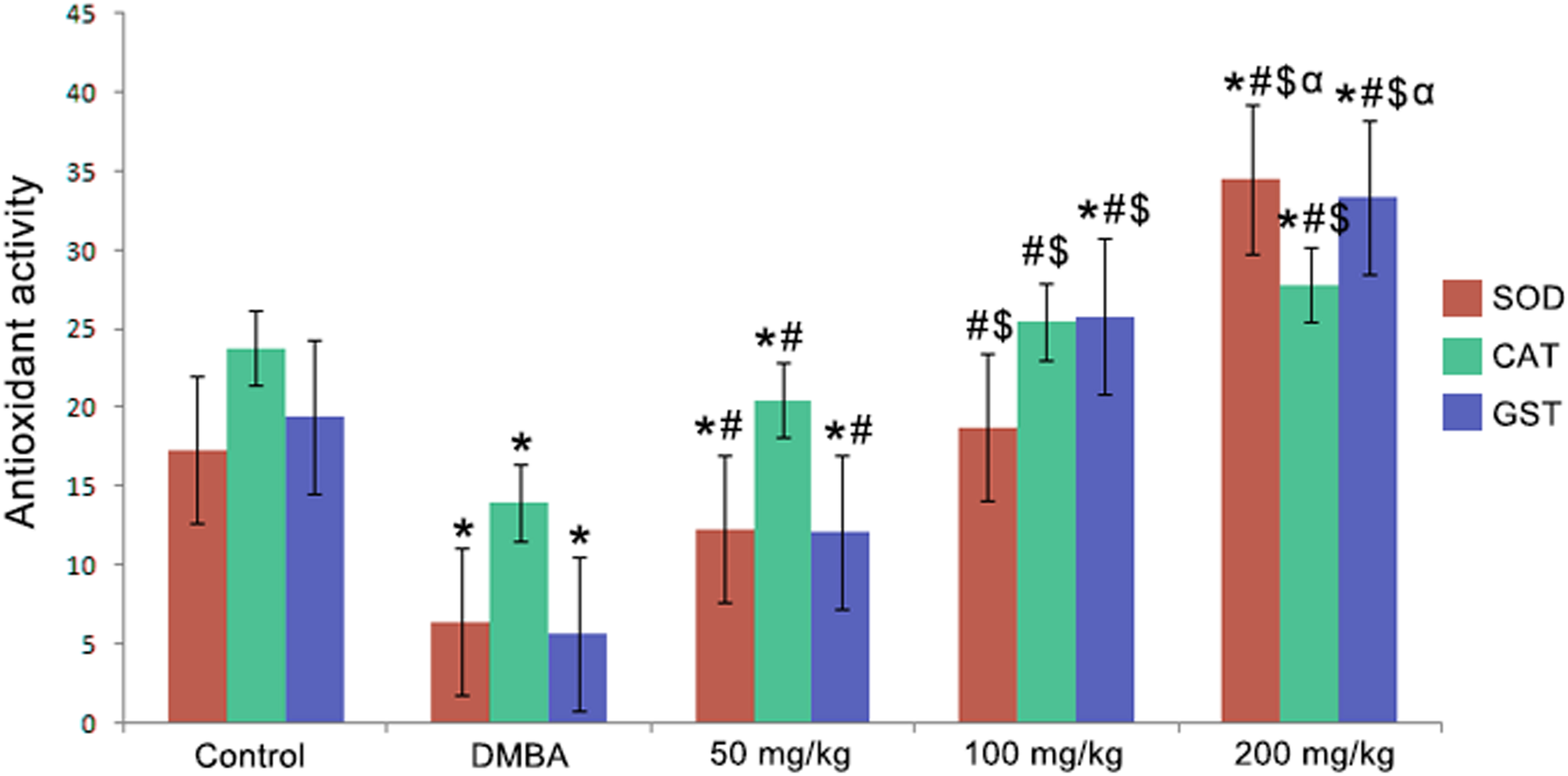
Histological analysis of mammary tissue
The histological evaluation of breast tissue has been represented in the (Figure 3). The normal architecture of mammary tissue was represented by the normal group (group I) which showcased the terminal duct lobular units (td), alveoli (a), alveolar septa (sg), acinus (ac), and serous gland (sg) of mammary tissue (Figure 3(a)). The carcinogen control group (group II) showed atrophy of glands with periductal stromal fibrosis and fatty tissue (psf), atrophy of glands (ag) surrounded with fatty tissue, atrophy of serous glands (asg) surrounded with stromal fibrosis, atypical hyperplasia (ah) (Figure 3(b)). The 50 mg/kg treated group showed atrophy of serous glands (asg), atrophy of glands (ag) and periductal stromal fibrosis and fatty tissue (psf) (Figure 3(c)). The 100 mg/kg group showcased hyperplasia of serous and mucinous glands (h) (Figure 3(d)). However, 200 mg/kg group depicted a significant downregulation of hyperplasia or cellular propagation in breast tissue (Figure 3(e)) and represented normal architectural profile of the mammary tissue. (a) Histological appearance of mammary tissue of normal control showing Terminal duct lobular units (td), alveoli (a), alveolar septa (sg), Acinus (ac), serous gland (sg) (b) DMBA control shows atrophy of glands with periductal stromal fibrosis and fatty tissue (psf), atrophy of glands (ag) with surrounding fatty tissue, atrophy of serous glands (asg) with surrounding stromal fibrosis, Atypical hyperplasia (ah). (c) Mammary tissue of DMBA induced group treated with 50 mg/kg alternariol showing atrophy of serous glands (asg), atrophy of glands (ag) and periductal stromal fibrosis and fatty tissue (psf) (d) Mammary tissue of DMBA induced group treated with 100 mg/kg alternariol showing hyperplasia of serous and mucinous glands (h) (e) Mammary tissue of DMBA induced group treated with 200 mg/kg alternariol having almost normal architecture.
Immunohistochemical analysis
The immunohistochemical analysis of the mammary tissue was carried out for assessment of the protein expression which includes tumor‐suppressor protein p53, proapoptotic protein Bax and caspase-3, the growth regulatory proteins PI3K, Akt (Figure 4). In the normal control group (group I) a high expression of tumor‐suppressor protein p53 was observed in the normal epithelial tissues along with terminal end bud and alveolar ducts (Figure 4(i) (a)). Whereas, in the carcinogen control group (group II) there was reduced expression of p53 (Figure 4(i) (b)). In the AOH treated group at the dose levels of 50, 100 and 200 mg/kg body weight a significantly (p < 0.05) high expression of p53 was detected at terminal ducts and alveolar region (Figure 4(i) (c)–(e)) as compared to carcinogen control group (Table 5). The expression of Bax was detected in the entire terminal end bud and cap cells for the animals of normal control group (Figure 4(ii) (a)). For the carcinogen control group the expression of Bax was significantly reduced (Figure 4(ii) (b)). Whereas, there was a significantly (p < 0.05) high expression of Bax in the alveolar duct region and terminal end buds for the AOH treated groups (Figure 4(ii) (c)–(e)) (50, 100, 200 mg/kg) in comparison to the carcinogen control group (Table 5). There was a significant expression of PI3K in the mammary tissue of normal control group (Figure 4(iii) (a)) at terminal end buds but a reduced expression was denoted at the cap cell layer. The expression of PI3K was significantly high for the carcinogen control group (Figure 4(iii) (b)) in the hyperplastic mammary tissue as compared to the normal control group. Conversely, a significant (p < 0.05) reduction of PI3K expression was noted in the AOH treated group (Figure 4(iii) (c)–(e)) (50, 100, 200 mg/kg) (Table 5). The expression of Akt was detected in the entire terminal end bud and cap cells for the animals of normal control group (Figure 4(iv) (a)). For the carcinogen control group the expression of Akt was significantly increased (Figure 4(iv) (b)). Whereas, there was a significantly (p < 0.05) low expression of Akt in the alveolar duct region and terminal end buds for the AOH treated groups (Figure 4(iv) (c)–(e)) (50, 100, 200 mg/kg) in comparison to carcinogen control (Table 5). In the normal control group (group I) a high expression of caspase-3 was noted in the terminal end bud regions (Figure 4(v) (a)). Whereas, in the carcinogen control group (group II) there was reduced expression of caspase-3 (Figure 4(v) (b)). In the AOH treated group at the dose levels of 50, 100 and 200 mg/kg body weight a significantly (p < 0.05) high expression of caspase-3 was detected at terminal ducts and alveolar region (Figure 4(v) (c)–(e)) as compared to carcinogen control group (Table 5). The immunohistochemical analysis of the (i) p53, (ii) Bax, (iii) PI3K, (vi) Akt and (v) caspase-3 expressions in the breast tissues of different groups of rats (a) the normal control (b) carcinogen control (c) 50 mg/kg of alternariol treated (d) and (e) 100 and 200 mg/kg alternariol treated. All images at 40×. Effect of alternariol on the expression of Bax, PI3K, p53, Akt and caspase-3 in breast tissues. §Each score represents the results of 6 slides per rat and 6 rats per group, mean ± SE (n = 6). Each field were selected randomly for evaluation of percentage of immune-positive cells. The results were compared using ANOVA, followed by a Tukey’s multiple comparison post-hoc analysis. *Significant difference as compared to the control (p < 0.05). #Significant difference as compared to the DMBA control (p < 0.05). $Significant difference as compared to the alternariol 50 mg/kg (p < 0.05). αSignificant difference as compared to the alternariol 100 mg/kg (p < 0.05).

Cell proliferation assay
The effect of AOH treatment in the cellular proliferation of breast tissue has been represented in the (Figure 5(i)) which depicted the chemotherapeutic action of AOH in the DMBA induced mammary carcinogenesis. A distinct nuclear localization and brown stain due the chromogen treatment has been showed in the PCNA labeled cells which were used for the cellular quantification. The normal control group showed a low level of PCNA labeled cells (Figure 5(i) (a)). The percentage of PCNA labeled cells was measured by the labeling index (LI) and the DMBA control group (Figure 5(i) (b)) showed the maximum LI (Table 6). However, a significant (p < 0.05) reduction in LI was noted for the AOH treated group (Figure 5(i) (c)–(e)) (50, 100, 200 mg/kg) (Table 6). [i] The immunohistochemical analysis of expression of PCNA of different group of rats (a) the normal control (b) carcinogen control (c) 50 mg/kg of alternariol treated (d) and (e) 100 and 200 mg/kg alternariol treated. All images at 40×. [ii] TUNEL assay of apoptotic (a) the normal control (b) carcinogen control (c) 50 mg/kg of alternariol treated (d) and (e) 100 and 200 mg/kg alternariol treated. All images at 40×. Cell proliferation and apoptosis in breast tissue. LI = Labelling index, PCNA-LI = percentage of PCNA labelled cells/total number of cells counted, AI = Apoptotic index. R = PCNA-LI/AI. AI was calculated as the percentage of TUNEL positive cells/total number of cells counted. Values represent mean ± SE. The results were compared using ANOVA, followed by a Tukey’s multiple comparison post-hoc analysis. §Total number of six slides were evaluated per rat. Each field consisted of approximately 500 cells. *Significant difference as compared to the control (p < 0.05). #Significant difference as compared to the DMBA control (p < 0.05). $Significant difference as compared to the alternariol 50 mg/kg (p < 0.05). αSignificant difference as compared to the alternariol 100 mg/kg (p < 0.05).

Apoptosis via TUNEL assay
The TUNEL assay was performed in order to identify the incidences of apoptosis in the cells of hyperplastic mammary tissue. The apoptotic cells were identified by their brown staining due to chromogen treatment (Figure 5(ii)). The carcinogen control group (Figure 5(ii) (b)) showed lowest number of TUNEL positive cells, on an average of 3–5 apoptotic cells, in a field of about 700 cells in comparison to the normal control group (Figure 5(ii) (a)). A significantly (p < 0.05) high expression of TUNEL positive cells were noted in the AOH treated group (Figure 5(ii) (c)–(e)) (50, 100, 200 mg/kg) in comparison to carcinogen control. An average of 8–9 apoptotic cells in a field of 700 cells were observed in the AOH treated group. In this case, the overlapping occurs between the brown stain and the condensed chromatin of the apoptotic body which correlates the TUNEL assay with the induction of apoptosis.
The percentage of TUNEL positive cells were measured by the apoptotic index (AI) (Table 6). The R value is denoted as the ratio of cell proliferation to apoptosis. In the carcinogen control group the increased R value designated the enhanced proliferative activity of the cancer cells but treatment with AOH significantly decreased R value due to induction of apoptosis.
Discussion
In recent years the traditional chemotherapeutic medications have become obsolete due to their severe side effects and the emergence of drug resistance which mandating the development of novel compounds to diminish cancer incidence while avoiding the associated adverse effects. In this instance, the natural products would be the most viable option because of their potential anticancer activity and minimal side effects. 24 The plant associated endophytic microorganisms seem to be well producers of a wide range of bioactive chemicals with numerous biological activities with significant chemotherapeutic efficacy. 25 Thus, the chemotherapeutic effect of AOH against DMBA-induced mammary carcinogenesis in the Wistar rat model was investigated and the possible molecular mechanistic approach of AOH as an anticancer drug was further postulated.
In order to establish the safe dosage of AOH for the carcinogenic study the toxicity study was performed to determine LD50 of this novel agent. The LD50 dose of AOH was determined as 600 mg/kg body weight for Balb/c mice. In toxicity study, significant histopathological changes were observed in the vital organs of mice at 400 mg/kg AOH treated group. On the other hand, the 50, 100, 200 mg/kg AOH treated group was not associated with any histopathological alternations of their vital organs which indicates the non-toxic and safe doses of AOH that could be further utilized in the carcinogenic study.
The induction of cancer through chemical carcinogens is multiphase process which is associated with triggering of normal cells to malignant one followed by invasion into nearby tissue. 26 In this study the breast cancer was induced in the Wistar rats through the administration of DMBA which resembles the similar pathway as it was seen in human breast cancer. Along with that, the histopathology, hyperplastic progression with premalignant and malignant lesions are quite identical with human breast cancer incidence. 27 The administrations of DMBA in the rats cause the development of the hyperplastic lesions and alter the normal architectural organization of mammary tissue with increased cellular proliferation, which was further assessed through histopathological evaluation. The treatment with AOH fruitfully restored the cellular architectural changes to normalcy of the mammary tissue through the inhibition of hyperplastic lesions and decreased cellular proliferation which clearly indicates the chemotherapeutic action of the drug against breast cancer.
The development of animal experimental model systems for the evaluation anticancer activity of AOH against mammary carcinogenesis is highly valuable for the assessment of human breast cancer as rat breast has high tendency of acquiring neoplasm that resembles human mammary carcinogenesis. 28 Previously the role of AOH in the viability of HCT116 colon cancer cell was also been evaluated which demonstrated activation of apoptosis through the upregulation of proapoptotic markers such as p53, caspase-3, and Bax. 29 The present study depicts the anticancer potential of AOH against DMBA induced breast cancer through the inhibition of cellular propagation, triggering of apoptosis and the reduction of breast cancer incidences via downregulating the expression of PI3K and Akt and the upregulation of apoptotic markers including p53, caspase-3 and Bax in rat breast cancer model. In addition, the research provides an overview of anticancer mechanism for this novel chemotherapeutic medication.
The immunohistochemical analysis of breast tissue was performed to investigate the expression of p53, caspase-3, Bax and cellular growth and survival proteins PI3K, Akt to establish the signaling pathway through which AOH shows its chemotherapeutic activity against breast carcinoma. Several studies reported that, the mutation of p53, a tumor suppressor protein, is directly associated with cancerous lesions due to alteration of tumor suppressive pathways. 30 Increased expression of p53 decreased the number of cells through the induction of intrinsic apoptotic pathway which further initiates the caspase mediated downstream events, eventually causing cell death.31,32 The findings of this research instigated the upregulation of p53, caspase-3 and Bax expression which confirms the stimulation of apoptosis in breast carcinoma cells. Furthermore, PI3K signaling pathway stimulates the cellular growth and prevents the cancer cell from apoptosis through several cell survival pathway mediated by PI3K. In this study the carcinogen control group showed upregulation of PI3K and Akt expression while the AOH treated group demonstrated a significant downregulation of these proteins in the breast tissue. As a result of the AOH treatment, cancer cells experience a considerable reduction in cellular proliferation and become more sensitive to apoptosis.
The healthy cells are able to protect themselves from the harmful effects of oxidants by the action of a complex system of antioxidant enzymes such as SOD, CAT and GST. 33 From the experimental findings, it was confirmed that the carcinogen control group showed a decreased level of SOD, CAT, and GST but the treatment with AOH caused an upregulation of antioxidant levels in breast tissue which may contribute to the prevention of ROS generation as well as inhibits the cellular growth and proliferation.
A significant hallmark of cancer incidence is uncontrolled cell proliferation. The control over the cell proliferation depends on the regulation of several mechanistic pathways which drives the machinery of cell cycle. The cell proliferation can be controlled through various checkpoint pathways. In this study the treatment with AOH causes a significant decrease in the number of proliferating cells which demonstrated its anti-proliferative activity against breast cancer. The cell proliferation activity of the cancer cells in breast tissue was assessed PCNA or proliferating cell nuclear antigen marker. The total percentage of PCNA labeled cells was measured by the labeling index in which the distinct nuclear localizations were showed by the cells labeled with PCNA. For the carcinogen control group a greater labeling index was detected which was reduced followed by the AOH treatment as seen in the AOH treatment group.
In the primary stage of breast cancer the apoptosis possesses an important role in the suppression of carcinogenesis. In this research the TUNEL assay was performed for the assessment of apoptosis induction in the cancer cells due to AOH treatment. Treatment with AOH (50, 100, 200 mg/kg) showed a greater percentage of cells undergoing through apoptosis while a significantly low apoptotic index was detected in carcinogen control.
Considering all the findings from this research, the chemotherapeutic activity of AOH against DMBA induced rat mammary carcinogenesis through the inhibition of cellular growth and proliferation as well as the induction of apoptosis by the upregulation of p53, caspase-3, Bax and downregulation of PI3K, Akt is strongly established. Moreover, this study strongly provides an insight of potential chemotherapy at a considerably low dose of drug therapy regimen in order to inhibit, reverse and prevent the development of breast carcinoma through the identification of potential biomarkers responsible for apoptosis in breast cancer cells by stimulating intrinsic apoptotic pathway. Despite the positive therapeutic outcomes, there are some limitations associated with this study that need to be further addressed for the development of this novel molecule as an effective chemotherapeutic agent to its full potential. The chemotherapeutic efficacy of this molecule on human breast cancer cell lines need to be investigated as well as the expression of several other carcinogenic markers should be assessed for the better establishment of this molecule as an anticancer agent in preclinical stage. Moreover, the absence of calculation and justification of the sample size (animals) is another limitation of this study which requires further assessment for implementation of this compound in clinical trial.
Conclusion
In conclusion, the novel chemotherapeutic approaches in the combat against breast carcinoma is urgently mandate as in the last decade a very few advancements has been achieved in this field, despite the availability of newer and better chemotherapeutic interventions. Our current study demonstrated the chemotherapeutic efficacy of alternariol against DMBA induced breast carcinoma through the alternation of p53/caspase3/PI3K/Akt pathway in rats. After a satisfactory assessment of the limitations of this current study, anticancer efficacy of alternariol should be validated in the preclinical stage, and it might then be evaluated as a possible medication alternative in clinical trials in the near future.
Footnotes
Author contributions
Xin J designed, planned and executed the experiments. Shang Y analyzed the data and wrote the manuscript.
Declaration of conflicting interests
The author(s) declared no potential conflicts of interest with respect to the research, authorship, and/or publication of this article.
Funding
The author(s) disclosed receipt of the following financial support for the research, authorship, and/or publication of this article: This work is supported by Taizhou Municipal Bureau of Science and Technology approved the project, Contract No.: 1901ky63.
Animal welfare
All the animal experimentations were performed according to the Institutional Animal Ethical Committee of Taizhou First People’s Hospital (Approval no. 2021KYB420) and all the procedures performed throughout the research have complied with ethical guidelines and were included in institutional ethical policies.