
Abstract
Inflammation is a physiological event that protects tissues from infection and injury. Chronic inflammation causes immune cell over activation and sustained release of inflammatory cytokines and chemokines cause pathologic conditions including autoimmune diseases. Heavy metals exposure affects innate and adaptive immune systems through triggering inflammatory responses. It seems that extended inflammatory responses could accelerate heavy metal-induced autoimmunity. In the present review we discuss the exposure route and toxicity of Cadmium (Cd), Lead (Pb), Mercury (Hg), Vanadium (V) and Platinum (Pt) and their effects on inflammatory responses by innate and adaptive immune system and autoimmunity.
Introduction
Heavy metals are naturally occurring elements that are non-biodegradable and possess a high atomic number and density. 1 While other metals are essential cofactors for enzymes (trace elements such as cobalt, manganese, copper, iron, molybdium, zinc and selenium), other are non-essential (cadmium, lead, mercury, vanadium, plutonium and tungsten) and regarded as potent toxins that can trigger harmful inflammation, accompanied by organ dysfunction and disease. 2 The essential heavy metals found in diets provides protection against diseases while the non-essential heavy metals may have negative impacts on human health: exacting a variety of toxic effects ranging from mutagenic, carcinogenic, nephrotoxic, neurotoxic to those affecting immune system causing a potential health problem including cancer, allergy or autoimmunity. 3
Heavy metals may affect immune system through reduction in numbers and/or functional impairment of immune cells (neutrophils, macrophages, natural killer cells, B and T lymphocytes). Industrial workers and environmentally exposed individuals (via drinking water, dust aerosols or contaminated food stuffs, tobacco, industrial fumes) have shown to have abnormal antibody levels or impaired proceeding inflammatory reactions. 4 Prevalence of immune related diseases and susceptibility to infections because of xenobiotic exposure is high in prenatal and younger ages due to their immature immune system while posing a high threat to autoimmunity and a higher incidence of tumor diseases in the aged. 5
This article aims to review the exposure of Cadmium (Cd), Lead (Pb), Mercury (Hg), Vanadium (V) and Platinum (Pt) as well as its effects on immune system leading to the inflammatory response and autoimmunity.
Methods
A comprehensive search up to the 15th of September 2021, and limited to articles written in English, was performed using PubMed, Web of Science, and Scopus databases. The used keywords were “Cadmium, Lead, Mercury, Vanadium, Platinum, Heavy metal, Autoimmunity, Inflammation.” The search was conducted using these terms in the keywords, titles, and abstracts of articles. The full-text of each included paper was assessed for eligibility criteria. Two authors independently extracted the data from the included studies.
Mechanism of heavy metals-induced inflammation and autoimmunity
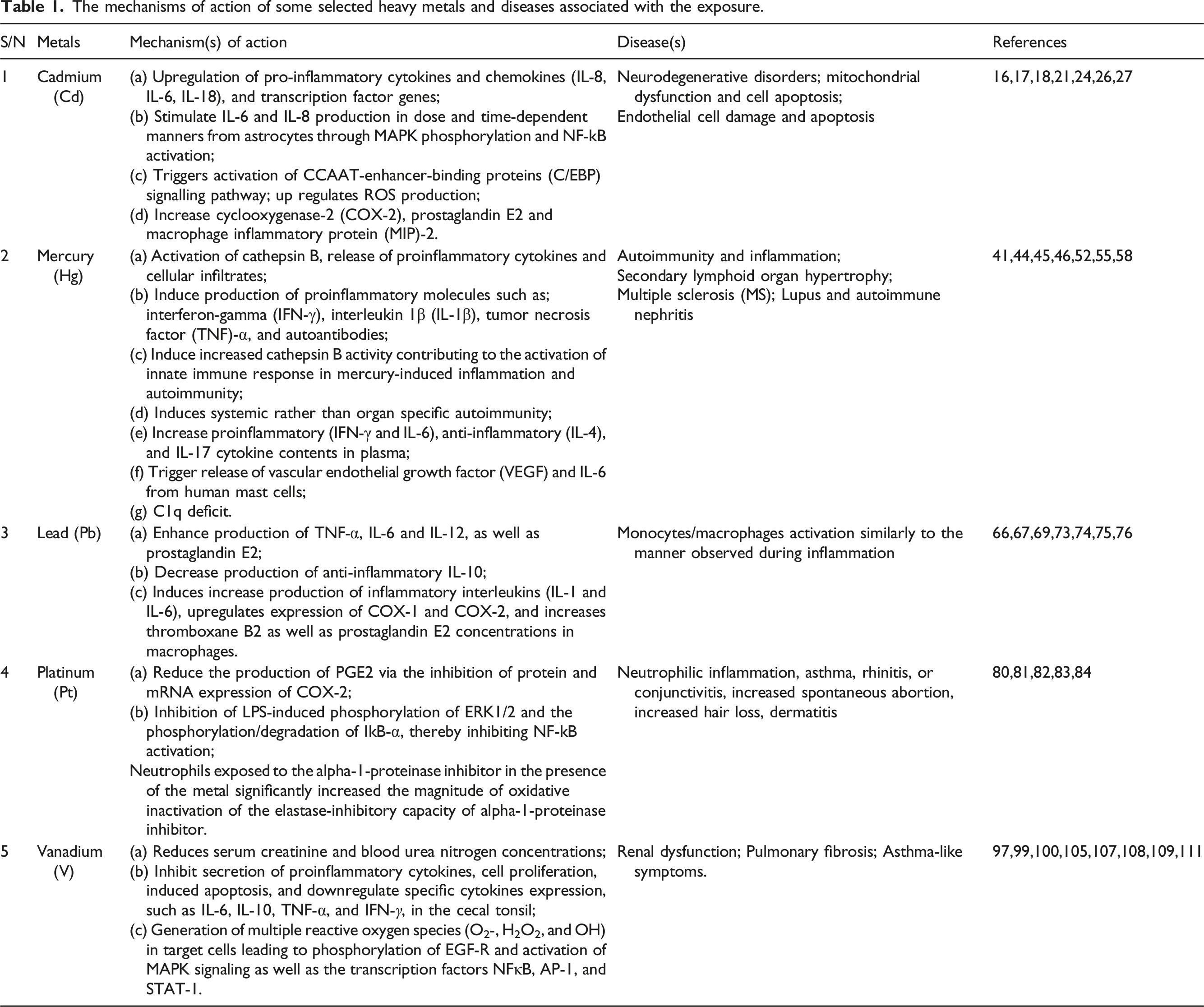
The mechanisms of action of some selected heavy metals and diseases associated with the exposure.

The effects of Lead, cadmium and Mercury exposure on the induction of autoimmunity and inflammation.
Cadmium
Exposure routes and toxicity of Cadmium
Eight common isotypes of cadmium exist as compound but never in elemental forms. 12 The main sources of cadmium (Cd) to the environment are the industrial and domestic environment. The industrial and domestic activities may include fossil fuels combustion (such as gasoline, coal, diesel etc.), industrial wastes incineration (Cd-containing plastics and batteries), metal alloy production, electroplating and manufacturing of phosphate fertilizer. Cadmium oxide (CdO), cadmium carbonate (CdCO3) and cadmium sulfide (CdS) are the widely abundant cadmium compounds. Improper handling of the metal itself, ingestion of contaminated food including shell-fish, inhalation of cigarette smoke (each cigarette containing 1–3 μg of Cd) and drinking of cadmium-contaminated water are the main exposure routes of cadmium to humans. 13
One of the most prevalent cadmium toxicity symptoms is impairment of the proximal convoluted tubule, which is thought to be linked to mitochondrial malfunction and, as a result, renal damage. 14 Cadmium exposure in children may cause osteoporosis. 15 While in adult it increases the risk of developing cardiovascular diseases.
The roles of Cadmium in inflammation and autoimmune disorders
Recent studies concluded that exposure to Cd could induce inflammation by one or more of the following proposed mechanisms; innate immune responses modulation in terms of susceptibility to microbial infections, cytokines and chemokine release and gene expression. 16 However, the exact mechanism through which Cd causes inflammatory reactions is unknown. Cd absorption by porcine epithelial cells causes upregulation of pro-inflammatory cytokines and chemokines (IL-8, IL-6, IL-18), as well as transcription factor genes in a time and concentration-dependent manner. 16 The study also showed that subtoxic quantities of cadmium from astrocytes stimulated IL-6 and IL-8 production in a dosage and time-dependent manner via mitogen-activated protein kinase (MAPK) phosphorylation and NF-kB activation, without impacting cell shape or viability. Inflammation is triggered by increased production of these pro-inflammatory cytokines, which may be linked to neurodegenerative illnesses. 16
Both increase in reactive oxygen species (ROS) and endoplasmic reticulum (ER) dysfunction could be induced by Cd. 16 In addition, activation of CCAAT-enhancer-binding proteins (C/EBP) signaling pathway can also be triggered by Cd exposure. 17 The expression of several genes involve in different cellular activity are altered including inflammatory reactions and oxidative stress. 18 Cd increases the generation of ROS, which causes mitochondrial malfunction and cell death. 17
Cyclooxygenase-2 (COX-2), prostaglandin E2 and macrophage inflammatory protein (MIP)-2 have been reported to increase due to Cd exposure.19,20 Mitogen-activated protein kinase pathway is proposed to be activated due to Cd exposure, resulting from the upregulation of the enzyme, COX-2. This elevated level of COX-2 eventually leads to the increased production of inflammatory PGE2 and cause endothelial cell damage and apoptosis.20,21 It has been successfully demonstrated that Cd exposure induces the activation of phosphatidylinositol 3-kinase (PI3K)/Akt signalling pathway and subsequently upregulates MIP-2 and COX-2 in macrophages.17,22
Another study on the epithelial cells of the throat and trachea demonstrates that cadmium induces Erk1/2 signaling pathway. 23 The activation of Erk, leads to over expression of proinflammatory mediators such as IL-8, suggesting respiratory airways inflammation occurring in an Erk1/2 dependent and NF-kB-independent manner due to Cd exposure.
Exposure to Cd has been implicated in some disorders such as osteoporosis, diabetes, and cancer.24,25 It is reported to have brought activation of numerous intracellular signaling pathways (especially NF-kB and AP-1) in micro molar (μ
Renal damage, osteoporosis in children, increased risk of developing cardiovascular illnesses, diabetes, cancer, and airway inflammation have all been linked to Cd exposure, either directly or indirectly. As a result, due control and care should be taken when handling foods, drinks, and other items that are suspected to be at the risk of Cd contaminated. This could only be accomplished by enacting sound public health policies regarding public smoking, which continues to put the environment and nonsmokers at risk of secondary smoke exposure.
Mercury
Mercury exposure routes and toxicity
Mercury is found in three fundamental forms: elemental (mercury[I] chloride, Hg2Cl2; mercury[II] chloride, HgCl2), organic (methylmercury, CH3Hg+; dimethylmercury, C2H6Hg; methylmercury chloride, CH3HgCl), and inorganic (mercuric sulphide, HgS; mercuric oxide, HgO). 32 Natural degassing of the earth’s crust give rise to the environmental distribution of mercury, large tons of mercury being released into the environment which occurs because of human activities. 33 The atmosphere is the precursor passageway of global transport of the pollutant (mercury vapor) and turns into water-soluble forms after a year and consequently release back to the earth surface. Micro-organisms, especially bacteria reconvert the water-soluble mercury to mono-methyl mercury compounds or mercury vapor, the later form of mercury can enter aquatic food chain via aquatic organisms such as plankton and fish. Other sources of mercury are categorized into occupational and environmental sources. The Chloralkali plants, production of lamps, and batteries and gold mining are examples of the occupational sources whereas dental amalgams and food are examples of the environmental sources. Humans get exposed through consumption of these animals, other routes of exposure include eye and skin contact. 33 Toxicity of mercury in children may include impaired neurological development and impaired nervous cognitive development. 34 Growth disorders, epilepsy, excessive salivation, deformity of limbs, chorea, body organ impairment, athetosis, dysarthria, damaged cerebellum, misalignment of the eyes and primitive reflexes, and the recently linked increased cases of type 2 diabetes are just some of the effects of mercury toxicity.35–38
The roles of Mercury in inflammation and autoimmune disorders
Recent studies on Mercury exposure in humans, has unveiled its effect on induction of autoimmunity and inflammation, by causing production of proinflammatory molecules, such as tumor necrosis factor (TNF)-α, interleukin 1β (IL-1β), interferon-gamma (IFN-γ), and autoantibodies.39–41 A substantial increase of our knowledge on Hg-dependent autoimmunity and inflammation is because of studies on murine models. 42
The mechanism of mercury-induced autoimmunity can be said to be initiated by development of an essential inflammatory reaction at the site of exposure; sequentially it starts with; activation and clonal expansion of T-cell dependent humoral response, hypertrophy of draining lymph nodes with new germinal centers, IgG production; that is generation of IgG ANAs, and deposition of immune complexes in glomeruli and blood vessels. 43 The inflammation that occurs after mercury exposure is thought to be caused by tissue damage, which leads to the expression of damage associated molecular patterns , which leads to the activation of innate immune sensors such as nucleic acid sensing TLRs, the production of inflammatory cytokines, and finally the induction of chronic inflammation. 44 Mercury-induced neutrophil extracellular trap (NET) activation and release (NETosis) as a source of TLR agonists supports this theory, 45 mercury modifies TLR expression, 46 and the requirement for TLR trafficking and signaling. 39 Mercury buildup in lysosomal compartments causes mercury-induced inflammation and autoimmune 47 and increased cathepsin B activity 48 contributes to the activation of immune response. 39 Chronic tissue injury and inflammation caused by mercury accumulation leads to secondary lymphoid organ enlargement and the creation of ectopic lymphoid structures, which are temporary structured clusters of lymphoid cells. 49 Worthy of note, those early inflammatory events including activation of cathepsin B, 48 release of proinflammatory cytokines 48 and cellular infiltrates 50 are linked to subsequent autoimmune responses. Similar to beryllium CBD, adaptive immunity mediated by mercury exposure also requires components of the innate immune system. 46 In contrast to beryllium, mercury exposure is however, an important distinction that induces systemic rather than organ specific autoimmunity. 51 By and large, the fundamental role of mercury in the induction of inflammation and autoimmunity is supported by these findings.
Accumulation of mercury in brain induces nerve cell damage and increases risk of developing multiple sclerosis (MS). 52 Specific loss of tolerance to self-antigens induced by mercury autoimmunity has been reported.53,54 In a study after exposure to subtoxic Hg doses; inflammatory reaction, specific antibodies to nucleolar antigens and glomerulonephritis with immunoglobulin deposits were suddenly observed in the susceptible mice. 55 Schmidt et al. 56 observed that susceptible MHC- class II genes are overexpressed, thereby mimicking the scenario seen in many autoimmune disorders.
Inflammatory pathways have also been shown to be valuable as biomarkers of Hg-stimulated autoimmunity, as evidenced by the up-regulation of proinflammatory cytokines reported in people following exposure. 57 Observations in rodents show this response as dependent on IFN-γ-associated genes. 40 Nyland et al. reported that exposure to Methyl mercury (MeHg+) could increase proinflammatory (IFN-γ, IL-17 and IL-6) and anti-inflammatory (IL-4) cytokine plasma contents. 43 According to the authors, alterations in serum cytokine profiles differed depending on the antinuclear autoantibody reaction. Low anti-inflammatory cytokines (IL-4) levels and low proinflammatory (TNF-α, IL-6, IL-1β, and IFN-γ) levels are associated with high antinuclear autoantibody levels in MeHg-exposure subset.43,58 These findings are in tandem with previous study which assessed human immune cell response to low Hg exposures in vitro,59,60 these findings are recently validated further by studies on proinflammatory cytokine responses to MeHg and Ethyl-Hg in vitro.57,61
In terms of human coexistence, the dispersion of mercury in the environment is a natural phenomenon. As a result, everyday human activities like gold mining, aquatic foods, Hg-containing cosmetics, chloralkali plant culture, and battery manufacturing must be regulated. Because even low levels of Hg can cause inflammation and autoimmunity, effective waste disposal systems should be readily available in regions where such activities are taking place. Because failing to do so predisposes individuals to a variety of health problems, including neurological defects in children, growth disorders, epilepsy, excessive salivation, limb deformity, chorea, body organ impairment, athetosis, dysarthria, damaged cerebellum, and type 2 diabetes, to name a few.
Lead
Exposure routes and toxicity of Lead
Inhalation and ingestion of lead contaminated particles through burning of gasoline (example of compounds are tetraethyl lead, C8H20Pb; and tetramethyl lead, C4H12Pb) and residential paint [example of compounds are lead carbonate, PbCO3; lead chromate, PbCrO4; lead monoxide, PbO; lead tetraoxide, Pb3O4; lead acetate; Pb(C2H3O2)2] are the main routes of lead exposure to humans. 62 However, Pb is also present in tobacco, and therefore, tobacco smokers are also at the risk of Pb exposure. 62 Pb toxicity is dependent on the severity of exposure; high levels of exposure in children are linked to decreased attention span, increased dullness, increased irritability, and a shorter attention span in the central nervous system, which can lead to headaches, seizures, coma, and even death. 63 The Agency for toxic substances and disease Registry’s has reported Pb to be the second most dangerous environmental poison. It is present in the environment in abundance, contributing 0.6% of the global burden of diseases. 64 Also worthy of mentioning that there is no lowest safe concentration exist for Pb. 65
The roles of Lead in inflammation and autoimmune disorders
Enhanced production of TNF-α, IL-6 and IL-12, as well as prostaglandin E2 as a result of Pb exposure at a concentration of 0.2–20 μ
Lead has been associated with negative effect to the immune system resulting to inflammation. Example is the effect of Pb on IL-2 cytokine, which is indispensable to proper growth, proliferation, and differentiation of T lymphocytes. Additionally, Pb also has negative effect on IL-4 cytokine, which acts extensively on B lymphocytes. Lavicoli et al.
69
looked into the effects of Pb on the levels of IL-2 and IL-4 in mouse blood serum, where they demonstrated that cytokine production can be reversed depending on blood Pb levels. Pb also has a negative effect on IL-8, which is a potent chemotactic agent for neutrophils,
70
and also associated with angiogenesis and metastasis.
71
Lin et al.
72
conducted a research where they stimulated human gastric adenocarcinoma cells (AGC cells) using 0.1 μM Pb(NO3)2, which induced activation of the IL-8 gene. Same group of researchers also earlier demonstrated that the promoter region of gene CXCL8 (gene responsible for encoding IL-8 protein) comprises of transcription factors, such as nuclear factor kappa B (NF-κB), activator protein 1 (AP-1), and nuclear factor for IL-8 expression (NF-IL6).
73
Therefore, Lin et al.,
73
and Hoffmann et al.,
74
decided to investigate these transcription factors (NF-κB and AP-1). Both confirmed that association of transcription factor AP-1 with the activation of the Pb-induced IL-8 gene, while minor role is contributed by NF-κB transcription factor. Furthermore, even a 0.1
Lead has no safe concentration as the second most harmful environmental poison. As a result, safety precautions such as wearing face masks and gloves when painting, mask when in a smoking area should be followed. Because Pb intoxication has been related to negative health effects in children, such as increased dullness, irritability, and a shorter attention span, which can lead to headaches, convulsions, coma, and even death.
Platinum
Exposure routes and toxicity of platinum
Sulfide and arsenide minerals are the main sources of platinum. Examples of some compound of platinum include cisplatin, cis-[Pt(NH3)2Cl2]; carboplatin, C6H12N2O4Pt; and oxaliplatin, C8H14N2O4Pt. Environmental platinum is mostly released from vehicle catalysts, therefore, urban cities and near highways tend to have high concentration of platinum. Exposure is mainly through inhalation, contact with eyes and or skin. 78 Toxicity of platinum compound is determined by its solubility; soluble platinum salt is more toxic than other compound with low solubility (oxides). Sensitization is the main health effect of platinum, manifesting as asthma, rhinitis, or conjunctivitis. 78
The roles of Platinum in inflammation and autoimmune disorders
Platinum is a member of the Platinum Group Metals, a group of elements that are also powerful allergens and sensitizers. They are also associated with nausea, asthma, increased spontaneous abortion, increased hair loss, dermatitis, and other health problems in humans. 79 However, there are contradicting studies regarding the roles of platinum in inflammation and autoimmune diseases. Cisplatin is a platinum-based type of chemotherapy used as cytotoxic agent in cancer chemotherapy. However, its role in inflammation has been linked to is ability to reduce the production of PGE2 via the inhibition of protein and mRNA expression of COX-2. 80 Nanoparticles are the newly introduced therapeutic approaches in cancer, inflammation, and aging research. Nano-Pt has recently been shown to have catalase mimetic activity for superoxide dismutase (SOD), which could be effective in the prevention of oxidative stress-related diseases such as inflammatory reactions and cellular transformation. 81 Platinum nanoparticle (Nano-Pt) has been implicated in the inhibition of LPS-induced phosphorylation of ERK1/2 and the phosphorylation/degradation of IkB-α, thereby inhibiting NF-KB activation. 82 The antioxidant and anti-tumor properties of nano-Pt have been documented. However, the mechanism by which they can cause inflammation has not been fully understood. 80 Platinum has been demonstrated to cause oxidative stress and inflammation, which are interconnected by amplification loops. 82 Pro-inflammatory cytokines may induce enormous production of free oxygen radicals which in turn may control the release of inflammatory mediators by triggering different transcription factors. 83
However, Theron et al. 2 demonstrated that Pt can potentiate the reactivity of human neutrophils and ciliated respiratory epithelial cells, as opposed to the generation of neutrophil-derived oxidants. They demonstrated that Pt pro-oxidative interactions with neutrophils have a potential health risk. In the study, activated neutrophils in the absence or presence of Pt was exposed to alpha-1-proteinase inhibitor, the major antagonist of neutrophil elastase. The extent of oxidative inactivation of the alpha-1-proteinase inhibitor’s elastase-inhibitory ability was dramatically increased in neutrophils exposed to the alpha-1-proteinase inhibitor in the presence of the metal. If these pro-oxidative interactions between Pt and neutrophils in the airways are functional in vivo, they may predispose people to pulmonary dysfunction, especially those who are occupationally and environmentally exposed. 2 Additionally, Schins et al. 84 also looked at the role of platinum in sino-nasal inflammation in people who were exposed to a lot of traffic and discovered that platinum levels in the nasal lavage were linked to neutrophilic inflammation.
People who live in metropolitan areas or near highways have a higher concentration of Pt in their nasal lavage. This puts patients at risk for rhinitis, conjunctivitis, nausea, asthma, spontaneous abortion, hair loss, and dermatitis among other things. As a result, precautions such as wearing a mask while driving are recommended, as are government policies that discourage highway occupancy. Although the concerns surrounding chemotherapy in cancer patient treatment are tough to examine, other possible remedies that do not contain the cisplatin chemical should be investigated in order to improve the odds of survival following chemotherapy.
Vanadium
Exposure routes and toxicity of Vanadium
Vanadium (V) does not exist as a metal in nature, but rather as a compound (ammonium vanadate, V; vanadium pentoxide, V2O5; oxytrichloride, VOCl3; oxytrifluotide, VOF3).
2
Several studies reported an incidence increase of innate and adaptive immune cells in numerous lung diseases (such as bronchitis, pneumonia, asthma).85,86 Grabowski et al.
87
reported that exposure of a much higher concentration of rat alveolar macrophages to a vanadium compound (sodium metavanadate) resulted in a general, increased dose-dependent (from 50 μ
Skin, on the other hand, does not appear to allow for a significant amount of Vanadium import. Dietary uptake is thus the main natural source for the body’s V supply, a very ineffective procedure because up to 99% of dietary V is normally eliminated with the feces. 90 Vanadate (H2VO - 4) is the dietary form of V, which is found mostly in drinking water. In the gastrointestinal system, H2VO - 4 is quickly absorbed. However, vanadate (V) is partially reduced in the stomach and precipitates in the intestines as VO(OH)2. Vanadium can also enter the bloodstream through injection or infusion, either on purpose when given intravenously (or intraperitoneally), or by mistake when present as a contaminant in infusion solutions. 91 Depending on the oxygen tension and the presence of redox-active substances, vanadium compounds entering the bloodstream via the gastrointestinal tract, the lungs, or infusion/injection are vulnerable to redox interconversion between VV and VIV. Transferrin is the primary transporter for both anionic vanadate (V) and cationic VO2+. 92 Transferrin (Tf) generates binary complexes (VOL-Tf) and (VOL′-Tf), in which L is a ligand that was originally coordinated to VO2+ and L′ is a low molecular mass (1 mm) ligand supplied by the bloodstream, such as lactate. 93 The VO2+ ion attaches to the same region in the protein as the Fe3+ ion. 90
Vanadium also has the combination ability with transferrin either in the form of vanadate-transferrin or vanadyl transferrin in blood. These can lead to further tissue damage (including liver). Additionally, tetravalent vanadium releases ferritin iron which stimulates vanadium-dependent lipid peroxidation. 94
The roles of Vanadium in inflammation and autoimmune disorders
The mechanism for vanadium induced pulmonary inflammation is hypothesized to be through generation of multiple ROS (O2-, H2O2, and ·OH) in target cells.95,96 Production of ROS relates to phosphorylation of EGF-R and activation of MAPK signaling96,97 as well as the transcription factors NFκB,98,99 AP-1,97,99 and STAT-1. 100 Additionally, vanadium is a phosphatase inhibitor 101 and therefore it will likely prolong phosphorylation and signaling along ROS-sensitive pathways. These series of events, in turn can affect synthesis and release of pro-inflammatory cytokines and chemokines inducing acute lung injury.100,102 Success recorded in pre-treatment of vanadium-induced inflammation of human bronchial epithelial cells with metal chelators has provided support to this hypothesis. The levels of vanadium-induced ROS, MAPK activation and cytokine release all subside. 103
Contrary to most heavy metals discussed above, there is significant evidence that showed anti-inflammatory activity of vanadium (V) both in vitro and in vivo studies. 103 For instance, Vanadium pentoxide (V2O5) is the most common commercial form of vanadium. It has been reported to inhibit secretion of proinflammatory cytokines, cell proliferation, induced apoptosis, and downregulate specific cytokines expression, such as IL-6, IL-10, TNF-α and IFN-γ, in the cecal tonsil.104,105 Furthermore, administration of vanadium has also been reported to ameliorate renal dysfunction by reducing serum creatinine and blood urea nitrogen concentrations. 106
Primates and rodents exposed to vanadium compounds particularly V2O5, has been reported to suffer from oxidative lung injury,107–109 with symptoms including coughing, wheezing, chest pain, bronchitis, and asthma-like symptoms. 110 Inhalation of V2O5 particles in primates, increases bronchoalveolar polymorphonuclear neutrophils (PMNs) and impaired pulmonary function, 111 and in rodents, it induces PMN influx, synthesis of proinflammatory mediators, as well as pulmonary fibrosis.102,112 Generally, occupational, or environmental exposure to vanadium has been associated with an increase in biological markers for oxidative DNA damage.113,114
Vanadium pentoxide should be used in a controlled manner to ensure a safe and healthy living environment. To combat the increased rates of bronchitis and asthma, which are thought to be indirectly linked, a safe waste disposal method for the chemical should be investigated and made available to the public.
Conclusion
Although the exact mechanisms of heavy metal induced autoimmunity in not fully understood, valuable investigations show that heavy metal toxicity might dramatically affect immune system in terms of auto-inflammatory responses, immune cell activation, and autoantibodies production. These metals induce autoreactive responses through activation of inflammatory signaling pathways and expression of inflammatory cytokines and chemokines. These events alter immune cell activation and finally cause loss of the immune tolerance and autoimmunity. It seems that heavy metal toxicity could be a major public health problem. Therefore, there is the need to intensify research on heavy metal toxicity and how to ensure a safe and conducive environmental atmosphere. This can only be achieved through public awareness of the harmful tendencies of our daily activities and day-to-day consumables such as residential paints, batteries, cd-containing plastics, phosphate fertilizers, aquatic animals such as fish, vehicle catalysts, etc., as well as a much more robust healthcare policy that prioritizes and ensures environmental protection.
Footnotes
Ethics approval
The study was reviewed and approved by the ethics committee of Alborz University of Medical Sciences, Karaj, Iran (Code No: IR. ABZUMS.REC.1400.320).
Declaration of conflicting interests
The author(s) declared no potential conflicts of interest with respect to the research, authorship, and/or publication of this article.
Funding
The author(s) received no financial support for the research, authorship, and/or publication of this article.