
Abstract
A computed tomography dose index can be used to quantify the radiation dose received during a CT scan and it is an indicator of the radiation dose to the polymetaylenmetaAcrylate (PMMA) standardized phantom. The objective of this study was 2-fold. The first was to measure the computed tomography (CT) radiation dose for the head and body polymetaylelenmetaAcrylate (PMMA) phantoms and to determine the accuracy of the CT radiation dose parameter displayed on the CT scanner console; these were measured in this investigation and compared with the dose displayed on the CT scanner console. The dose was calculated using the formalism described in the American Association of Physics in Medicine (AAPM) Report 96. The second was to compare the dosimetric results of the head and body polymetaylelenmetaAcrylate (PMMA) phantoms with dose reference levels published in international journals, as well as to measure the central cumulative dose (DL′ (0)), as recommended by the American Association of Physics in Medicine (AAPM) report 111. This is a new, cutting-edge methodology for estimating the CT radiation dosage provided by the abdomen, thorax, and head of a PMMA phantom. We used a Philips Big Bore CT scanner with 16 slices. A CT dosimeter head phantom with a diameter of 16 cm, a CT dosimeter body phantom with a diameter of 32 cm, a 100 mm pencil chamber (PC), and a 20 mm short chamber (SC) were employed. These were coupled to an electrometer and a dosimetric readout device. The measured volume computed tomography dose index (CTDIvol) values were in good agreement with the CT radiation dose displayed on the corresponding CT scanner console. The percentage disagreement was less than 10%, with a maximal difference of 1.7% and 5.5% for the body and head phantom, respectively. The central cumulative dose (DL (0)) measurements (for L′ = 100 mm) also matched nominal or the corresponding computed tomography dose index (CT) scanner console volume computed tomography dose index (CTDIvol) values. In this case, the agreement is always below 3% for abdomen scans and 1.0% for head examinations. This result implies that the radiation dose supplied by the 16-slice computed tomography (CT) system was in good agreement with the international dose reference level and we observed something different.
Keywords
Introduction
Computed tomography (CT) is a highly effective tool used by radiologists to detect illness in the human body. It was introduced in the early 1970s and was the first computer-based medical imaging modality.
1
The computed tomography dose index (CTDI) is used to calculate the radiation dosage during a CT scan.
2
The CTDI is measured in the radiation dose of the X-ray source using a pencil chamber (PC) with a length of L = 100 mm. It is defined as follows:
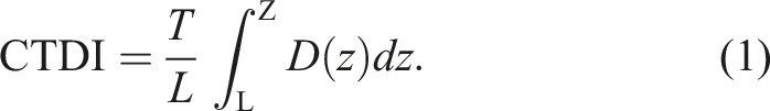
The dose profile in the crania-caudal direction is denoted by −D (z). 1
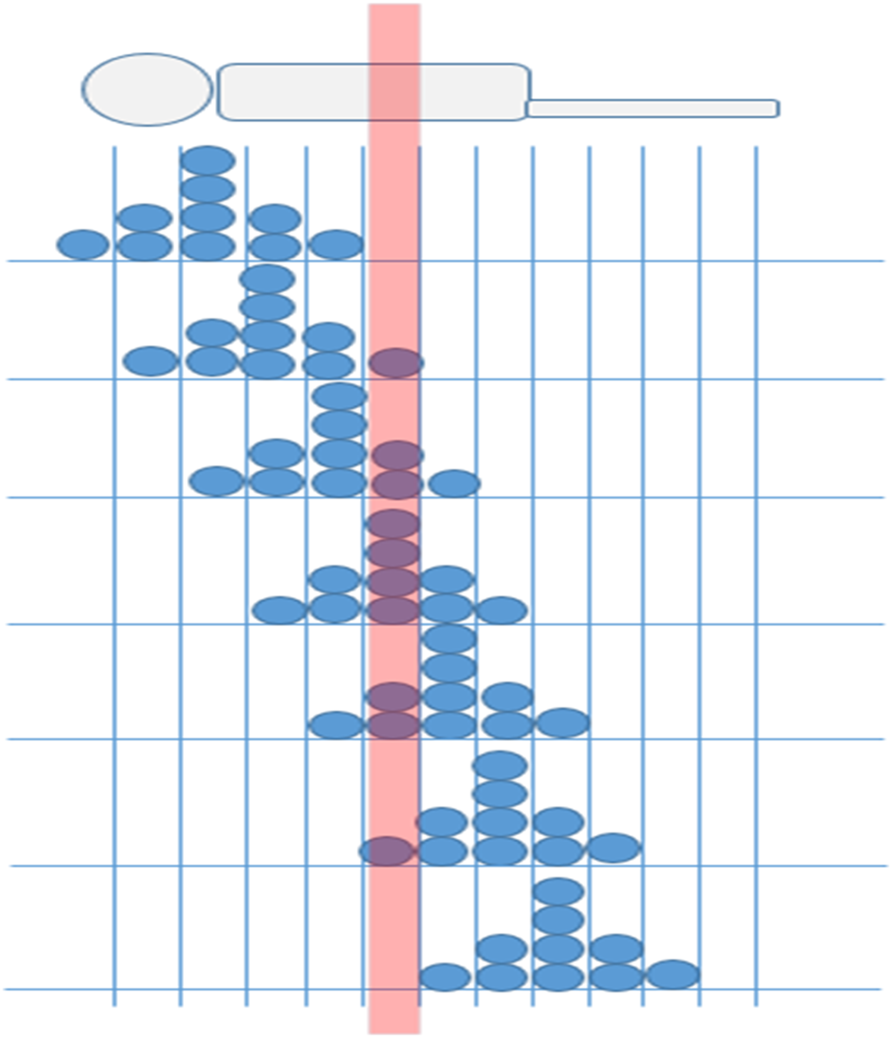
The general irradiation of a patient undergoing sequential computed tomography dose index (CT) examination to illustrate the CTDI approach in the patient dosimeter is shown in Figure 1,
3
where each circle represents a unitary dose expressed in arbitrary units. The dose absorbed in the patient slice of interest (highlighted in the figure) is given by the five central CT irradiations in the figure. The figure also shows that the first and final irradiations (placed at a distance from the slice of interest) do not contribute to the dose in this anatomical region. Consequently, the dose in the patient slice of interest is given by adding the contributions in the slice of interest (highlighted in the figure). It is equal to 10 in arbitrary units (Figure 1). The typical dose profile in a sequential computed tomography (CT) examination is moving along the z-axis. The dose in the slice of interest is calculated by adding the contributions in the slice.
4
The relationship between the CTDI and the dose in the patient slice is defined in equation (1). For simplicity, the patient slice was assumed to be 10 mm (x-ray) slice thickness (T = 10 mm). As shown in Figure 2, the pencil chamber (PC) averages Rx (X-ray) contributions to the dose over its length, L. As a result, in Figure 2, a final reading M = 1 in arbitrary units was obtained. The pencil chamber (PC) reading averages the Rx (X-ray) contributions over its length, L.
This reading M is also expressed by the relation
Equation (2) yields a CTDI of 10 (au) for L = 100 mm and T = 10 mm. This is the same value of the dose absorbed in the patient slice obtained above by adding the dose contributions as shown in Figure 1. This numerical equivalence is crucial because it indicates that the CTDI value of a clinical CT scan is a dosimetric indicator estimating the dose in a patient slice. The weighted CTDI is the average dose over the central slice of a series of contiguous slices obtained during a head or body CT examination.5,6 The CTDI measured using dosimetric phantoms is also referred to as the weighted computed tomography dose index (CTDIw). It is given by the following equation:

For helical CT examinations, the parameter estimating the dose in a patient slice is the CTDIvol.
7
It is defined as follows:

The central cumulative dose DL′ (0) was introduced by the new modern American Association of Physics in Medicine (AAPM) to address the shortcomings of the conventional CTDI-based dosimetric approach.
8
This dosimetric indicator is defined as the dose in a patient slice caused by a CT examination with a scan length of L′. The method is highly practical because it allows for direct measurements of all clinical helical scans using short camber (SC) in the typical dosimetric head and body PMMA phantoms. DL′ (0) is the equilibrium dose (Deq) that is comparable to the CTDIvol for a scan length of L′ = 100 mm.4,9 utilizing dose measurements obtained from the phantom’s central hole (Deq, c) and those obtained from the phantom’s peripheral hole (Deq, p)1,10 DL′ (0) c=is refers to the DL′ (0) measured in the central position of the dosimetric phantom and DL′ (0)p = is the mean value of DL′ (0) measured in the peripheral locations of the same phantom.
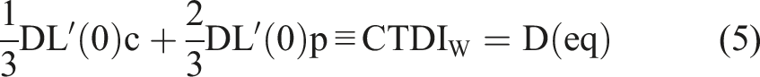
Equation (5) explains one of the advantages of the American Association of Physics in Medicine (AAPM) dosimetric approach. It enables the assessment of (CTDIvol) by performing central cumulative dose (DL′ (0)) measurements for L′ = 100 mm on clinical scans. 10 The purpose of the present study was to estimate the CT dose index for head and body PMMA phantoms and to determine the accuracy of the CT radiation dose parameter displayed on the CT scanner console. 9 The CT dose index was estimated using the American Association of Physics in Medicine (AAPM) Report 96 formalism. The study was performed on a Philips 16-slice CT scanner with a 100 mm pencil ionization chamber. The measured body and head phantom doses were compared to selected international dose reference levels and varying deviations were observed. The role of the medical physicist in this study is to track and measure the dose to patients using appropriate indicators.11,12
Materials and Methods
The measurements were performed on a Philips Big Bore 16-slice CT system installed at the Radiotherapy Department of the S. Chiara Hospital, Trento, Italy. The study involved the use of a standard CTDI head phantom, a standard CTDI body phantom, and a Radical 3CT pencil chamber (PC) with a length of 100 mm and a short 10 × 5 ionization chamber (SC) with a charge collection length of 20 mm. Both the chambers were coupled with a Radical 9010 electrometer (Figure 3). Set-up for CTDI determination.
The characterization of the computed tomography (CT) scanner involved preliminary measurements of the CTDI in free air, and the PC (pencil chamber) was placed at the CT center. The selected scan parameters of the series of single-rotation CT protocols were 90-120-140 kV, 100-200-400 mAs, and a slice thickness ranging from 3 mm to 24 mm.
The second series of measurements assessed the CTDIvol with head and body phantoms. These provided a direct comparison between measured CTDIvol values and the nominal value displayed on the CT scanner console and compared international dose reference levels.4,13-15 The scan parameters for both types of phantom head and body were 120 kV, 100 mA, and a slice thickness of 24 mm.
The final dosimetric evaluations involved the measurement of the central cumulative dose DL′ (0) of the helical head, abdomen, and thorax scans with a scan length L′ of 100 mm and different scan parameters. In these conditions, we obtained DL (0) = CTDIvol. Therefore, we estimated equation (5). DL′ (0) was then compared with the corresponding nominal volume CTDIvol and to the CTDIvol international reference levels. The CT dosimetric values were estimated in the American Association of Physics in Medicine (AAPM) report 96. 3
Results
Normalized Measured CTDI in free air for Different Voltages (kV), Tube Charges (mAs), and X-Ray Slice Collimations (mm).

This result increased CTDI values and the Rx (X-ray) slice thickness will be decrease as shown in Figure 4. Normalized computed tomography dose index (CTDI) in free air (mGy) for different Rx (x-ray) slice thicknesses.
Percentage Disagreement Between CTDIvol Measurements And The Reported Result Of The Corresponding Dose Displayed on the console (Nominal CTDIvol on the CT Console).

Comparison of the Measured Central Cumulative Dose (DL′(0)) to the Dose Displayed on the Console.

Comparison of the Dosimetric Results of the Head and Body Phantom with International Dose Reference Levels.

Discussion
The American Association of Physics in Medicine (AAPM) Report 96 formalism and the comprehensive methodology for the evaluation of radiation dose in the CT report of the American Association of Physics in Medicine (AAPM) task group 111 were used to evaluate CT radiation dose exposures.3,8 The CTDI value was calculated in free air at tube potential (90.120, 140) kV, tube current (100, 200, and 400) mAs and X-ray slice thickness (3, 6, 12, and 24) mm were estimated in this study. The X-ray slice thickness increases the radiation dose for free air decrees and vice versa. The present study was to estimate the CTDI for head and body phantoms and to determine the accuracy of the CT radiation dose parameter displayed on the CT scanner console using a 16-slice CT scanner, short ionization chamber, and pencil ionization chamber. The result of the CTDI in the present study was a good agreement for the corresponding CT scanner console value and also the selected international does reference level and varying deviations were observed. The measured CT radiation dose for head and body phantoms was less than the international dose reference level. We advised radiologists to utilize there must be carefully selection of technical parameter of CT scanner that control exposure of patient radiation dose and regular checking of scanner performance with measurement of the CT dose index parameter. The purpose of the present study was computed tomography. The CT dose measurement is a very important measurement in the acceptance of any CT scanner after installation. The CT radiation dose parameter is accepted.
Conclusions
Computed tomography (CT) is a highly effective tool used by radiologists to detect illness inside the human body and deliver a high dose to patients compared with other imaging modalities. The CT radiation dose reported by the computed tomography console during a CT scan examination is based on the CTDI stated in reports by the American Association of Physics in Medicine (AAPM). The CTDI is a standardized measure of the dose output of the CT system. The present study showed a deviation of 1.7
Footnotes
Acknowledgments
The author expresses gratitude to the International Center for Theoretical Physics (ICTP), Addis Ababa University’s Physics Department, and Wachumo University for their assistance.
Declaration Conflict of Interest
The author(s) declared no potential conflicts of interest with respect to the research, authorship, and/or publication of this article.
Funding
The author(s) received no financial support for the research, authorship, and/or publication of this article.
Ethical Approval
This article does not report any studies performed on animals.