
Abstract
Quantification of scattered photons in addition to unscattered primary particles, under realistic exposure scenario, is best dealt with a parameter called “Buildup factor”. The aim of this work is to simulate the transmission buildup factor (BUF) of gamma-ray in the energy range .15–15 MeV for 20 human tissues and organs using the Geant4 (version 10.5) Monte Carlo simulation followed by a geometrical progression (GP) parameterization procedure. Firstly, we verified the accuracy of Geant4 ability to predict the effective transmitted dose according to published data. Also, a comparison of simulated BUF for different geometrical configurations was carried out for some tissues and source energies. Then, we checked out the linear dependency of the K parameter (BUF is function of K) function of mean free path (mfp). Finally, we developed a fitting procedure according to GP method for BUF corresponding to 20 tissues and organs and different mfp (from 1 to 8) for energy range .15–15 MeV. We found a good agreement with previous published data. Proper comprehension of BUF for tissues leads to carefully controlling the energy absorption in the human body. Consequently, provided BUF could be of great interest for estimating safe dose levels in medical imaging and radiation therapy.
Introduction
Nowadays, gamma-ray is commonly used for medical imaging and radiation treatment over the world. 1 Thus, the ultimate need to accurately investigate and to enhance our knowledge on radiation protection is obvious. Especially, the proper knowledge of the interaction of ionizing radiation with human tissues and organs is necessary to avoid unsafe outcomes. For studying such interaction, the buildup factor (BUF) was introduced to improve the prediction of transmitted beam, through a given medium, for realistic cases, using the modified Beer-Lambert law.2,3 Such factor estimates the contribution of primary and scattered photon beam along its attenuation within a given barrier.
Tissue equivalent materials respond to incident ionizing radiation in the same manner as human tissues. These phantoms are commonly used in medical applications including radiation therapy, diagnostic radiology, radiation protection, and radiobiology for calibration purposes and depth‐dose estimation. On the other hand, there are 3 main categories of BUFs: absorbed dose, exposure, and dose-equivalent buildup factors, which are based on energy absorption, kerma, and equivalent dose responses, respectively. Hence, the importance of BUF factors for biological tissues is needed.
Gamma-ray BUF accurately measured is not always allowed. Therefore, studies of gamma-ray buildup factors were carried out using Monte Carlo simulation,4,5 invariant embedding, 6 geometric progression (GP) fitting,7-9 and other methods.
However, on the basis of our knowledge and the available literature, there is no more precise dataset of buildup factors than those tabulated by ANSI/ANS-6.4.3-1991. 10 This dataset provides five GP fitting parameters of BUF for 23 elements and three compounds in the energy range of .015–15 MeV at penetration depths up to 40 mean free paths (mfp). Although, generally, proposed data concern the specific case of unidirectional parallel beam impinging on planar stratified shielding and a correction factor was given to take into account spherical geometries. Moreover, there is no data base including BUF values directly derived from Monte Carlo simulations for all human body corresponding to isotropic point source with stratified spherical layers medium, which can be considered valuable to the scientific research community, as it could be easily included into point kernel-based programs. Furthermore, the Geometry ANd Tracking version (Geant4) Monte Carlo toolkit11,12 has not been used to fully simulate buildup factors, except for recently published work in which the mass energy attenuation coefficients were simulated and used with the GP parameters 2 and our previous work attempt. 4 In order to address previous enumerated questions, we provide a BUF database and its corresponding parameterization for photon point isotropic sources and realistic penetration through selected materials. At our knowledge, this is the first time using the Geant4 Monte Carlo simulation to fully compute the buildup factors for 20 human body tissues followed by parametric search of the well-known geometric progression formula and thus providing necessary materials for point kernel calculation applied to the High-Definition Reference Korean-Man (HDRK-Man).13-15 Our previous work mainly focused on using GP parameters to compute BUFs, for all human tissues and organs, with a simple verification of Geant4 ability to predict the exposure buildup factor of water for a photon energy of .1 MeV and penetration depths up to 10 mfp. 4 Also, the work carried out by Jarrah et al. 16 focused on the energy absorption buildup factor simulations of some ICRU tissue-like using MCNP5 code. Moreover, Kurudirek et al. 8 estimated the energy absorption buildup factors of some human tissues using GP parameters with a benchmark of MCNP6.1 simulation of EABF for soft tissue and water at .662, 1.173, and 1.25 MeV photon source, against GP fitting method. Also, the work carried out by Rafiei et al. 17 simulated the EABF for water and some tissue-equivalent materials for 356, 662, 1173, and 1332 keV gamma-ray energy up to depths of 10 mfp, using MCNPX code. Hence, this work can be considered as a continuation to those efforts, as we used Geant4 simulations of BUF for all HDRK-man tissues and the proposition of an in-house GP-like fitting method.
Our goal can be summarized as follows: (i) benchmark of Geant4 against EGS4 results for a given case study, (ii) perform BUF simulations, and (iii) search and propose of GP fitting parameters. Thus, after checking the ability of our computation to reproduce the transmission beam through Pb material previously published by Kato et al., 18 we carried out the simulation of BUF for 3 standard photon energies .15, 1.5, and 15 MeV for some selected penetration depths (1–8 mfp, with a step of 1mfp). For each case study, we adopted and isotropic point source located at the center of 10 mfp sphere layered into 9 shells (the outer one has a thickness of 2 mfp to minimize backscattering effects). Then, we started by checking the linear behavior of the geometric progression factor function of mfp and terminated by a mathematical minimization procedure to search for the other GP fitting parameters. Finally, a verification of the ability of the fitting procedure to reproduce directly simulated data was carried out. The results of this study can be considered as an enhancement, to the large community of radiation physics researchers, of current point kernel-based techniques used to estimate the transmitted dose other than the dose distribution mapping for radiotherapy treatment planning and radiation diagnosis purposes based on the energy range and the medium thickness investigated.
As this study focused on simulating (Geant4) and computing (C++ fitting procedure) BUFs for HDRK-man tissue-like at only three photon energies of .15, 1.5, and 15MeV and for penetration depth up to only 8mfp, a straightforward extension of this work to cover energy and depth ranges, to be consistent with ANS standard tables, is feasible unless having the appropriate computing facilities (parallel works, large CPU workstations).
Methods
Elemental composition (%), Atomic Density (g/cm3) and Equivalent Atomic Number (Zeq), at Different Energies in MeV, for Studied HDRK-Man Organs/Tissues (* stand for organ contents). RBM Means Red Bone Marrow.

After showing a brief introduction of BUF theoretical basics, we will describe the simulation and the derived fitting procedures carried out, as follows:
BUF Theoretical Frame
The effective transmitted dose, for an incident photon with energy E, through an absorber medium with x thickness in cm, is defined as the ratio of the kerma measured in presence of the absorber, 

Also, we can rewrite T(x,E) in the following way
Thus, using previous equations, we can deduce BUF value for a given E and x as follows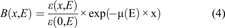
Simulation Procedure
Mainly managed and developed by the CERN organization (European Center for Nuclear Research), the versatile Monte Carlo simulation platform Geant4 has well known and verified capabilities able to mimic the transport of the majority of particle types (photons electron, ion, neutron, short-lived particle, etc…), covering large range of particle source energy (from eV to TeV) and modeling complicated and realistic setup configurations. It was used to compute mass attenuation coefficients leading to indirectly benchmark ANSI data based on calculated GP fitting parameters. 25 In our previous work, 4 we attempted to initiate a direct computation of buildup factors of water medium. Here, we advanced such computation for 20 human tissues and organs of the HDRK-man anthropomorphic phantom. Moreover, Table 1 illustrates their density and elemental composition as fully described elsewhere.4,13 During this work, Geant4 version 10.5 was used to simulate the transport of gamma rays isotropically emitted from a point source through a given medium. The geometry of the problem consists of a monoenergetic point source located at the center of ten concentric spheres, separated by one mfp for each step. As, we were limited to only eight mfp thickness, for all studied media and three photon energy of .15, 1.5, and 15 MeV and in order to minimize the boundary effect, we added an outer sphere with two mfp for each case study. We tracked gamma rays through the setup to compute the photon flux crossing each shell/surface of the medium. Then, to be more consistent, we internally (within SteppingAction class) converted each crossed photon energy into kerma using the equation (2) and based on the Log-Log interpolations of k(E) and μ(E), as described above. Such procedure was repeated with replacing the medium with air in order to accomplish the ratio formula given by equation (4). Finally, we can determine the overall BUF database for all studied cases including data extracted (ε(x,E) and ε(0,E)) numbered as 20 × 3 × 8 × 2 = 960 values. In this study, the buildup factor was computed for 109 gamma rays by run. We activated all the physical processes for electrons and photons (Photoelectric, Compton, Pair production effects for photon and Ionization, Bremsstrahlung for electron). We used the G4EmStandardPhysics_option3 built-in physics library with a cutoff of 1 keV for electrons and photons. The overall statistical uncertainty does not exceed 1 % for all runs.
There are three geometrical configurations used for this work. As seen in Figure 1, the left one describes the setup used to verify again our simulation regarding to the data found by EGS4 code
18
to compute the effective transmitted dose through different thickness of lead of photon with different energies. Also, a visualization of the actual BUF simulation scenario by Geant4 is shown in the middle of Figure 1. Finally, the right part of Figure 1 shows the monodirectional parallel-plane source impinging on stratified shields, used for comparison purposes. Visualization of Geant4 simulated setup using HepRAPP
26
package for different typical geometries used to acquire transmitted beam: (left) isotropic point source in planar configuration; (middle) isotropic point source in stratified spherical layers; (right): parallel unidirectional plane beam in planar shielding.
Fitting Procedure
According to the GP fitting method, we have
X is expressed in mean free paths: 
As described by Harima,7,27 for X ≤ X
k
we can limit our study to the first part including the linear behavior of Log(K) as a function of Log(x), as we have
Moreover, as we limited our study to penetrations up to 8 mfp (medical applications) and due to the fact that the second term of the right side of equation (6) was included in order to take into account the Log(K) deviation from linearity beyond 15 mfp up to about 40 mfp with a tangent-hyperbolic function, we are concerned with only 3 fitting parameters (a, b, and c) along this procedure. Therefore, the buildup factor for a given isotropic point source located at the center of spherical material geometry will be fitted using the following formula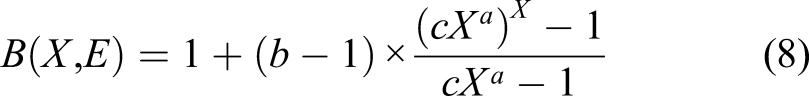
Thus, our main goal is focused on searching the 3 fitting parameters: a, b, and c, for 20 human tissues and organs, in spherical geometry setup and for isotropic point source of gamma-ray with energy: .15, 1.5, and 15 MeV.
Results and Discussion
Geant4 Verification
Figure 2, shows a global good agreement between actual results and previously published data by Kato et al.
18
for the effective transmission dose through 1, 2, and 3 mm Pb shield thickness, as function of photon source energy. Also, the effect of 88 keV transmission corresponding to the characteristic peak of lead, is clearly seen, as we have the corresponding linear attenuation coefficient of lead material at 87.9 and 88.1 keV of 21.7 and 87.2 cm−1, respectively. Geant4 simulated effective transmitted dose through lead material against EGS4
18
data for different thickness of 1, 2, and 3 mm.
Furthermore, we confirmed previous conclusion conducted by Rasouli et al. and Lin et al.28,29 claiming that the effect of changing the geometry setup from parallel plane to spherical layer and the source from unidirectional plane parallel beam to isotropic point source can be explained by the boundary effect.
Figure 3, shows a large deviation decreasing from .15 to 15 MeV photon energy for tissues having low, medium, and high equivalent atomic number (Lung, Adipose, and Bone) as function of mfp. Such decrease can be explained by the close behavior of both setups at large thickness, as the mfp becomes larger at higher photon energy. Also, the effect of Zeq, for each energy, on such deviation as function of penetration, is seen resulting to the variation of bremsstrahlung and multiple scattering effects. Difference of BUF simulated using Geant4, for isotropic source and spherical layers (extracted from ANSI/ANS-6.4.310,30) and for parallel beam and parallel sheets, for Bone, Adipose, and Lung tissues for .15, 1.5, and 15 MeV photon source function of mean free path. (Delta(%) = 100*(1-Parallel/Isotropic)).
Fitting Parameters
Deducing of the three fitting parameters from tabulated BUF was carried out using an in-house C++ program able to proceed with the multidimensional nonlinear fitting opportunity of the GNU Scientific Library (GSL).
31
Moreover, Figure 4 confirms the hypothesis of the linear dependency of the K parameter function of mfp, for the range in concern. From the same figure, we can see that such parameter continues to be more and more unique for all tissues and organs when the energy increased, which is obvious. Linear dependency of the K factor function of mfp for Bone, Adipose, and Lung tissues for .15, 1.5, and 15 MeV isotropic spherical photon source.
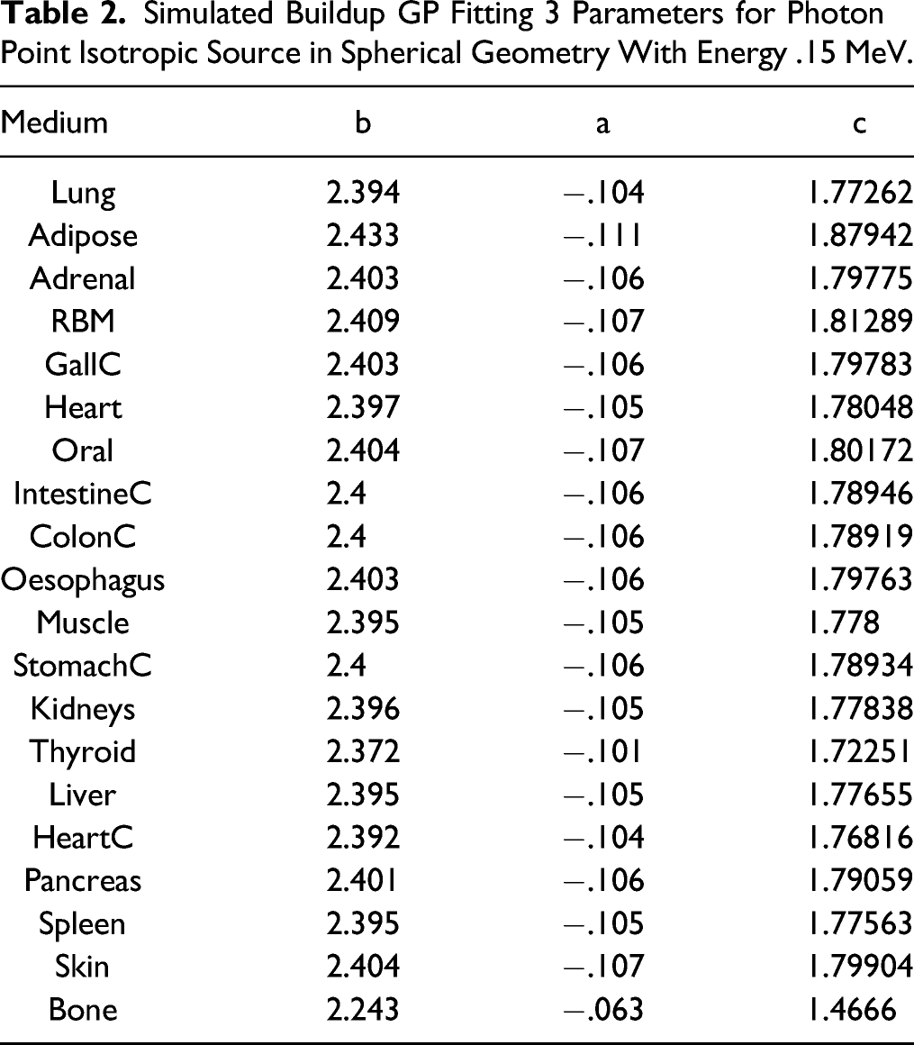
Simulated Buildup GP Fitting 3 Parameters for Photon Point Isotropic Source in Spherical Geometry With Energy .15 MeV.
Simulated buildup GP fitting 3 Parameters for Photon Point Isotropic Source in Spherical Geometry With Energy 1.5 MeV.

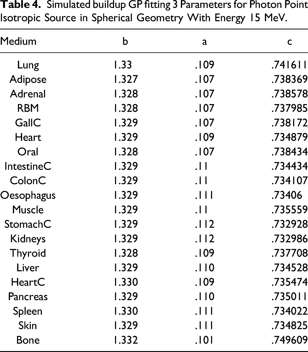
Simulated buildup GP fitting 3 Parameters for Photon Point Isotropic Source in Spherical Geometry With Energy 15 MeV.
In order to check the fitting procedure, we plotted, as an example, fitted and directly simulated BUF for Heart organ. As an overall deviation of BUF fitted to directly simulated values, for all studied cases, we found an average value of .5 ± .6 % (Figure 5). Simulated and fitted BUF for Heart tissue for .15, 1.5, and 15 MeV isotropic spherical photon source.
Finally, we can consider that the actually provided parameters database can be of great interest to medical physicist community, however, we were limited to only a thickness of eight mfp and photon particles and energy no more than 15 MeV. Also, our next work will be focused on the simulations of BUF for spherical multilayers.
Buildup Correction Factor For Plane Parallel and Plane Isotropic Distribution Accordingly to Isotropic Point Source for Water Medium.
Conclusion
Transmission photon buildup factor for 20 human tissues and organs has been simulated by Geant4 Monte Carlo toolkit for point isotropic source included within concentric spherical layers with different thickness. Covered photon source energy and thickness range were .15–15 MeV and 1–8 mfp, respectively. Among the analysis of simulated BUF values, it was verified that the K parameter behaves linearly as function of the thickness (in Log-Log scale) allowing us to derive a 3 fitting parameters according to the GP method for all studied cases. Such parameterization accurately reproduced the directly simulated BUF with an error less than 1 %. Nevertheless, the current study is limited to previous studied cases, the straight forwardly extension to other particles (neutron, beta, alpha…) and other source geometry and energy can be carried out. It is the main goal of this work to propose a BUF data base able to improve the accuracy of practical tools for medical imaging and treatment planning based on point kernel codes.
Footnotes
Declaration of Conflicting Interests
The author(s) declared no potential conflicts of interest with respect to the research, authorship, and/or publication of this article.
Funding
The author(s) disclosed receipt of the following financial support for the research, authorship, and/or publication of this article: This work was supported by the authors extend their appreciation to the College of Applied Medical Sciences Research Center and the Deanship of Scientific Research at King Saud University, Saudi Arabia.