
Abstract
Selenium is a fundamental trace element of the living system. Microorganisms play a crucial role in the selenium cycle, both in the environment and in life. Biogenic selenium nanoparticles have shown promising prospects for use in medicine as an antioxidant and anticancer agent. In this study, SeNPs were biosynthesized by Penicillium citrinum. The spore suspension which was previously prepared was exposed to different doses of gamma radiation (10, 20, 30, 50, and 60 Gy). SeNPs were then produced by an irradiated P citrinum. UV spectroscopy, transmission electron microscopy, X-ray diffraction, and GSH content were assayed to evaluate the probability of P citrinum synthesizing SeNPs. In conclusion, irradiation of P citrinum by gamma ray enhances the mycosynthesis of SeNPs.
Introduction
Nanotechnology is an evolving technology with a broad variety of uses involving the synthesis and processing of nanomaterials (1-100 nm). Recently, there has been a great deal of focus on nanoparticle synthesis methods, including chemical, physical, and biological methods.1,2,3 There is a growing use of biological approaches for nanomaterial synthesis, involving high-yield, non-toxic, low-cost, eco-friendly, and biocompatible.2,4,5
Selenium (Se) is an important trace element for the proper and healthy life of humans, animals, bacteria, and other living systems and has an irregular distribution in the earth’s crust. 6 Selenium is well known today to play a crucial role in a variety of physiological functions in different organisms. Some of the methods used to synthesize SeNPs are physical and chemical processes. Biogenic synthesis for the preparation of SeNPs is an interesting and exciting process which is non-toxic, clean, and eco-friendly. It also has additional benefits over chemical methods, such as higher efficiency and lower costs. 7
There are many attempts to synthesize SeNPs from microorganisms such as bacteria, fungi, 8 and yeast. 9 Furthermore, few fungi can synthesize SeNPs such as Alternaria alternata 10 and Aspergillus terreus, 8 whereas Fusarium oxysporum can generate SeNPs with an average size of 78 nm. 11 Many fungi have the advantage of producing very high yields of secreted proteins, which can increase the rate of nanoparticle synthesis because they have mycelia, which has a much larger surface area than bacteria and can be used to facilitate the interaction of metal ions and fungal-reducing agents. 12
Gamma radiation is a short wave of high-energy electromagnetic radiation; it may trigger certain mutations. Tallentire 13 found that exposure of microbial cells to ionizing radiation caused a series of reactions leading to physiological changes and antimicrobial activity. These modifications are based on the absorbed dose. Thus, radiation adds additional stress to the cells, which tends to interrupt their organization.
As a result of this biochemical disruption, a wide range of changes can be observed in irradiated cells, some of which are transient and some are permanent. The specific dose of radiation is determined by changes in cell’s radiosensitivity or radioresistance. Approved by this result, the physically induced mutations of the Aspergillus terrus strain isolated and identified from Saudi Arabia were examined by exposing the conidia of this fungus to various doses of gamma rays, resulting in autotrophic and color mutants of conidia A. terrus. 14 Haggag and Mohamed 15 also concluded that induced mutagenesis using gamma rays was shown to be effective in enhancing the production of antifungal metabolites in 3 Tichoderma species. SeNP synthesis by gamma-irradiated Bacillus laterosporus cells at different radiation doses was reported by El-Batal et al. 16
Low doses and low dose rates of ionizing radiation have been shown to stimulate the induction of antioxidant defense systems such as glutathione (GSH), glutathione peroxidase (GSH-Px), superoxide dismutase (SOD), and catalase (CAT).17,18 Thus, the present investigation aims at studying the possible synthesis and characterization of SeNPs through mutating Penicillium citrinum by gamma radiation.
Material and Methods
Fungal Species
A fungal organism, namely, P citrinum (kindly provided by the Regional Center for Mycology and Biotechnology (RCMB)) was screened for production of selenium nanoparticles, SeNPs.
Preparation of Sample for Irradiation
The culture was maintained in the Czapek dox agar medium, incubated at 28°C for 7 days. The conidial suspension was prepared from the freshly raised seven-day-old culture of P citrinum on Czapek dox agar slants by suspending in 10 mL of .85% sterile saline solution. 19
Gamma Irradiation
The spore suspension which was previously prepared was exposed to different doses 10, 20, 30, 50, and 60 Gy of gamma radiation using cesium 137 as a source of gamma radiation (Gamma cell −40 Canadian, Activity 3032 Ci, Dose rate: .675 rad/second at the time of experiment at room temperature), At NCRRT, Cairo, Egypt (where, 1 Gy = 100 rad).
Biomass Preparation and Biosynthesis of SeNPs
P citrinum was grown on malt extract broth at 28°C in a rotary shaker (120 r/min) for 96 hours. The biomasses were harvested by filtration using Whatman filter paper No. 1, followed by washing with distilled water to remove any components of the medium. The biomass (25 gm) wet weight was placed in individual flasks containing 100 mL water. Then, for the synthesis of SeNPs, 10 mM sodium hydrogen selenite was added into 100 mL water. This reaction solution was allowed to react completely for 48 hours at 37°C in a rotatory shaker (120 r/min). 6
Characterization of SeNPs
After 48 hours of synthesis, the biomasses were harvested by filtration using Whatman filter paper No. 1, followed by washing with distilled water. The red SeNPs in the pellet of collected tubes were dissolved in 10 mL of 1M Na2S and after centrifugation to remove fungal cells. The production of SeNPs in aqueous solution was monitored at the RCMB using:
UV-Visible Spectroscopy Analysis
Change in color was visually observed over a period of time. Absorption measurements were carried out using UV-visible spectrophotometer (Milton Roy Spectronic 1201) at the RCMB, Al-Azhar University.
Transmission Electron Microscopy (TEM)
For TEM analysis, a drop of the solution was placed on the carbon-coated copper grids and dried by allowing water to evaporate at room temperature. Electron micrographs were obtained using a JEOL JEM-1010 transmission electron microscope at 70 kV. 20
Energy Dispersive Analysis of X-ray (EDX)
The presence of elemental selenium was confirmed through EDX. The EDX microanalysis was carried out by the X-ray micro-analyzer (Oxford 6587 INCA) attached to the JEOL JSM-5500 LV scanning electron microscope at 20 kV at the RCMB, Al-Azhar University. The EDX spectrum recorded in the spot profile mode from one of the densely populated silver nanoparticles’ region on the surface of the film. 6
Electron Microscopy
For TEM preparation, the samples were fixed in 3% glutaraldehyde in .1 M sodium cacodylate buffer (pH 7.0) for 2 hours at room temperature, rinsed in the same buffer, and post-fixed in 1% osmium tetroxide for 2 hours at room temperature. The samples were dehydrated in an ethanol series ranging from 10% to 90% for 15 minutes in each alcohol dilution and finally with absolute ethanol for 30 minutes. Samples were infiltrated with epoxy resin and acetone through a graded series till finally in pure resin. Ultrathin sections were collected on copper grids. Sections were then double-stained in uranyl acetate followed by lead citrate. Stained sections were observed with a JEOL JEM 1010 transmission electron microscope at 70 kV at the RCMB, Al-Azhar University.21,22
Determination of Reduced Glutathione (GSH)
Harvested fungus is grinded by mortar in phosphate buffer saline 1:10 and then centrifuged. GSH was assayed in the resulted solution based upon the development of a relatively stable yellow color when 5,5′-dithiobis-(2-nitrobenzoic acid) is added to sulphdril compounds according to Beutler et al. 23
Results and Discussion
Effect of Gamma-Irradiated Penicillium citrinum on the Biosynthesis of SeNPs
The fungal cells were exposed to different gamma rays by doses 10, 20, 30, 40, 50, and 60 Gy. The fungal cells were exposed to radiation doses from 10 to 30 Gy, and formed reddish cells were observed after the growth of the irradiated cells with 10 mM sodium hydrogen selenite; the reddish color decreased with increase in radiation from 40 to 60 Gy. However, there is no change in the color of un-irradiated cells after growth with 10 mM sodium hydrogen selenite (Figure 1). From the above results, it was observed that the exposure of P citrinum to gamma radiation enhanced the biosynthesis of selenium nanoparticles. Blatchley et al
24
reported that when the bacterial cells were exposed to various doses: .5, 1.5, 2, 3, 4, 5, and 6 kGy, it was found that by increasing the dose of radiation, the concentration of SeNPs increased by a maximum of 10.01 ppm at a dose of 1.5 kGy, but by increasing the radiation dose, the production decreases. Dhanjal and Cameotra
25
concluded that Bacillus laterosporus produced a reddish cell suspension that reflected its ability to reduce the toxic, colorless, soluble selenite SeO3(2-) ions to non-toxic, red elemental, insoluble SeNPs. The characteristic red color of the generated SeNPs is due to the excitation of the surface plasmon vibrations of the selenium particles and presented a convenient spectroscopic signature for their production.
26
Studies have also shown that active metal efflux is a widely used technique for generating tolerance by reducing intracellular concentrations to subtoxic levels.
27
However, El-Batal et al.
16
suggested that efflux pumps are not expected to mediate the metalloid tolerance process in the strain B laterosporus, while the tolerance of selenite is associated with the intracellular decrease of these oxyanions and then by their accumulation within the cytoplasm or periplasm of the bacterial cell and subsequent exudation by the bacterial cell. Mycosynthesis of SeNPs after exposure to different doses of gamma radiation. (A) Un-irradiated fungal pellets after growth with 10 mM of sodium hydrogen selenite. (B) 10 Gy. (C) 20 Gy. (D) 30 Gy. (E) 40 Gy. (F) 50 Gy. (G) 60 Gy.
Measurement of Elemental Selenium Produced by Fungal Reduction of Selenite Using Na2S Solution
A red-brown liquid without any turbidity resulting from the addition of Na2S to the red SeNPs was subject to a different characterization analysis for selenium nanoparticles. Intracellular SeNPs can be determined by Na2S, as the alkalinity of the 1-M Na2S solution can dissolve cell membranes and allow for the evaluation of intracellular SeNPs deposits as well as SeNPs bound to extracellular protein. 28
UV-Visible Spectroscopy Analysis
From the results in Figure 2, it appeared that the absorption spectra of nano-Se after exposure of P citrinum to different doses of gamma irradiation (10, 20, 30, 40, 50, and 60 Gy), 295 nm for nano-Se after 10 Gy, 290 nm for nano-Se after 20 Gy, 284 nm for nano-Se after 30 Gy, 285 nm for nano-Se after 40 Gy, 286 nm for nano-Se after 50 Gy, and 300 nm for nano-Se after 60 Gy. According to Chen et al.,
29
the SeNPs’ absorption spectra under irradiation (0, 2, 4, or 6 Gy) showed specific absorption spectra, 204 nm and 203 nm for nano-Se after 2 Gy, 202 nm for nano-Se after 4 Gy, and 201 nm for nano-Se after 6 Gy, which were consistent with the average diameter change. Worrall et al.,
30
Moaveni et al.,
31
and Zare et al.
8
defined the SeNPs’ absorption spectra at 300 nm and the other value at 540 nm as well. The absorption spectra of biosynthesized SeNPs by bacteria were obtained at 260 nm. Bacterial synthesis of SeNPs is widespread and shows a significant variation in the absorption peak due to differences in NPs’ sizes.
32
Fesharaki et al.
33
and Praharaj et al.
34
detected strong, maximum-absorption bands (218 and 248 nm) between 200 nm and 300 nm due to the formation of selenium nanoparticles during selenium ion reduction (Se+4) as reported previously by Shah et al.
35
and Simona and Cristian.
36
UV-visible absorption spectrum obtained for selenium nanoparticles synthesized by Penicillium citrinum after exposure to different doses of the γ-ray.
Microscopic Characterization by TEM
The data obtained from transmission electron-micrograph of biosynthesized SeNPs by P citrinum after exposure to different radiation doses showed distinct shapes and sizes of nanoparticles. The particles were spherical (Figure 3). The diameter of nano-Se after exposure of P citrinum to 10 Gy was about 65.95 ± 12.957 nm; with further increase in the radiation dose, the diameter was decreased to 48.90 ± 2.205 nm at 20 Gy and 19.76 ± 6.177 nm at 30 Gy. However, after exposure to 40, 50, and 60 Gy, the diameter was increased to 22.85 ± 5.332 nm, 25.41 ± 7.561 nm, and 35.17 ± 3.221 nm but still less than that of 10 and 20 Gy. Thus, from the TEM results, it was observed that there is a wide range in the particle size distribution for biosynthesis of SeNPs. Moreover, the irradiation dose of γ-rays is the controlling factor in determining the particle size. TEM micrograph of the biosynthesized selenium nanoparticles after Penicillium citrinum exposure to different radiation doses. (A) 10 Gy, (B) 20 Gy, (C) 30 Gy, (D) 40 Gy, (E) 50 Gy, and (F) 60 Gy.
These results indicate that the prepared particle size gets smaller and the distribution of particle size is enhanced with irradiation dose, increasing until 30 Gy, and the diameter of particles’ size increased with increasing dose from 40 to 60 Gy. The present finding is consistent with the findings of past studies which concluded that the diameter of nano-Se after exposure to 2 Gy decreased to 25.8 ± 5.2 nm, 4 Gy decreased to 22.4 ± 5.8 nm, and 6 Gy decreased to 19.82 ± 4.7 nm. These findings suggest that small doses of irradiation do not destroy the selenium nanoparticle structures but may induce higher selenium ion concentrations. 29 The AuNPs were biosynthesized by Chenopodium murale leaf extract after c-ray irradiation process (1 MR and 6 MR), and particle size reduced to 214 nm with an average size of 7 nm after irradiation increased from 1 MR to 6 MR. 37
Energy Dispersive X-ray (EDX) Analysis
Energy dispersive x-ray (EDX) spectroscopy showed typical absorption for SeKα peaks approximately at 11.45 keV for all samples after exposure of P citrinum to different radiation doses, but EDX peaks for K, O, S, and Na were also produced, suggesting that these elements were in the cytoplasm of cells. Also, the percent of selenium nanoparticles concerning other elements present in samples was about .1, .2, 1.8, 1.3, .9, and .04 for 10, 20, 30, 40, 50 and 60 Gy, respectively. From the previous results, a high percent of SeNPs was observed at 30 Gy in comparison to other exposure doses.
The present finding is consistent with the findings of past studies which concluded that the typical absorption of SeLα and SeKα peaks by electron-dense selenium nanoparticles was approximately 1.37 keV and 11.22 keV, respectively, that indicates bacterial uptake and transformation of selenite into intracellular SeNPs.8,16,25,38 The presence of SeKβ nanoparticles that had a peak at 12.49 keV is also recorded by Dhanjal and Cameotra
25
(see Figure 4). Histogram showing the percent of the biosynthesized SeNPs and EDX graphs of the biosynthesized SeNPs after Penicillium citrinum exposure to different radiation doses. (A) Un-irradiated control, (B) after exposure to 10 Gy, (C) after exposure to 20 Gy, (D) after exposure to 30 Gy, (E) after exposure to 40 Gy, (F) after exposure to 50 Gy, and (G) after exposure to 60 Gy.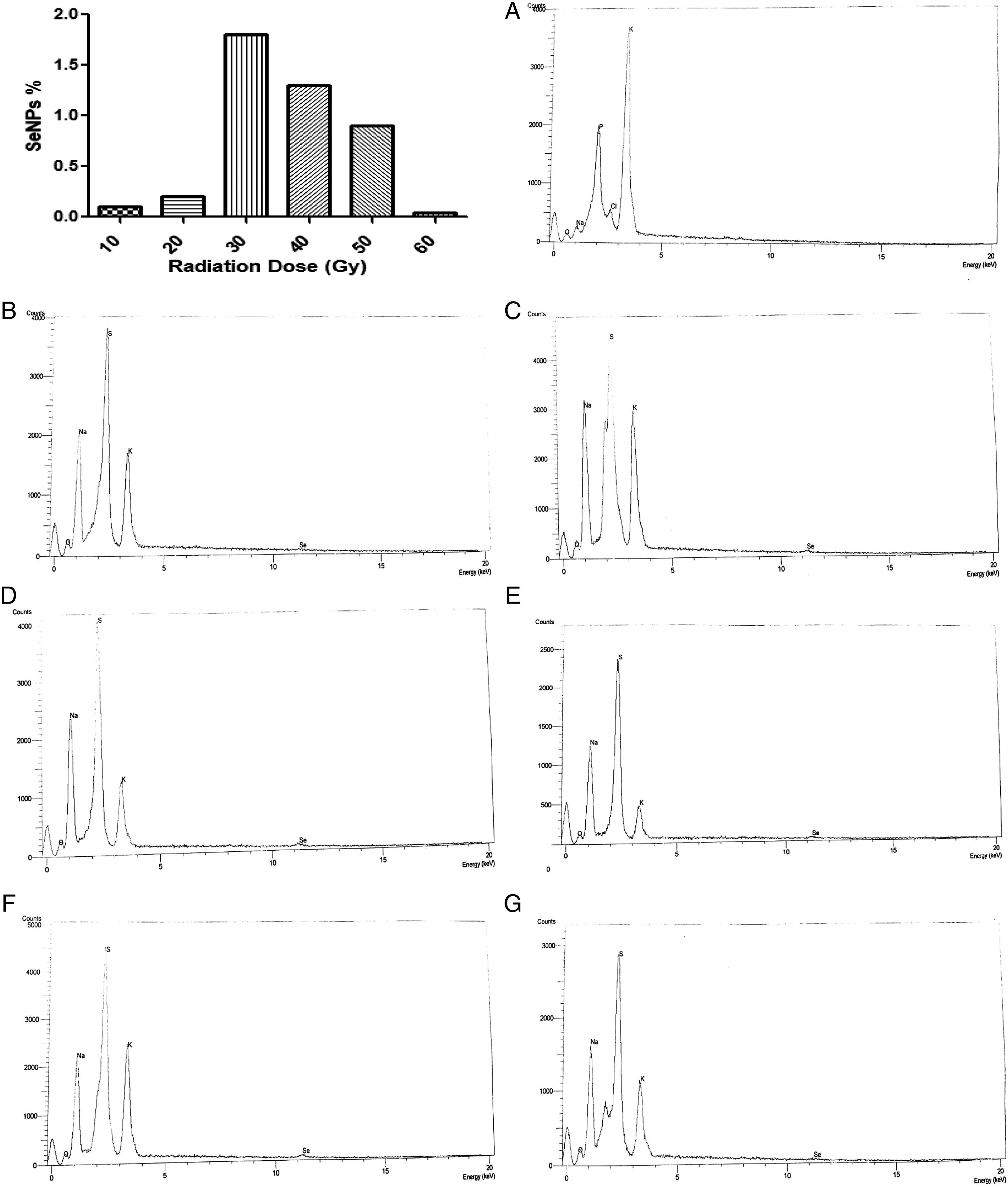
Electron Microscopic Observations
TEM was used to determine the location of SeNPs produced by P citrinum and exposed to γ-ray (30 Gy). TEM analysis found that most of the electron-dense nanoparticles were located in extracellular spaces and were observed in the cytoplasm (Figure 5B) compared with that of the control fungal mycelium (Figure 5A). Dhanjal and Cameotra
25
and Lortie et al.
39
also observed precipitation of SeNPs intracellularly and extracellularly in Bacillus cereus; the particles were spherical with mean diameter size, 19.76 nm.
40
Our results revealed that the γ-ray (30.0 Gy) was playing a significant role in SeNPs’ synthesis; these data matched with those reported by Mosallam et al.
41
Transmission electron micrographs of elemental selenium nanoparticles produced by the biomass of P. citrinum. (A) Control (without γ-ray effect) and (B) distribution of electron-dense selenium nanoparticles of different sizes located extracellularly, and intracellular fungal biomass (arrows) was exposed to the γ-ray (30.0 kGy).
Reduced Glutathione Content (GSH)
GSH increased by increasing the radiation dose until 30 Gy but decreased again with the higher doses as shown in Figure 6. Selenite may be reduced to SeNPs by reaction with protein/peptide reactive thiol groups (activated at the plasma membrane), suggested as a general microbial detoxification reaction to oxyanions. Among these protein/peptides is the glutathione (GSH)/glutathione reductase (GR) system which is responsible for the formation of SeNPs. These proteins/peptides may act as oxido-reductase enzymes or as protons antitransporters.
42
Glutathione (GSH or reduced glutathione) is a gamma-glutamyl-cysteinylglycine tripeptide and the major intracellular antioxidant in several organisms. GSH effectively protects the cells against a variety of free radicals including reactive oxygen species. It has 2 forms, the reduced form of glutathione (GSH) and the oxidized form (GSSG), where sulfhydryl bonds bind 2 GSH moieties. Glutathione peroxidase (GPx) and glutathione-s-transferase (GST) conduct the detoxification reactions effectively using GSH, transforming it to GSSG. Glutathione reductase (GR) runs the recovery pathway by transforming GSSG to GSH with the loss of NADPH and restoring the cellular GSH reservoir. Therefore, GSH and GSH-dependent enzymes are essential to preserve the body’s normal redox balance and assist in cell survival under stress conditions. GSH and dependent enzymes give living cells a survival advantage against radiation.
43
Reduced glutathione content after adding 10 mM of sodium hydrogen selenite to irradiated fungus exposed to different doses of gamma radiation.
Footnotes
Declaration of Conflicting Interests
The author(s) declared no potential conflicts of interest with respect to the research, authorship, and/or publication of this article.
Funding
The author(s) received no financial support for the research, authorship, and/or publication of this article.