
Abstract
Nano-fertilizer(s), an emerging field of agriculture, is alternate option for enhancement of plant growth replacing the synthetic fertilizers. Zinc oxide nanoparticles (ZnO NPs) can be used as the zinc source for plants. The present investigation was carried out to assess the role of ZnO NPs in growth promotion of maize plants. Biosynthesized ZnO NPs (using Bacillus sp) were characterized using Scanning Electron Microscope (SEM), Transmission Electron Microscope (TEM), X-ray diffraction (XRD) and Zeta potential. Different concentrations of ZnO NPs (2, 4, 8, 16 mg/L) were explored in pot culture experiment. Size of ZnO NPs ranged between 16 and 20 nm. A significant increase in growth parameters like shoot length (61.7%), root length (56.9%) and significantly higher level of protein was observed in the treated plants. The overall pattern for growth biomarkers including the protein contents was maximum at 8 mg/L of ZnO NPs. It was observed that application of biosynthesized ZnO NPs has improved majority of growth biomarkers including plant growth parameters, protein contents and leaf area. Therefore, biosynthesized ZnO NPs could be considered as an alternate source of nutrient in Zn deficient soils for promoting the modern agriculture.
Introduction
Nanotechnology has led to new revolutions in every field of science through the incorporation of nanoparticles (NPs) in various industrial and medical products such as ceramic materials, cosmetic products and food products. 1,2 Efforts are also made to understand the role of nanoparticles in the field of agriculture particularly for plant growth. Use of nanoparticles is becoming a promising strategy to enhance plant growth and productivity due to the presence of exceptional properties such as small size, high surface area/volume ratio, high adsorption, large number of reactive sites, high catalytic activity and high chemical stability as compared to bulk ions. 3 These properties make the nanoparticles highly reactive upon their exposure to biological systems. Many researchers focused on the bioenvironmental impact of nanoparticles particularly their effects on animals, plants and microbes. 4 These studies focused mainly on the toxic impact of nanoparticles to environment as they often used high doses of nanoparticles for short periods of time. 5 Mostly, these studies mentioned the negative impacts of nanoparticles to both environment and plants. While, relatively less studies examined the beneficial effects of nanoparticles on plants. 4,6 Recently, nanoparticles have gained a lot of attention for its usage in agriculture, particularly in the context of fertilizers with the emergence of novel technique as nanofertilizer. 7
The exceptional properties of metallic nanoparticles enhance the bioavailability and uptake of micronutrients to the plants, thus enhancing the overall growth of the plants. 8 The added advantage of nanofertilizers is the potential reduction in the loss of nutrients in the soil compared to conventional fertilizer application, which ultimately reduces the application rates of fertilizer.
Although, micronutrients are required in small quantity, but essential for plant development and yield. Among these nutrients, zinc (Zn) plays a significant role in the growth of the plants, animals and humans as its deficiency may lead to several disorders. 4 Plants generally require Zn for carbohydrate metabolism and for gene expression related to environmental stress. 9 Usually, Zn is applied, as fertilizers in the form of soluble salt, that help to assimilate the Zn which may cause environmental problems.
Maize is very sensitive to Zn deficiency and may lead to yellow stripe appearances on leaves within 2 weeks of growth. It may result in decreased photosynthesis, stomatal conductance, efficacy of photosystem, biomass, and Zn concentration in plants. 10 Therefore, it is very essential to determine the Zn deficiency carefully before heavy damage may occur to the crop. To cope with these challenges, use of nanotechnology could be a better substitute to chemicals. In this context, zinc oxide nanoparticles (ZnO NPs) have gained more attention for their use in agriculture as United States Food and Drug Administration (USFDA) recognizes it as a safe substance. 11 Moreover, antimicrobial properties of ZnO NPs and low cost of production have compelled the researchers to consider nanoparticles for application in food and agricultural industries. 12,13 To overcome the Zn deficiency problems in soil due to non-availability of Zn, ZnO NPs, could be a possible alternative of Zn fertilizers. ZnO NPs application is anticipated to fulfill Zn requirements of plants effectively due to their unique properties of small size. ZnO NPs uptake by the plants serve as Zn source to overcome nutrient deficiency in the crops. Moreover, ZnO NPs could easily be absorbed and transmitted to the plant and in comparison of chemical Zn fertilizers. 14 -16 Thus, the use of ZnO NPs could enhance the plant growth in Zn-deficient soil by providing the Zn nutrient.
There are several ways to synthesize ZnO NPs including physical, chemical and biological methods. Both physical and chemical methods are not only expensive but they also involve generation of toxic secondary metabolites. Biosynthesis of ZnO NPs would be preferred option to existing methods 17 as it is environmentally benign and less expensive. Therefore, the present study was conducted to investigate the potential of biosynthesized ZnO NP as source of Zn nutrient on the growth of maize (Zea mays L.). As a result, this study would offer valuable insight for the development of nanomaterial as micronutrient source for crop production.
Materials and Methods
Plant Materials
Seeds of maize (Zea mays L.) were obtained from Ayub Agricultural Research Institute, Faisalabad-Pakistan. Healthy looking and uniform sized seeds were surface sterilized with 1% sodium hypochlorite solution for 10 min, followed by repeated washing with sterile distilled water. 18
Biosynthesis of ZnO Nanoparticles
Bacillus subtilis inoculum (obtained from department of Microbiology, Government College University, Faisalabad-Pakistan) was inoculated in flask containing nutrient broth and incubated at 37°C for 24 h. 25 mL of this culture was taken and diluted 4 times in nutrient broth (75 ml) and again incubated for 24 h. Zinc nitrate was then dissolved in the bacterial solution under constant stirring using magnetic stirrer. After complete dissolution of the mixture, the solution was kept under vigorous stirring at 30°C for 5-6 h until white deposition starts to appear at the bottom then allowed to cool at room temperature and the supernatant was discarded. The pale white solid particles obtained were centrifuged at 4500 rpm for 15 min after thorough washing and dried at 80°C for 7-8 h. 19
Characterization of Biosynthesized ZnO NPs
The biosynthesized ZnO NPs were characterized through UV-Visible spectroscopy, X-ray diffraction (XRD), Scanning Electron Microscopy (SEM) and Transmission Electron Microscopy TEM as described by Wang et al. 20
UV-Visible Spectroscopy
For UV-Visible spectroscopy, ZnO NPs concentration (5 mg/20 ml) was prepared by diluting in de-ionized water and spectrum scans were performed in a wavelength range 300-700 nm using HACH DR5000 spectrophotometer to find wavelength for maximum absorbance.
X-ray Diffraction (XRD)
Dried samples were used for the XRD. The crystalline size of the ZnO NPs was measured with an analytical X′Pert, X-ray diffractometer using CuKα1 radiations (λ = 1.540598 Å), at 40 kV and 40 mA with a divergence slit of 10 mm. The 2θ range was acquired from 30° to 80° and JCPDS Cards were used as standards to find the respective phases of the particles. The crystallite size was calculated by Debye-Scherer equation. 21
Transmission Electron Microscopy (TEM)
Transmission electron microscopy was used to determine the size, shape and morphology of nanoparticles. ZnO nanoparticles dispersions were diluted to 100 μg/mL in distilled water and Dulbecco’s Modified Eagle Medium (DMEM) separately. The samples were prepared by dropping 10 µL aliquots of the particle suspensions onto a copper grid and then allowed to dry. 22 TEM was performed on JEOL JEM-1400 instrument (Jeol LTD, Tokyo, Japan).
Scanning Electron Microscopy (SEM)
SEM analysis was performed with a scanning electron microscope (JEOL JSM-6480). It is used to determine topology and observation of surface. 23 For SEM images, dried particles were mounted on aluminum stub and coated with gold to get better contrast.
Dynamic Light Scattering (DLS)
Additionally, the size distribution and zeta potential of biosynthesized ZnO NPs was determined using Malvern Zetasizer Nano-ZS zen 3600 (UK). 24
Experimental Design for Zea mays L
Seeds of Zea mays L. were primed through soaking in different concentrations (2, 4, 8,16 mg/L) of biosynthesized ZnO nanoparticles under optimized aeration conditions for 24 h. 25 Seeds priming was followed by washing with distilled water and air drying under shade. These seeds were germinated in Petri plates that were filled with soil and farmyard manure (in a ratio of 1:6) in germination chamber at 25 o C and watered daily. At 10 days after sowing, seedlings were transferred to pots filled with acid-washed sand allowed to grow under natural environmental conditions in Randomized Complete Block Design (RCBD) with triplicate of each treatment.
Determination of Growth Biomarkers
The growth biomarkers i.e. shoot and root length, shoot and root fresh mass (FM) and dry mass (DM) were determined by the method described by Drazkiewicz and Baszynski
26
The leaf area of the plant was measured using the formula described by Hunt et al.
27
LA = Leaf length x Leaf width x CF (0.75) Where LA: Leaf Area and CF: Correction Factor
Determination of Total Leaf Protein
Fresh leaves (1 g) were taken and crushed using the mortar and pestle mixed with extraction buffer (70 mM phosphate buffer; pH 7.0, 1 mM EDTA, 1 mM PMSF, 0.5% Triton X 100, and 2% PVP). The mixture was centrifuged at 12000 × g for 10 min at 4°C and the supernatant was used further for estimation of protein contents. The total protein content of leaves was determined using the method followed by Bradford. 28
Results
Characterization of ZnO NPs
UV-Visible spectroscopy
The UV-Visible spectroscopy revealed that tested ZnO NPs exhibited a well-defined plasmon band at the wavelength of 331 nm with an absorbance value of 1.89 (Figure 1). The symmetrical shape of Plasmon band indicated the sharp particle size distribution with smaller particle size.

UV-Vis spectrum of biosynthesized ZnO NPs.
SEM and XRD Characteristics
SEM studies were carried out to find out the surface morphology of synthesized ZnO nanoparticles. SEM studies exhibited that ZnO NPs are in pure form and white colored (Figure 2A). The XRD spectra of the biosynthesized ZnO NPs showed crystalline nature of the ZnO NPs. The average size obtained from the X-ray diffraction spectrum was 11.9 nm (Figure 2B).

A) SEM micrograph of ZnO NPs; B) XRD spectra of biosynthesized ZnO NPs.
TEM Characteristics
TEM was used to determine the morphology and size of the nanoparticles. ZnO NPs are spherical and without aggregate with the size of 16-20 nm of diameter when diluted in DMEM (Figure 3A and 3B).

A) Diameter of biosynthesized ZnO NPs calculated by TEM; B) TEM micrograph of biosynthesized ZnO NPs.
XPS Analysis
In XPS analysis, the percentage that each element represents is calculated from the peak area of the element using its relative and sensitive factors (RSF). To calculate the total amount of Zn and O we took the total area of the corresponding peak. Curve fitting analysis was made to determine the percentage of the element in each of the different links. The overview data shown in Figure 4A indicates that there is no contamination in the samples since only Zn, O and Au (substrate) were detected in the XPS analysis. The amount of each element cannot be extrapolated from these graphs since the RSF of Zn are higher than RSF of O (RSF Zn: 28.72 / RSF O: 2.93), that is why the signal of Zn appears to be very increased in XPS graph.
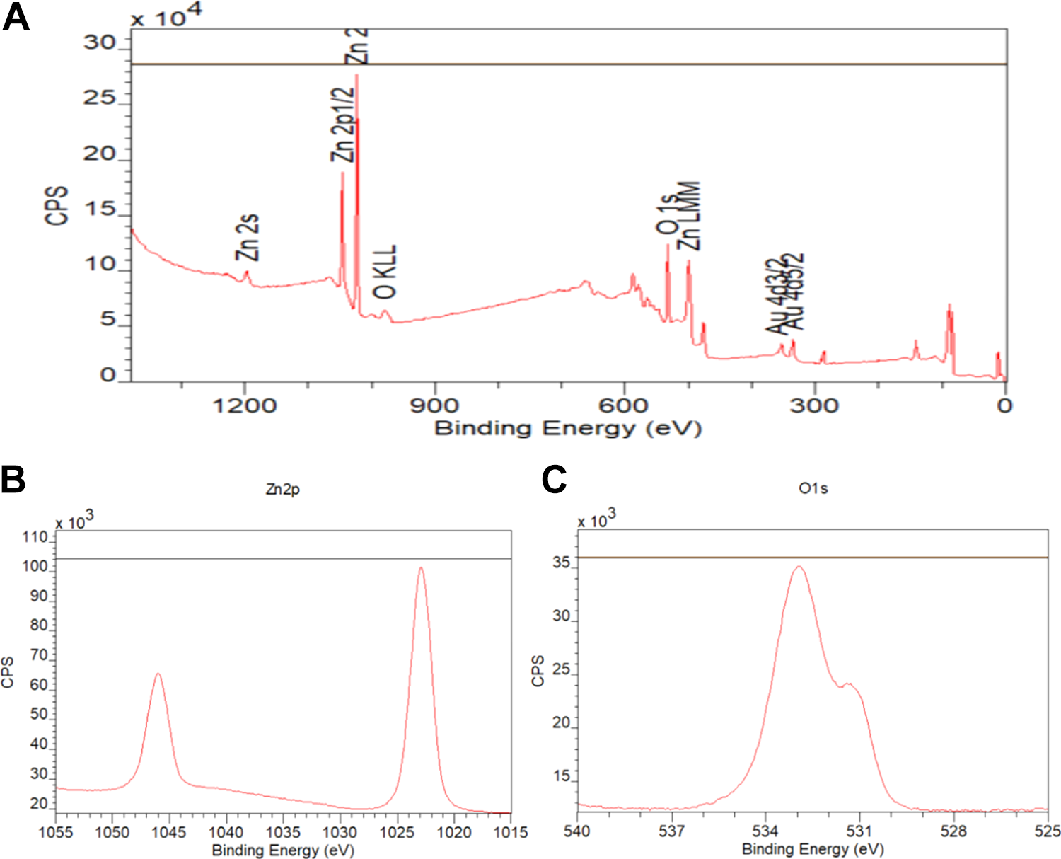
A) XPS wide survey spectra of biosynthesized ZnO NPs, (B, C) spectrum showing the data of Zn 2p (I) and O 1 s (II) of biosynthesized ZnO nanoparticles.
Size Distribution and Zeta Potential Analysis of ZnO NPs
Stability of ZnO NPs was monitored by zeta potential (Figure 5). The overall characteristics of biosynthesized ZnO NPs were represented in Table 1 which shows the acquired data of the different tested parameters. The table shows that the size of zinc oxide nanoparticles in both mediums is between 16-20 nm. The size of nanoparticles was well observed in the DMEM because water exhibit low Zn solubility and due to precipitation of Zn particles in the water the results vary little as compare to DMEM. 29
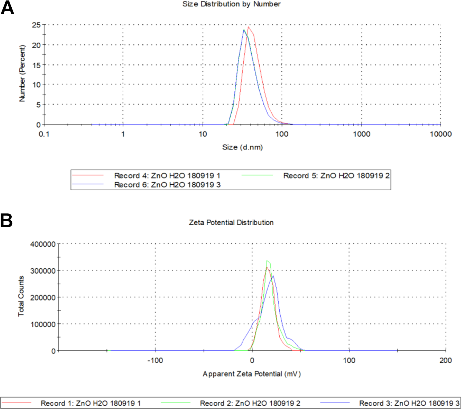
ZnO NPs. A) Size distribution and B) zeta potential.
The Average Size and Stability of Biosynthesized ZnO NPs.

Dispersant: The medium in which suspension made; ZnO NPs: Biosynthesized Zinc oxide nanoparticles.
While measurements taken from TEM images were consistent with the nominal size range for nano ZnO particles, sizes were significantly larg when the hydrodynamic radius was measured by DLS, especially for those samples diluted in DMEM. Interestingly, dispersion values show differences depending on the solvent used, as the Z-potential shows that dispersions in distilled water are moderately stable while particles diluted in DMEM were prone to aggregation.
Effect of ZnO NPs on Growth Biomarkers
The results demonstrated that biosynthesized ZnO NPs facilitated the increase in root and shoot length at 35 days after sowing as compared to control. The maximum increase was observed for 8 mg/L ZnO NPs and then at 16 mg/L ZnO NPs, decrease in root and shoot length has been observed as shown in Figure 6A. The maximum value for root length was 17.94 cm followed by 14.47 cm at 8 mg/L and 16 mg/L ZnO NPs concentration respectively. The overall pattern for growth parameters at different concentration was in the order 8 > 16 > 4 > 2 > 0 mg/L of ZnO NPs. Similar trend was observed for the root and shoot fresh and dry mass as shown in Figure 6B. However, in case of the root fresh and dry mass, no significant difference was observed at 4 mg/L and 8 mg /L concentrations of ZnO NPs.

Effect of seed priming of Zea mays L at different concentration of ZnO NPs at 35 DAS on growth parameters A) Root and shoot length; B) Root and shoot fresh mass (FM) and dry mass (DM) Values reported in the figures are means of triplicates with standard deviations.
The maximum leaf area per plant (56.12 cm2) was recorded when 8 mg/L ZnO NPs concentration was used followed by 43.69 cm2 in plants treated with 4 mg/L ZnO NPs. Statistically significant difference was observed in leaf area of plants treated with 8 mg/L ZnO NPs (P ≤ 0.05) when compared to that of control as shown in Figure 7A.

Effect of seed priming of Zea mays L at different concentration of ZnO NPs at 35 DAS. A) Leaf area; B) Total protein contents: Values reported in the figures are means of triplicates with standard deviations.
For the protein contents estimation of maize plant under various ZnO NPs concentrations, plant treated with 8 mg/L concentration of ZnO NPs showed increased in protein contents (77.3%) compared to that of control (Figure 7B). Moreover, the increase in protein content was related to applied concentration of ZnO NPs. The pattern for the protein content in the plants treated was in order of 8 > 16 > 4 > 2 > 0 mg/L.
Discussion
During the last 5 decades, an increase in cereals crops yield played an important role in world food requirements. With the increasing demand of nutritious food, chemical fertilizers efficiency is limited due to the loss of fertilizers by leaching. 30 In this regard, nanofertilizers (either nutrient themselves or nanomaterials as nutrients carriers) use is gaining attention among researchers. 31 This study is conducted to evaluate the ZnO NPs potential for the plant growth. Morphology and the texture of the ZnO NPs synthesized using biological agent were determined through different techniques. As biological synthesis of nanoparticles is clean, cost effective, nontoxic and environmental friendly 32 as compared to chemical synthesis, therefore ZnO NPs were synthesized using Bacillus sp. TEM image of ZnO NPs exhibited a small size and showed uniform crystal shapes. Moreover, close packing texture of ZnO NPs was revealed by the SEM image. It has been observed that the particles in the samples were compactly arranged and were almost spherical in shape. Motshekga et al. 33 also examined the morphology of ZnO NPs and found spherical shaped nanoparticles. In most of the studies, the size of ZnO NPs ranges between 40–75 nm but our study findings examined the ZnO NPs diameter in the range of 16-20 nm. Normally, the size of the nanoparticles depends on the experimental protocol used. As Khajeh and Golzary 34 synthesized ZnO NPs in hexagonal shaped with 11–25 nm size while Singh et al. 17 found spherical shaped ZnO NPs with 35-80 nm in size using Pseudomonas aeruginosa. Crystal structure of the biosynthesized nanoparticles was investigated using X-ray diffraction and obtained crystal patterns correspond to pure forms of ZnO. 23
In this study, increase in root length was observed with the increase in ZnO NPs concentration. One of the reasons is that there might be greater absorption of Zn in the root due to very small size of biosynthesized nanoparticles. Overall, the root and shoot length was higher (32%) in the plants treated with biosynthesized ZnO NPs as compared to the control. The growth in root and shoot length reached maximum at 8 mg/L concentration of biosynthesized ZnO NPs.
Zinc oxide nanoparticles potential to boost the growth and yield of crops has been studied. Prasad et al. 14 studied the different concentrations of zinc oxide nanoparticles (25 nm in size) using priming of peanuts seeds and showed that 1000 ppm concentration of ZnO NPs enhanced the seed germination and plant growth. This study demonstrated that treatment with 8 mg/L ZnO NPs concentration is most suitable for improving the root and shoot lengths. Batsmanova et al. 12 demonstrated the positive influence of ZnO NPs as micronutrient in cereal crops. Xun et al. 35 showed the increase in the plant growth and photosynthesis due to ZnO NPs as compared to control in Zea mays L plants. This improvement in root and shoot length could be attributed to smaller size of the nanoparticles due to possibility of higher Zn absorption by plants as compared to larger particles as described by Sabir et al. 9 Similarly, Tripathi et al. 36 observed higher absorption of Zn in the wheat seedlings treated with Zn NPs in comparison of larger zinc sulfate particles.
It has been studied that impact of nanoparticles on plants depends on method of their synthesis, dose, size as well as on the method of application to plants. For this study, seeds of Zea mays L were primed with ZnO NPs and enhancement in growth parameters has been observed. According to our results, seed priming may be an effective method to enhance the growth parameters. Seed priming with ZnO NPs has been reported by Munir et al. 37 depicted higher concentration of Zn in roots and shoots than control. While comparing the green synthesized ZnO NPs (35 nm) with chemically synthesized ZnO NPs (48 nm), Singh et al. 38 observed higher growth rate (root and shoot lengths) in green synthesized ZnO NPs relative to chemically synthesized ZnO NPs and ascribed it with smaller size of NPs.
Plant growth response to nanoparticles was dose dependent as observed by different studies. Hazeem et al. 39 reported lower Zn concentration in cucumber shoot with 125 mg/kg concentration of Zn NPs as compared to 25 mg/kg which could be due to higher Zn levels that deform the plant cells. While, contrary to this, Moghaddasi et al. 40 reported higher Zn concentrations in plants treated with high nanoparticles concentration and correlated it with higher penetration of NPs into plant cells. In another study, increased root and shoot lengths of wheat plant has been observed upto 60 mg/kg of TiO2 NPs while at higher concentration, decrease in root and shoot lengths was observed. Rafique et al. 41 However, more studies are required to understand the absorption mechanism within the plant cells. Positive influence of nanoparticles to plant depends on specific concentration of nanoparticles with specific plants. While, studying the ZnO NPs effect on tomato plants, Faizan et al. 18 concluded that a significance increase in growth parameters depends on concentration and time of the treatment. Similar findings in terms of the increased growth biomarkers in maize plants (exposed to ZnO NPs) were observed in present study. Although, we discussed here the beneficial effects of nanoparticles, but these may be phytotoxic as well. The positive and toxicity effects depend on dose, nature, duration and conditions of exposure. 42,43 Normally, nanoparticles effects plant physiological traits i.e. germination, root elongation and biomass 44 -46 The nanoparticles may cause reduction in seed germination, plant elongation and sometime cause plant death. 47 Due to phytotoxicity, plant growth may be slow, decrease in plant hormones, change in transcriptional genes profile and decreased photosynthetic rate had been observed. 48 -50 Yusefi-Tanha et al. 51 found that small sized CuO nanoparticles (25 nm diameter) were more phytotoxic than large sized CuO nanoparticles (50 nm and 250 nm) to Soybean. Moreno-Olivas et al. 52 reported that nanoparticles interaction with plant cell could lead to change in plant genes expression. Higher concentration of nanoparticles is toxic to plants as López-Moreno et al. 53 reported that high dose of CeO2 nanoparticles damaged the DNA structure in soybean. Thus it is important to study nanoparticles effect carefully for each of the plant species before application in field for commercial use.
However, significant increase in plant shoot and root growth and root area in Solanum lycopersicum, Vigna radiata and Cicer arietinum was observed by Mahajan et al. and Raliya et al. 54,15 by treating the plants with ZnO NPs. In this study, ZnO NPs significantly impacted the leaf protein contents of maize plant. The trend for protein contents was toward the higher concentration of the ZnO NPs. Rizwan et al. 55 suggested that wheat plants treated with ZnO NPs had higher protein contents that accelerate the photosynthetic activity. In contrast, total protein contents in foxtail millet were not statistically different in ZnO NP-treated plants as compared to control. 56
Hence, the current study clearly demonstrated positive influence of ZnO NPs on plant growth. Nano-priming could be an efficient technique for the better production of crops. So, the privilege to use nutrients such as Zn at the level of nanoscale might be a revolutionary step in agriculture. Overall, the results of this study could be helpful to the fertilizer industries to make a decision about the nanofertilizers production especially ZnO NPs that could be used as nutrient source to reduce the Zn deficiency in plants.
Footnotes
Acknowledgments
The Authors would like to acknowledge Dr Ricard Marcos Dauder, Departament de Genètica i de Microbiologia, Universitat Autonoma de Barcelona, Spain for his great help in nanoparticles characterization.
Declaration of Conflicting Interests
The author(s) declared no potential conflicts of interest with respect to the research, authorship, and/or publication of this article.
Funding
The author(s) received no financial support for the research, authorship, and/or publication of this article.