
Abstract
The objective of our study was to investigate the effects of heavy metals on the fertilizing capacity of exposed zebrafish sperm, on embryonic survival, and on occurrence of embryonic deformities following fertilization with exposed sperm. It is important to test heavy metals because they are well-known pollutants. Sperm of externally fertilizing species can get in contact with pollutants found in aquatic environment. Zebrafish sperm, despite its advantages, has seldom been used in in vitro toxicological studies and no reports are available regarding the fertilizing capacity of exposed sperm. Zebrafish sperm was stripped and exposed to concentrations of the tested heavy metals (Zn2+, Cd2+, Cr3+, Cu2+, Ni2+, Hg2+, As3+) for 30 or 120 minutes. Calculated half-maximal effective concentration (EC50) values do not differ significantly from those calculated for motility for any of the tested heavy metals, which means fertilization rate can indicate the toxicity of the given substance following exposure of sperm. Thus, its application as in vitro toxicological end point is reasonable. The survival of embryos and embryonic development have not been affected by the exposure of spermatozoa, which means all alterations in spermatozoa caused by heavy metals have been expressed before 24 hours post fertilization.
Keywords
Introduction
In vitro test systems are becoming widespread in toxicology and ecotoxicology in the last decades, mainly due to ethical concerns. Fish sperm has several positive features such as the noninvasive collection of sperm, no long-term maintenance needed such as in the case of cell and tissue cultures, and it provides measurable end points. The mentioned advantages make fish sperm a suitable in vitro toxicological model as an alternative for in vivo tests. Numerous studies have recently been published that used fish sperm as toxicological test system. Mostly motility parameters and antioxidant response of exposed sperm were tested in these reports. Furthermore, species with large body size had been used due to the greater volume of individually produced sperm in most cases, including rainbow trout (Oncorhynchus mykiss) 1 -3 and common carp (Cyprinus carpio). 4 -12 Zebrafish (Danio rerio) has the following advantages compared to large-bodied species: easy and cheap maintenance in recirculation systems, short generation time, its full genome map is known, year-round spawning and sperm collection with constant quality without hormonal induction. Furthermore, this species is specified in the Organisation for Economic Co-operation and Development guidelines as a feasible model species in ecotoxicological studies, and its sperm has been used in in vitro toxicological-aimed studies only in 3 cases. 13 -15
Heavy metals are known as ubiquitous aquatic pollutants due to their widespread use (processing industry, agriculture, manufacture, mining, smelting activities, etc). The environmental concentrations of these heavy metals are different due to the industrial activities of the given territories (eg, in the case of Zn, the aquatic concentration is between 0.92 and 70.81 mg/L, and considering As, it is between 55 and 195 μg/L). 16,17 Many publications deal with the harmful effects of metals on aquatic organisms, such as mortality of fish species, 18 -21 motility parameters of fish sperm, 4 -6,9,15,22,23 and enzymatic activities of exposed sperm. 10,24 -26 Progressive sperm motility (PMOT) is the most sensitive parameter of all. The toxic substance can also have an effect on straight line velocity as well as on curvilinear velocity but not to an extent as on PMOT. 15 The effect of heavy metal exposure was examined on the fertilizing capacity of sperm only in 4 fish species: common carp (Cyprinus carpio), 12 rainbow trout (Oncorhynchus mykiss), 27 -29 topsmelt silverside (Atherinops affinis), 30 and African catfish (Clarias gariepinus). 31 However, it has been reported that damage can form in spermatozoa (eg, oxidative DNA damages) due to the exposure to toxicants, which effects do not manifest on the motility, morphology, and antioxidant parameters of sperm, yet they can impair embryonic development. 32,33 These damages can only be investigated on spermatozoa directly by staining methods (eg, Feulgen staining, Halomax test) or by special equipment (eg, flow cytometer). However, fertilization experiments are simpler, less costly, and do not require the use of expensive devices. Fertilization and hatching rates are robust quality biomarkers as they allow to observe if viable and healthy offspring can be obtained, which is the main objective and final outcome of in vitro fertilization. Despite the mentioned advantages, the investigation of fertilization following exposure of fish sperm to different toxicants is not a widespread end point in these studies. There are no reports on zebrafish sperm in this context; furthermore, only a few publications exist regarding other fish species. A possible reason of this is that in the case of motility studies an immediate result is gained, while in the case of fertilization tests, the effect can only be observed after some hours. Fertilization is also a very important end point as DNS damage can not be observed by motility studies and toxic exposure is also able to reduce not only fertilization but also hatching rate. 34 -38
Consequently, the objective of our study was to investigate the effects of heavy metals on the fertilizing capacity of zebrafish sperm in order to estimate whether the motility or the fertilizing capacity of exposed sperm is the more sensitive parameter of toxic exposure. Furthermore, embryonic survival and development of embryonic deformities were examined following fertilization with exposed sperm in order to determine whether heavy metals cause impairments in spermatozoa which affect these parameters during embryogenesis.
Materials and Methods
Animals
All experiments were carried out in the zebrafish holding facility of the Department of Aquaculture, Szent István University (Gödöllő, Hungary). Mature, wild-type zebrafish males of the AB line were used for the experiments. Fish were kept in 3-L polycarbonate tanks, in a ZebTEC (Tecniplast, Buguggiate, Italy) recirculating zebrafish housing system, under constant water quality parameters (25°C ± 2°C; pH 7.0 ± 0.2; conductivity 525 ± 50 µS; hardness: <0.5 dH; DOC: >90%; from here onward referred to as system water) and photoperiod (14-hour light:10-hour dark cycle). Zebrafish were fed 2 times a day with ZEBRAFEED diet (Sparos Lda, Olhão, Portugal) supplemented with live Artemia salina nauplii every second day.
Sperm Collection
Sperm was collected from the males by stripping. Before stripping, fish were anesthetized in 0.06% MS-222 (Acros Organics, Geel, Belgium), buffered to pH 7.0, and rinsed in system water to prevent contamination of sperm with the anesthetic solution. 39 Fish were fixed in a wet sponge with the abdomen facing upward, then gentle pressure was applied to the abdominal wall by a forceps. The outflowing sperm was collected by a calibrated glass capillary to evaluate its quantity. Sperm was pooled directly into 50 µL of cyprinid immobilizing solution (200 mM KCl, 30 mM Tris, pH 8) 40 until a total of 10 µL of sperm was collected in the pool. This volume was collected from 19 ± 8 males containing 16 ± 8 × 10⁶ spermatozoa/pool. The samples were stored on crushed ice until use. After stripping, fish were placed back into system water. Fish were stripped maximum once a week.
Assessment of Sperm Concentration and Motility Parameters
Sperm concentration was determined using a Bürker-Türk-type hemocytometer (Marienfeld Superior, Paul Marienfeld GmBH & CO. KG, Lauda-Königshofen, Germany). Sperm quality was evaluated using a microscope (Olympus BX41, AgroLegato, Budapest, Hungary) with a negative phase-contrast objective (20× magnification) and connected to a computer-assisted sperm analysis (CASA) system (Sperm Vision v. 3.7.4.; Minitube of America, Verona, Wisconsin). Progressive sperm motility (the percentage of cells performing forward movement, %) was measured immediately after stripping to evaluate the fresh sperm quality. The CASA was used with the following settings: (1) threshold limit of the PMOT: straight line distance between the initial and the end point of moving (DSL) >5 µm; (2) pixel/µm ratio: 151/100; and (3) size of the head area: 1 to 100 µm2. Spermatozoa were activated in a Makler chamber (SEFI Medical Instruments, Haifa, Israel). System water supplemented with bovine serum albumin (Albumin Bovine Serum Cohn Fraction V; VWR International Ltd, Radnor, Pennsylvania) was used for the activation at a ratio of 5:1 (vol/vol). 41 At least 2 activations were carried out in the case of each sample (thus each measurement was conducted in duplicate). Samples having more than 60% progressive motility were used for the fertilization experiments.
Applied Chemicals
Seven heavy metal compounds were tested as follows: zinc nitrate hexahydrate [Zn(NO3)2 × 6 H2O], cadmium nitrate tetrahydrate [Cd(NO3)2 × 4 H2O], chromium (III) nitrate nonahydrate [Cr(NO3)3 × 9 H2O], copper nitrate trihydrate [Cu(NO3)2 × 3 H2O], nickel nitrate hexahydrate [Ni(NO3)2 × 6 H2O], mercury (II) nitrate monohydrate [Hg(NO3)2 × H2O], and arsenic trioxide [As2O3] (each from Sigma Aldrich, St Louis, Missouri). Each stock solution was prepared by dissolving the compounds in cyprinid immobilizing solution. In the case of arsenic trioxide, sonication was required to dissolve the compound completely (Branson Digital Sonifier, Models 250, 40% amplitude, 4 × 4 minutes, VWR Hungary, Budapest, Hungary). The stock solutions were stored at −80°C for up to 7 days. The final solutions were prepared from stocks immediately prior to the experiments. Exposure concentrations refer to immediate concentrations of heavy metal ions.
Sperm Dilution and Exposure
Sperm pool samples (10 μL) were exposed to 10 µL of double-concentrated heavy metal solution in order to reach the final concentration of the substance following dilution. Consequently, each test solution contained 3 ± 1 × 10⁶ sperm cells. Four concentrations of each heavy metal were tested on sperm from the same pool. The applied concentrations were as follows: 0.5, 1, 2.5, and 5 mg/L in the case of Hg2+; 1, 5, 25, and 50 mg/L in the case of Cu2+ and Cd2+; 50, 100, 150, and 200 mg/L in the case of Cr3+, Zn2+, and As3+; 600, 800, 1000, and 1200 mg/L in the case of Ni2+, according to the previously tested effective concentrations on zebrafish sperm motility parameters. 15 In parallel, a control group from the same pool was exposed only to the cyprinid immobilizing solution at the same dilution ratio. Exposure duration was 30 or 120 minutes. All experiments were carried out using 3 independently collected pools of sperm in the case of both exposure durations; thus, for statistical analysis, the number of independent samples was N = 3. Samples were stored on crushed ice during the exposure.
Egg Collection and Fertilization
For inducing maturation, females were placed together with males into double-walled breeding tanks (1 L volume) in the afternoon prior to the experiments. Fish were separated according to sex, and the final sex ratio was 1:1. Before the stripping of eggs, the females were anesthetized in 0.06% MS-222 buffered to pH 7.0 and were placed on a wet paper tower. The genital papillae was wiped dry to prevent the activation of eggs by water. Eggs were stripped into a dry Petri dish until 50 to 100 eggs were collected in a pool. Five pools of eggs (in 5 Petri dishes) were created for the 4 concentrations and 1 control group per heavy metal. The fertilization was conducted maximum within 10 minutes after the stripping of eggs to prevent their drying. During the fertilization, 20 µL of total exposed sperm sample was added to one batch of eggs reaching an average sperm:egg ratio of 30 000:1. The gametes were activated with ∼1 mL of system water. Five minutes after fertilization, system water was replaced on the batches to prevent the toxic exposure of eggs by the treated sperm, then the eggs were incubated at 26°C for 48 hours. After stripping, females were placed into system water to recover. The same females were stripped again minimum 1 week later.
Investigation of the Fertilization Rate, the Embryonic Survival, and the Rate of Embryonal Deformities
Fertilization rate was detected at 24 hours-post fertilization (hpf) expressed in the percentage of live embryos. The rate of embryonic survival was determined 48 hours after fertilization expressed in the percentage of live embryos compared to the number of fertilized eggs. The rate of embryonic deformities (yolk edema, pericardial edema, deformities of head and eyes, tail and spinal deformations, disintegration) was examined at 24 hpf as well as at 48 hpf, expressed in the percentage of living embryos.
Statistical Analysis
In the case of the tested exposure durations (30 and 120 minutes), the number of tested pools was considered as the number of independent samples (N = 3). The results were evaluated by the statistical software GraphPad Prism 6.01 (GraphPad Software Inc, San Diego, California) and R 3.3.2 (R Foundation, Vienna, Austria). Homogeneity of variances and normality of distribution were verified prior to the test. Two-way analysis of variance with Bonferroni post hoc test was used to determine which concentrations had a major effect on the fertilization rate at 24 hpf and on embryonic survival at 48 hpf at various exposure durations of sperm. Kruskal-Wallis test with Dunn post hoc test was applied to examine the effect of sperm exposure on the rate of embryonic deformities. The significance level was P = .05 in each case. At those points of time when the fertilization rate reduced beyond half of the controls, median effective concentrations (EC50 values) with standard deviation were calculated by fitting dose–response curves. 42 A 2-sample t test was used to compare the calculated EC50 values with those previously published on other parameters of sperm (PMOT). The PMOT values of fresh sperm samples were not included in statistics, and its measurement served as indicator of fresh sperm quality.
Results
The PMOT of fresh sperm samples was 70% ± 6% in the fertilization experiments of the 7 heavy metals (N = 3 at testing of both exposure durations in each metal). In cases of Zn2+ and Cd2+, neither the concentration nor the exposure duration of sperm had a significant main effect on the fertilization rate. In cases of Cr3+, Cu2+, Ni2+, and Hg2+, a significant main effect of the concentration (but not of exposure duration) was observed on fertilization rate (Cr3+: P = .0044, Cu2+: P = .0002, Ni2+: P = .0103, Hg2+: P = .0004). After 30 minutes of exposure, the effect of Ni2+ was observed at the lowest concentration, while Cu2+ and As3+ produced toxic effect at the highest tested concentration. The effect of Cr3+ was only documented after 120 minutes of exposure. Only in the case of As3+, a significant main effect of concentration (P = .0232) as well as of exposure duration of sperm (P = .0356) was observed (Figure 1). There was no interaction between the 2 examined variables in any of the tested heavy metals. Due to this, calculation of EC50 values was possible only in the case of 5 heavy metals (Table 1). Embryonic survival at 48 hpf (Table 2) and the rate of embryonic deformities (Figure 2) have not been affected by the exposure of sperm to any of the tested heavy metals.
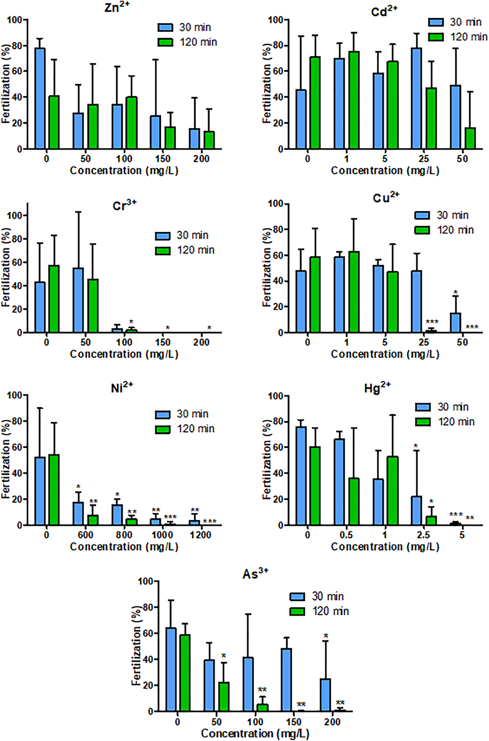
Average fertilization rate with standard deviation at 24 hpf (in percentage, N = 3) fertilized with sperm exposed to various heavy metal concentrations next to different exposure duration in zebrafish. Blue bar signs the 30-minute exposure of sperm, green bar signs the 120-minute exposure of sperm. Significant differences compared to the control value next to the given exposure duration are labeled with an asterisk (*) at *P < .05, **P < .01, ***P < .001 (2-way ANOVA with Bonferroni post hoc test). ANOVA indicates analysis of variance; hpf, high-power field.
Median Effective Concentrations (EC50 Values in mg/L, Where It Can Be Estimated) of Heavy Metals on the Fertilizing Ability of Zebrafish Sperm at 30 and 120 Minutes of Exposure.a

a The values of R 2 are in Table (df = 10 in each case).
Average Embryonic Survival With SD (in Percentage) Examined 48 Hours Followed by Fertilization With Sperm Exposed to Various Heavy Metal Concentrations Next to Different Exposure Duration (30 and 120 Minutes) in Zebrafish.a
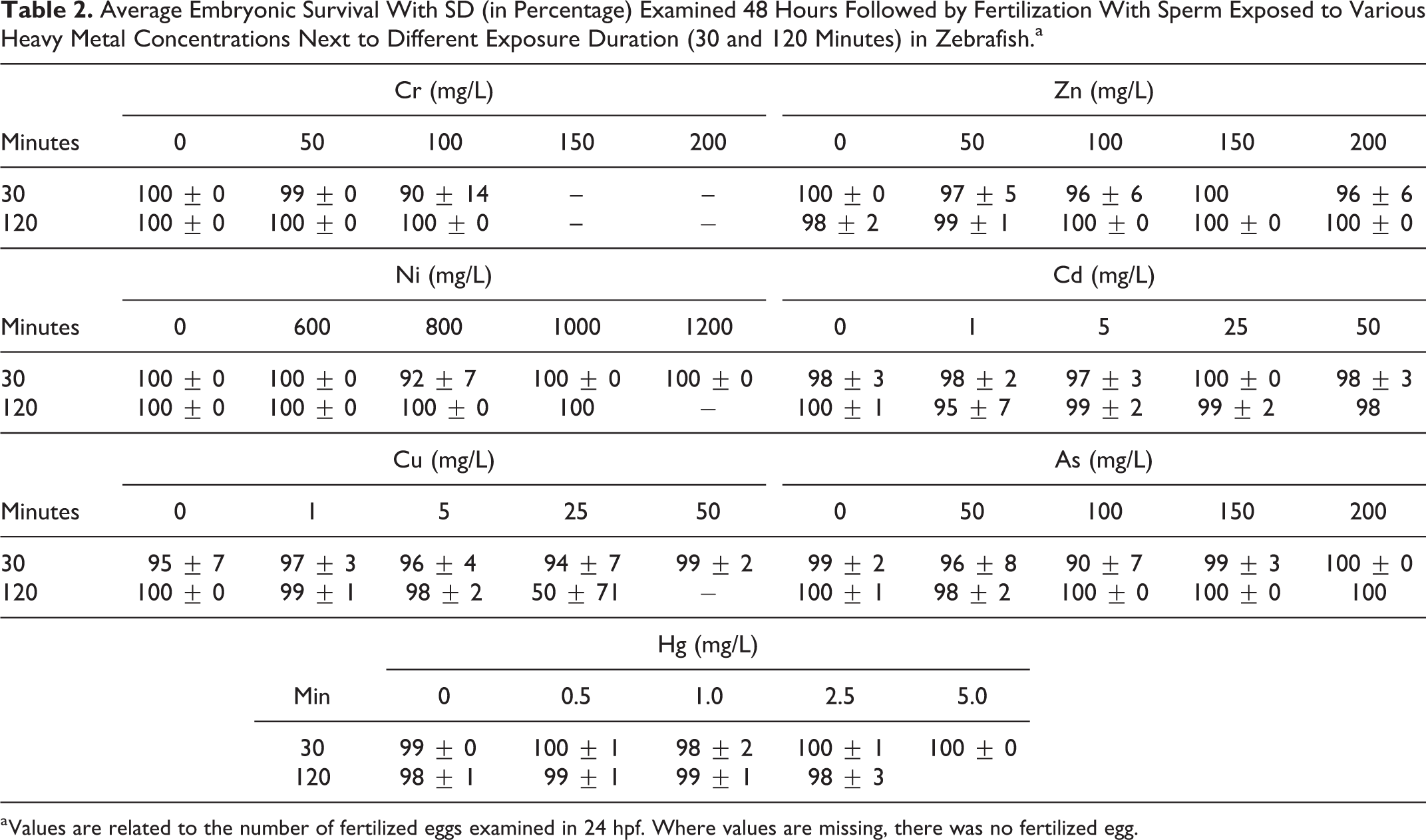
a Values are related to the number of fertilized eggs examined in 24 hpf. Where values are missing, there was no fertilized egg.
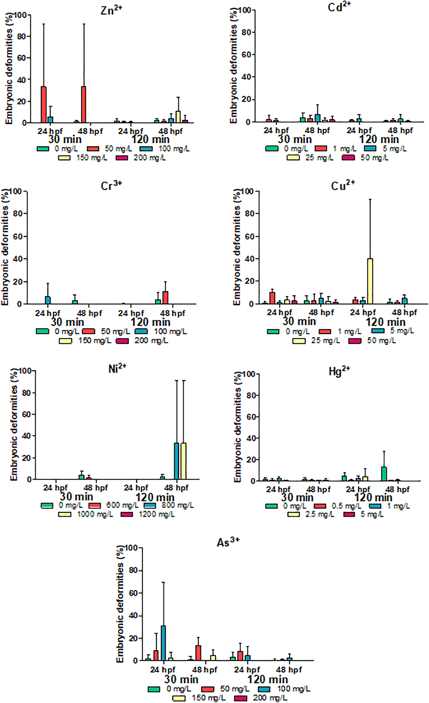
Average rate of embryonic deformities with standard deviation (in percentage, N = 3) examined 24 and 48 hours followed by fertilization (24 and 48 hpf) with sperm exposed to various heavy metal concentrations next to different exposure duration (30 or 120 minutes) in zebrafish (Kruskal-Wallis test with Dunn post hoc test). hpf indicates high-power field.
Discussion
Heavy metals are widely used toxic aquatic pollutants that are essential to investigate. They are capable to get in contact with the sperm of externally fertilizing species and can have an effect on their fertilizing capacity. Zebrafish sperm is a suitable in vitro model to test the effects of heavy metals as sperm can be collected with constant quality without hormonal induction.
The effects of 7 heavy metals on fertilization rate, on embryonic survival, and on the development of embryonic deformities have been examined following the exposure of zebrafish sperm to different concentrations and exposure durations. Until now, none of these metals was tested on these parameters of zebrafish sperm. Motility of fish sperm is controlled by molecular mechanisms which are very sensitive and can be easily affected by the composition of water. The ionic composition of water can be modified by aquatic toxicants 9 : they can change the regulation of water and ion channels of spermatozoa, 43 its mitochondrial activity, 44 the dynamic of cytoskeleton, and the structure of axoneme by oxidative stress. 25,26,45 Moreover, chemicals can damage the structure of cell components directly, as well, for example, the plasma membrane, which can disrupt the membrane potential and impair the ability for the motility. 14,46,47 The reduction in sperm motility can lead to impaired fertilization, increase in abortion rates, and increase in incidence of embryonic deformities. 48 Toxicants can also cause mutations in the DNA of spermatozoa which can affect the survival of embryos and the embryonic development. 32
In this study, in the case of Cd2+ (EC50: 6.5 mg/L) 19 and Zn2+ (EC50: 44.48), 19 the applied concentrations had no significant effect on the fertilization rate at the tested exposure durations. In common carp, only 100 mg/L of Cd2+ caused a reduction in fertilization rate following fertilization immediately after exposure, whereas 50 mg/L of Cd2+ did not. 12 In our study, we experienced that 50 mg/L of Cd2+ did not reduce fertilization even after 120 minutes of exposure. However, 100 mg/L of Cd2+ has not been examined in the present study; thus, it can be speculated that this concentration can cause the reduction in fertilization rate also in zebrafish. Despite this, analysis of the PMOT value of zebrafish sperm after Cd2+ exposure revealed that already 0.5 µg/L after 10 minutes of exposure 14 and 5 mg/L after 30 minutes of exposure can reduce motility values. 15 In the case of Zn2+, fertilization has not been examined after exposure of sperm in any fish species. However, 200 mg/L of Zn2+ can reduce the PMOT of zebrafish sperm 15 ; thus, sperm motility is more sensitive to Cd2+ and Zn2+ exposure than the fertilizing capacity of sperm. 35,38 The reason for this is that there is no maternal effect in the case of motility, while regarding fertilization, it also influences the results.
In the case of the other 4 tested heavy metals (Cr3+, Cu2+, Ni2+, and Hg2+), the applied concentrations affected the fertilizing capacity of exposed sperm significantly. These effects have not been investigated in the case of any cyprinid species previously, but only on sperm of species belonging to other taxa.
In the case of Cr3+ (EC50: 54.57 mg/L), 21 this effect was tested on sperm of rainbow trout, where 0.5 µg/L of Cr3+ resulted in a significant decrease compared to the control after 40 minutes of exposure. 28 Despite this fact, only 100 mg/L of Cr3+ affected the fertilization after 120 minutes of exposure in our study, which shows that salmonids have more sensitive spermatozoa than cyprinids. This can be due to several reasons: different morphology (elongated head and mitochondrial ring), different activation mechanism (a cyclic adenosine monophosphate–dependent process), and there is a deviation in the mode of energy storing (lipid is stored instead of fructose, and energy is gained from lipids). 49,50
Comparing our results with the motility of zebrafish sperm, where 100 mg/L of Cr3+ reduced PMOT already after 30 minutes of exposure, 15 it can be concluded that the fertilizing capacity of sperm is less sensitive to Cr3+ exposure than motility.
A 40-minute exposure to 0.5 mg/L of Cu2+ (EC50: 0.17 mg/L) 19 had a significant effect on the sperm of rainbow trout, 28 while only 25 mg/L caused reduction in this study. It confirms that the sperm of salmonids is more sensitive. Furthermore, already 1 mg/L affected the motility of zebrafish sperm exposed to Cu2+, 15 and thus, it can be concluded that motility is more sensitive to Cu2+ exposure than the fertilizing capacity of sperm. However, toxic effects of the sperm exposure to heavy metals can have consequences in later life stages, and future studies should investigate this matter.
In the case of Ni2+ (EC50: 9.07-18.92 mg/L), 18 fertilization has been examined after a maximum 1 mg/L exposure of sperm in rainbow trout, which has not been affected after a 5 minute-exposure. 27 In our experiments, a different range of concentrations was tested: 600 mg/L of Ni2+ caused reduction in the fertilization rate, at 30 minutes of exposure. However, regarding the motility of Ni2+-exposed sperm, only 800 mg/L caused reduction after 30 minutes of exposure. 15 This means that the fertilizing capacity of zebrafish sperm is more sensitive to Ni2+ exposure than the motility, unlike any of the previously discussed heavy metals. Probably, a DNA damage occurred, which is not present while testing the motility of sperm but it can appear during embryonic development. Investigating only the motility of the sperm would not show this effect of the tested substance.
In the case of Hg2+ (EC50: 0.14 mg/L) 19 , Billard and Roubaud 28 observed that reduction in the fertilization rate occurred only above 1 mg/L, at 40 minutes of exposure in rainbow trout, which is almost the same as experienced in our study, where 2.5 mg/L reduced the fertilization after 30 minute-exposure of zebrafish sperm. Comparing this result to the motility of Hg2+-exposed zebrafish sperm, where a similar effect was observed, 15 the sensitivity of motility and fertilizing capacity is homologous following Hg2+ exposure of zebrafish sperm.
In the case of As3+ (EC50: 6.75 mg/L) 20 , applied concentrations as well as exposure durations had an effect on the fertilization rate: 100 mg/L caused reduction after a 120-minute exposure of sperm. The effect of As3+ exposure has not been examined on the fertilizing capacity in any fish species previously. However, the PMOT of zebrafish sperm showed a significant reduction already at 50 mg/L, after 30 minutes of exposure, 15 which means that motility reacts more sensitively to As3+ exposure of sperm than fertilizing capacity, as in the cases of all the other heavy metals except for Ni2+ and Hg2+.
The calculated EC50 values ranged from 1.7 mg/L (Hg2+) to 472 mg/L (Ni2+) at 120 minutes of exposure, and the order of toxicity expressed in rate of fertilized embryos are Hg2+ > Cu2+ > As3+> Cr3+ > Ni2+ > Cd2+ > Zn2+. In cases of Zn2+ and Cd2+, calculating EC50 values was not possible. When these results are compared with those obtained on motility of zebrafish sperm, where the EC50 values at 120 minutes of exposure were between 1.2 mg/L (Hg2+) and 696 mg/L (Ni2+) at the same exposure duration, the order of toxicity was Hg2+ > As3+ > Cd2+ > Cr3+ > Cu2+ > Ni2+ > Zn2+. 15 Thus, it can be concluded that despite the fact that there are differences in the orders of toxicity and the sensitivity of the measured end points, the calculated EC50 values do not differ from each other significantly in the case of any tested heavy metals, where it can be estimated.
Regarding the embryonic survival, none of the tested heavy metals caused significant reduction in this parameter in the 48th hour compared to the number of fertilized eggs. There is only one publication dealing with the effect of heavy metals on fish sperm. According to this study, 1 mg/L of Cu2+ did not cause any reduction in the fertilization rate of rainbow trout after 5-minute exposure of sperm; but the hatching rate has been affected significantly. However, in the case of Ni2+, 1 mg/L did not cause any reduction neither in fertilizing capacity nor in the hatching rate. 27 The results obtained in Cu2+ overlaps ours in the case of fertilizing rate. Hatching rate should be investigated in the future. Considering also the rate of embryonic deformities, it can be concluded that it was not significant in the case of any tested heavy metals. No information is available regarding this effect of heavy metals, currently. Consequently, all damages induced by spermatozoa exposed to heavy metals have been expressed before 24 hpf and can lead to the prevention of fertilization and/or to early embryonic mortality. However, effects on the survival of embryos and in the embryonic development have not been observed. However, the toxic effects of the sperm exposure to heavy metals can have consequences in later life stages and future studies should investigate this matter.
The effect of investigated toxicants has not previously been examined on the fertilizing capacity of in vitro exposed zebrafish sperm nor on the rate of embryonic survival following fertilization with this sperm previously. However, it is practical to use zebrafish sperm for toxicological studies, as the rule of 3R (reduction, refinement, replacement) is also taken into account by reusing the same individuals for samplings. Moreover, embryonic deformities caused by sperm exposure have not been investigated in any fish species before. The developed protocol can serve as a simple and fast toxicology test system in order to investigate the possible toxic effect of chemicals in vitro.
Footnotes
Authors’ Note
Flóra Kerekes and Tímea Kollár contributed equally to this work.
Declaration of Conflicting Interests
The author(s) declared no potential conflicts of interest with respect to the research, authorship, and/or publication of this article.
Funding
The author(s) disclosed receipt of the following financial support for the research, authorship, and/or publication of this article: This work was supported by the ÚNKP-17-3 New National Excellence Program of the Ministry of Human Capacities, by the EFOP-3.6.3-VEKOP-16-2017-00008 project cofinanced by the European Union and the European Social Fund, by the NVKP_16-1-2016-0003, by the NVKP_16-1-2016-0023 projects, and by the Higher Education Institutional Excellence Program FEKUT 2019 (TUDFO/47138/2019-ITM) awarded by the Ministry of Human Capacities within the framework of water-related researches of Szent István University.