
Abstract
The aim of the current study is to assess the effectiveness of milk thistle seeds (Mth) in combination with Taraxacum officinale (Tof) and/or Camellia sinensis (Csin) against tetrachloromethane (Tcm) renotoxicity in rats. Tetrachloromethane was injected in a single dose, followed by 1-month treatments with Mth, Tof, and Csin alone or in combination. Serum urea, uric acid, and creatinine levels were significantly increased matched with the control group. Masson trichrome stain revealed increase in the deposition of fibrous tissue in the interstitium between the tubules and the renal corpuscles. Immunohistochemical analysis of kidney tissues revealed that Tcm induced an increase in the immune response of tumor growth factor β (TGF-β) and Janus kinase (JAK) protein expressions and cysteine–aspartic acid protease 3 (caspase 3), while B-cell lymphoma 2 (Bcl2) was downregulated. Treatment with the antioxidants in question either alone or in combination ameliorated all kidney function parameters and showed mild immune reactivity toward TGF-β and JAK protein expressions in blood vessels and glomeruli in the kidney tissues and downregulated caspase 3 and activated Bcl2 protein expression. The combination regimen of the 3 antioxidants showed the most significant renoprotective effect. This was also confirmed histopathologically. It was concluded that the antioxidant mixture is considered as a promising candidate toward renal dysfunction and immune reactivity induced by Tcm and other toxicants.
Introduction
Kidney is a harmonizing organ that gets rid of waste products from the blood and regulates the quantity of body fluids. Kidney diseases occur in every age-group with a rate of 3.0 per million in children. 1 Among the inducer of kidney diseases are a hereditary alteration in the urinary system, focal segmental glomerulosclerosis, hemolytic uremic syndrome, immune complex diseases, 2 and the intake of medication and xenobiotic. 3 The major cause for kidney diseases is the oxidative stress and lipid peroxidation due to ecological pollution from air, soil, water, foodstuff, and manufacturing pollution. Tetrachloromethane (Tcm) is a haloalkane that has many industrial applications as a solvent for oils, fats, varnishes, and resins. 4 Liver and kidneys are the major target organs for toxicity following acute inhalation or intake of Tcm. 5 Kidney injuries extend from 1 to 6 days including liver damage. Trichloromethyl radical (•CCl3) and trichloromethyl peroxide radical (CCl3O2•) are the reactive metabolites induced upon Tcm toxicity. These free radicals initiate the peroxidation of membranes that lead to cell necrosis, glutathione exhaustion, and reduction in antioxidant enzyme activities that are responsible for kidney diseases. 6
The role of phytotherapy as anti-inflammatory agents is being investigated, and newer hypotheses are focusing upon the balancing of proinflammatory and counterinflammatory mechanisms as important directions for novel remedies. 7
Milk thistle (Mth) is a detoxifying agent and has cell-regulating effects, the directive of cell membrane permeability, suppressive of the 5-lipoxygenase pathway, and uptake of reactive oxygen species (ROS). 7
Taraxacum officinale (Tof) has been used to treat hepatitis and digestive diseases, and it has also shown to have anti-inflammatory, antioxidant, and anticarcinogenic effects. 8
Catechin is derived from the leaves of Camellia sinensis (Csin) and contains alkaloids and polyphenols. It has been found to provide protection to the liver against a variety of toxins. It could be beneficial for the suppression of high-fat diet-induced obesity by modulating lipid metabolism. 9
Due to the fatality of kidney damage that affects optimal human functions, attention should concentrate on strategies to prevent the occurrence of kidney disease. This initiates our interest to investigate the beneficial role of the combination of aforementioned natural antioxidant agents that have no side effects compared to drugs commonly used in the treatment of kidney diseases and to elucidate novel mechanisms of actions and to evaluate whether these agents can attenuate the altered molecular signaling pathways and therefore serum urea and creatinine uric acid levels. Masson trichrome staining as well as protein expression using immunohistochemistry for tumor growth factor β (TGF-β) and Janus kinase (JAK) protein expressions and cysteine–aspartic acid proteases 3 (caspase 3) and B-cell lymphoma 2 (Bcl2) protein expressions using Western blot in renal tissues was performed.
Materials and Methods
Chemicals
All chemicals used were of high analytical grade, obtained from Sigma and Merck companies (USA). The Tcm, Mth, Tof, and Csin were all obtained from Sigma Chemical Co (St Louis, Missouri).
Animals and Treatments
Fifty-four male Wister albino rats, weighing 190 to 200 g, were obtained from the Experimental Animal Center, Faculty of Pharmacy, King Saud University, Riyadh, Saudi Arabia. Animals were housed in cages kept at standardized conditions (22°C ± 5°C, 55% ± 5% humidity). They were allowed free access to water and pelleted standard chow diet 1 week before the experiment. All procedures relating to animal care and treatments strictly adhered to the ethical procedures and policies approved by King Saud University (KSU, SE, 19-06).
Rats were fasted 12 hours before treatments and were divided into 9 groups, with 6 rats each: group 1—normal control rats; group 2—Tcm-intoxicated rats 10 ; group 3—Tcm intoxicated rats treated with Mth alone 11 ; group 4—Tcm-intoxicated rats treated with Tof 12 ; group 5—Tcm-intoxicated rats treated with Csin 13 ; group 6—Tcm-intoxicated rats treated with Mth and Tof; group 7—Tcm-intoxicated rats treated with Mth and Csin; group 8—Tcm-intoxicated rats treated with Tof and Csin; and group 9—Tcm-intoxicated rats treated with Mth, Tof, and Csin. A single dose of Tcm was injected intraperitoneally 1.1 mL/kg body weight in corn oil. Milk thistle seeds (200 mg/kg), Tof (200 mg/kg), and Csin (200 mg/Kg) were orally given for 24 hours post Tcm administration. After completion of all treatments, rats were fasted for 12 hours, subjected to carbon dioxide treatment with gradually increasing concentration, and killed by decapitation. Blood sample was collected; serum was separated by centrifugation at 3000 rpm. Right kidneys were collected in 10% formalin for histopathological and immunohistochemical studies;other parts were immediately frozen in liquid nitrogen and stored at −80°C for Western blot analysis.
Determination of Serum Urea, Creatinine, and Uric Acid
Serum urea, creatinine, and uric acid were determined using diagnostic kits by following the instructions of the manufacturer. Diagnostic kits were purchased from Randox Company Chemical Co (USA).
Histopathological Study
The excised kidneys were fixed in 10% buffered formalin at 4°C for 24 hours and processed to prepare 4-µm-thick paraffin sections. These sections were stained with Masson trichrome stains to detect abnormal deposition of fibrous tissue.
Immunohistochemistry Study of TGF-β and JAK Protein Expression
Immunostaining of liver paraffin sections for detection of abnormal immune reaction of different primary antibodies (TGF-β and JAK) was performed using streptavidin-biotinylated horseradish peroxidase method (Novalink Max Polymer detection system; Novocastra, USA). The procedure involved the following steps: Endogenous peroxidase activity was inhibited by 3% H2O2 in distilled water for 5 minutes and then the sections were washed in Tris-buffered saline (T 5030-100 TAB, pH 7.6; Sigma) for 10 minutes. Nonspecific binding of antibodies was blocked by incubation with protein block for 5 minutes (Novocastra). Sections were incubated with rabbit polyclonal or mouse antirat primary antibodies diluted at 1:500 for 1 hour at room temperature. Sections were washed in Tris buffer 3 times each for 3 minutes and then incubated with biotinylatedantirabbit immunoglobulin G (Novocastra) for 30 minutes. This was followed by washing in Tris buffer for 3 times, each for 3 minutes, and then incubated with Novolink polymer (Novocastra) for 30 minutes. Then, the sections were washed in Tris buffer for 3 times, each for 3 minutes. Peroxidase was detected with working solution of diaminobenzedine substrate (Novocastra) for 10 minutes. Finally, the sections were washed in distilled water for 10 minutes, and nuclei were stained with Mayer hematoxylin and sections were mounted in a mixture of Distyrene, a plasticizer, and xylene. For negative control sections, the same procedure was followed with omission of incubation in primary antibodies. 14
Western Blot Analysis for Caspase 3 and Bcl2
Western blots were performed to determine the protein expression of caspase 3 and Bcl2. Proteins bands were visualized using the ECL-Plus detection system (Amersham Life Sciences, Little Chalfont, Buckinghamshire, United Kingdom) according to the manufacturer’s instructions. Positive immunoreactive bands were quantified densitometrically and compared to control. 15
Statistical Analysis
Data were expressed as means ± standard error of the mean. The results were analyzed statistically by 1-way analysis of variance using SPSS (version 16.0.1; Chicago, Illinois) software. Individual treatment means were compared post hoc by the Scheffe test. The level of significance was set at P < .05, P < .01, and P < .001. Statistical analysis was performed using GraphPad Instat 3 software (GraphPad Inc, San Diego, California).
Results
Group 2 rats administered Tcm showed significantly greater serum urea, uric acid, and creatinine levels than group 1 rats recieving no Tcm (P < .001), while the supplementation of Mth either alone or in combination with Tof and/or Csin significantly downregulated these elevations in the serum kidney injury biomarkers compared to Tcm-treated group (Table 1). Coadministration of the combination of the 3 antioxidants was the most effective treatment.
Serum Uric Acid, Creatinine, and Urea Levels in Control, Tcm-Intoxicated Rats, and in All Treated Groups.a
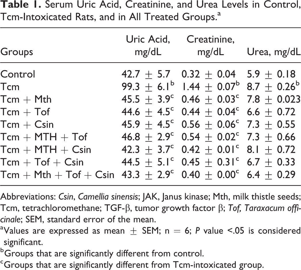
Abbreviations: Csin, Camellia sinensis; JAK, Janus kinase; Mth, milk thistle seeds; Tcm, tetrachloromethane; TGF-β, tumor growth factor β; Tof, Taraxacum officinale; SEM, standard error of the mean.
a Values are expressed as mean ± SEM; n = 6; P value <.05 is considered significant.
b Groups that are significantly different from control.
c Groups that are significantly different from Tcm-intoxicated group.
Figure 1 revealed that Masson trichrome–stained kidney sections with Tcm showed that Tcm increased the deposition of fibrous tissue in the interstitium in between the tubules and the renal corpuscles, while those that received Mth or Tof showed diminution of the abnormal fibrous tissue surrounding renal tubules. On the other hand, Csin treatment showed marked decrease in the abnormal deposition of fibrous tissue.

Masson trichrome–stained kidney sections of rats that (A) received Tcm showed increased deposition of fibrous tissue in the interstitium between the tubules and the renal corpuscles (arrow). B, Kidney tissue from normal control rat with normal distribution of the fibrous tissue restricted to the wall of the blood vessel (arrow). C, Kidney from rat exposed to Tcm and Mth and (D) from rats exposed to Tcm and Tof, showing diminution of the abnormal fibrous tissue surrounding renal tubules (arrows). E, Kidney section from rat exposed to Tcm and Csin showing marked decrease in the abnormal deposition of fibrous tissue (arrow). ×200. Csin indicates Camellia sinensis; Mth, milk thistle seeds; Tcm, tetrachloromethane; Tof, Taraxacum officinale.
Sections of a kidney from a rat exposed to Tcm and administered Mth and Tof or received Mth and Csin as well as Csin and Tof or that received a mixture of Mth, Tof, and Csin showed disappearance of the abnormal fibrous tissue with prominent vascular wall connective tissue fibers (Figure 2).

Masson trichrome–stained sections of kidney from rat (A) exposed to Tcm and administered Mth and Tof. (B) Rats exposed to Tcm, Mth, and Csin; (C) rats exposed to Tcm and Csin and Tof; and (D) rats exposed to Tcm and received mixture of Mth, Tof, and Csin showing disappearance of the abnormal fibrous tissue with prominent vascular wall connective tissue fibers (arrows). ×200. Csin indicates Camellia sinensis; Mth, milk thistle seeds; Tcm, tetrachloromethane; Tof, Taraxacum officinale.
Figure 3 shows anti-TGF-β immunostained kidney sections of rats that received Tcm revealed sporadic focal areas of strong intense immune positivity of the nuclei of many tubular epithelium and nuclei of some glomeruli, while sections from rats that received Mth, Tof, and Csin, each one alone, showed decrease in the abnormal immune reaction of the renal glomeruli, and the tubules showed immune reactivity and less decrease in the immune reaction of both blood sinusoids and the nuclei showed a decrease in the immune reaction that became restricted to the sinusoids and few in the cytoplasm. Sections from a rat administered a mixture of Mth and Tof or Mth and Csin as well as Csin and Tof besides rats that received a combination of Mth, Tof, and Csin demonstrated the moderate immunopositive reaction of nuclei of epithelium renal tubules, while the glomeruli have no immune reaction (Figure 4).

Anti-TGF-β-immunostained kidney sections of rats that (A) received Tcm sporadic focal areas of strong intense immune positivity of the nuclei of many tubular epithelium (arrow) and nuclei of some glomeruli (star). B, Kidney tissue from normal control rat showing moderate immune reaction of renal tubules (arrow), while there is immune positivity of the glomeruli (star). C, Kidney from rats exposed to Tcm and Mth. D, Kidney section from rat exposed to Tcm and received Tof. E, Kidney section from rat exposed to Tcm and Csin showing decrease in abnormal immune reaction of the renal glomeruli (stars), while the tubules show immune reactivity (arrows). Decrease in the immune reaction of both blood sinusoids and nuclei (arrow) was observed. Decrease in immune reaction that becomes restricted to the sinusoids and few in the cytoplasm, while the nuclei are not stained (arrow). Scale bar = 100 µm. Csin indicates Camellia sinensis; Mth, milk thistle seeds; Tcm, tetrachloromethane; TGF-β, tumor growth factor β; Tof, Taraxacum officinale.
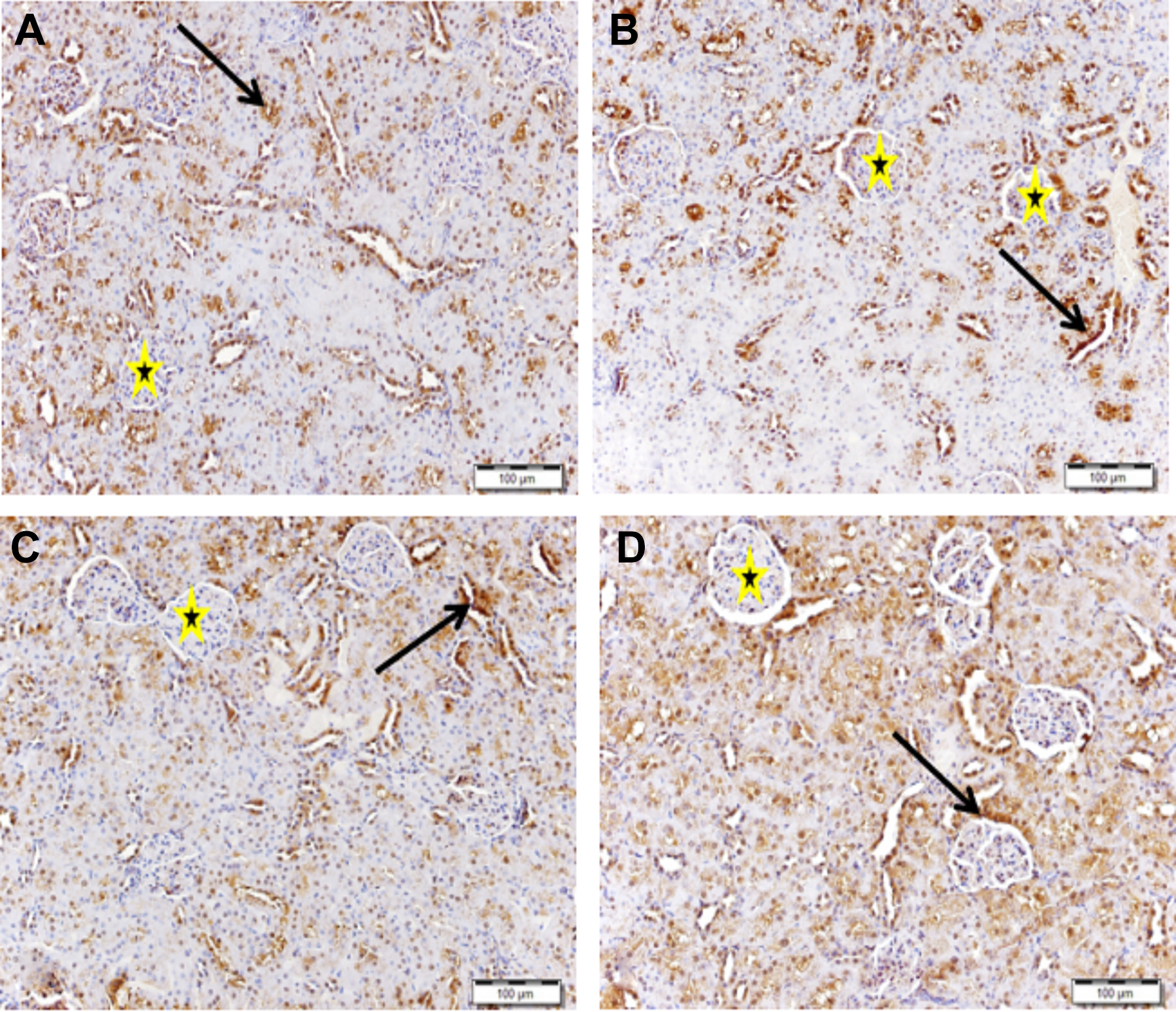
Anti-TGF-β immune-stained sections of kidney from rats (A) exposed to Tcm and administered Mth and Tof; (B) rats exposed to Tcm and Mth and Csin; (C) rats exposed to Tcm and Tof; and (D) rats exposed to Tcm and mixture of Mth, Tof, and Csin demonstrating moderate immunopositive reaction of nuclei of epithelium of renal tubules (arrow), while the glomeruli has no immune reaction (stars). Scale bar = 100 µm. Csin indicates Camellia sinensis; Mth, milk thistle seeds; Tcm, tetrachloromethane; TGF-β, tumor growth factor β; Tof, Taraxacum officinale.
Figure 5 shows anti-JAK immune-stained sections of a kidney from rats exposed to Tcm which demonstrated strong immune positivity of tubular epithelium. While there were few glomeruli with positive reaction, most of the glomeruli showed a negative reaction. Rats that received Mth showed many immunopositive glomeruli and immunonegative tubules. Kidneys from rats that received Tof showed many tubules with positive immune reaction, while the glomeruli showed no immune positivity. Kidney section from rat that received Csin shows many glomeruli and tubules with weak negative immune reaction in addition to glomeruli with a negative reaction. Kidney from rat administered Mth and Tof demonstrated an increase in tubular immunopositivity and the tubules showed no reaction, whereas kidney from rats that received Mth and Csin showed tubules with weak immune reaction: Some glomeruli showed weak immune reaction, while many glomeruli showed immune negative reaction. On the other hand, rats that received Csin and Tof showed very few glomeruli with weak immune positivity, while most of the glomeruli and the tubules showed no immune reaction. Rats that received a mixture of Mth, Tof, and Csin showed a moderate increase in the intensity of the immune reaction of the tubules. Few glomeruli showed segmental immune positivity, while many glomeruli have no immune positivity (Figure 6).

Anti-JAK immune-stained sections of kidney from rats (A) exposed to Tcm show strong immune positivity of tubular epithelium (arrow), while there are few glomeruli with positive reaction (star), and most of glomeruli show negative reaction (red triangle). B, Kidney from control rat shows negative immune reaction of both tubules (arrow) and glomeruli (red triangle). C, Rats exposed to Tcm and Mth show many immunopositive glomeruli (star) and immune negative tubules (arrow). D, Kidney from a rat exposed to Tcm and Tof shows many tubules with positive immune reaction (arrow), while the glomeruli have no immune positivity (red triangles). E, Kidney section from a rat exposed to Tcm and Csin shows many glomeruli (red triangle) and tubules (star) with weak negative immune reaction in addition to glomeruli with negative reaction (red triangle). Scale bar = 100 µm. Csin indicates Camellia sinensis; JAK, Janus kinase; Mth, milk thistle seeds; Tcm, tetrachloromethane; TGF-β, tumor growth factor β; Tof, Taraxacum officinale.

Anti-JAK immune-stained sections of kidney from rat (A) exposed to Tcm and administered Mth and Tof demonstrating increase in tubular immunopositivity (arrow), while the tubules show no reaction (red triangles). B, Kidney from rats exposed to Tcm and Mth and Csin shows tubules with weak immune reaction (arrow), some glomeruli with weak immune reaction (star), and many glomeruli with immune negative reaction (red triangle). C, Rats exposed to Tcm and Csin and Tof show very few glomeruli with weak immune positivity (star), while most of glomeruli (red triangle) and the tubules (arrow) show no immune reaction. D, Rat exposed to Tcm and mixture of Mth, Tof, and Csin showing moderate increase in the intensity of the immune reaction of the tubules (arrow). Few glomeruli show segmental immune positivity (star), while many glomeruli show no immune positivity (red triangle). Scale bar = 100 µm. Csin indicates Camellia sinensis; JAK, Janus kinase; Mth, milk thistle seeds; Tcm, tetrachloromethane; TGF-β, tumor growth factor β; Tof, Taraxacum officinale.
Protein expression of caspase 3 was upregulated, whereas Bcl2 was downregulated upon Tcm administration. Treatments with the aforementioned antioxidants separately or together ameliorated the previous protein expression (Figure 7).

Serum levels of Bcl2 and Caspase-3 in all groups. Note. Data are shown as mean ± standard error of the mean (SEM; N = 6). Csin indicates Camellia sinensis; Mth, milk thistle seeds; Tcm, tetrachloromethane; Tof, Taraxacum officinale.
Discussion
Kidneys are essential organs for the removal of several xenobiotics, such as drugs and ecological pollutants, as well as endogenous metabolites. Kidneys have developed transport systems to avoid urinary loss of filtered nutrients, including glucose, oligopeptides, and inorganic ions, as well as to facilitate the elimination of several xenobiotics. 16
The ROS are generated throughout the detoxification of xenobiotics and drugs and exhibit oxidative stress (OS). Oxidative stress has been shown to be linked to kidney toxicity and diseases. Hence, it is associated with harm to an extensive range of macromolecular species, such as lipids, proteins, and nucleic acids. 17 Chronic renal diseases are rapidly increased all over the world and frequently lead to the entire destruction of functional kidney tissue, and hence, the affected population depends mainly on dialysis along with a life or renal transplantation. 18
The Tcm is metabolized by cytochrome P450 in endoplasmic reticulum and mitochondria with the formation of trichloromethyl peroxyl radicals, a reactive oxidative free radical that initiates lipid peroxidation. 19 It was reported that the intake of Tcm induced acute and chronic renal damage as well as OS in rats. Moreover, severe corruption of renal function was observed in rats as assessed by increased serum creatinine and urea compared to the control rats. Kidney sections of Tcm-treated group revealed changes in microanatomy. 20 The results of the current study were in accordance with that of Elsawy et al, 20 as Tcm exhibited an increase in serum creatinine, urea, and uric acid levels matched with the control values, and treatment with Mth or Tof or Csin alone or in combination improved all the previously measured parameters; the combination regimen achieved the best renoprotective results. Moreover, Masson trichrome–stained kidney sections of rats that received Tcm showed increased deposition of fibrous tissue in the interstitium in between the tubules and the renal corpuscles, whereas administration of Mth or Tof or Csin diminished the abnormal fibrous tissue surrounding renal tubules. The mixture of Mth, Tof, and Csin exhibited disappearance of the abnormal fibrous tissue with prominent vascular wall connective tissue fibers.
Caspase3 and Bcl2 are well-known pro- and antiapoptotic regulatory genes in eukaryotes. 21 Regular function and development of the kidney have a confirmable reliance on apoptosis. 22 Apoptosis, a morphological form of programmed cell death required for the control of cell populations, has been shown to have a role in the cell deletion associated with renal scarring. 23 Tetrachloromethane induced kidney injury as evidenced by elevation in serum biochemical markers and apoptosis induced by overexpression of caspase-3 activity in the kidney. In addition, it decreased the levels of antiapoptotic Bcl-2 protein and increased levels protein of Bax and the release of cytochrome C from mitochondria in the kidneys. 24
In the present study, the protein expression of caspase 3 was upregulated and Bcl-2 protein expression was downregulated upon the exposure to Tcm. The administration of natural products in question attenuated the previous protein expressions, and the combination of the 3 antioxidants achieved the most pronounced antiapoptotic activity.
The cytokine TGF-β signaling cascade is implicated in the chronic kidney diseases that are attributed to the mechanism of fibrogenesis. Excessive renal tissue fibrogenesis is the dominant pathomechanism induced by TGF-β in the kidney, leading to glomerular and tubulointerstitial scarring (apoptosis), which plays a role in the development of renal disease. 25
The role of epithelial–mesenchymal transdifferentiation (EMT) in the progression of glomerular and tubulointerstitial diseases is due to overexpression of TGF-β which mediates apoptosis. Tumor growth factor β is closely linked to SMAD family proteins; these complexes are deposited in the nucleus, where they directly activate the transcription of target genes. 25
In the present study, TGF-β in immunostained kidneys of rats that received Tcm showed sporadic focal areas of strong intense immune positivity of the nuclei of many tubular epithelium and nuclei of some glomeruli. Milk thistle seeds, Tof, and Csin solely showed a decrease in the abnormal immune reaction of the renal glomeruli, while the tubules of TGF-β less decrease the immune reaction of both blood sinusoids and nuclei showed a decrease in immune reaction that becomes restricted to the sinusoids, while the nuclei were not stained. The administration of Mth and Tof or Mth and Csin or Csin and Tof or a mixture of Mth, Tof, and Csin demonstrated the moderate immunopositive reaction of nuclei of the epithelium of renal tubules, while the glomeruli have no immune reaction.
The activation of JAK/STAT signaling pathways can be involved in tubulointerstitial fibrosis and EMT in several conditions, including diabetes, in animal models. 26 –28 Similarly, JAK/STAT activation is reported in rat glomerular cells exposed to high glucose 29 and may be important in the glomerular TGF-β activation and fibronectin accumulation critical for extracellular matrix deposition in early diabetic nephropathy. 30
In the present study, anti-JAK immune-stained sections of a kidney from a rat exposed to Tcm represented strong immune positivity of tubular epithelium, while there are few glomeruli with positive reaction and most of the glomeruli showed a negative reaction. Treatment with Mth shows many immunopositive glomeruli and immuneonegative tubules. Treatment with Tof showed many tubules with a positive immune reaction, while the glomeruli have no immune positivity; Csin treatment showed many glomeruli and tubules with weak negative immune reaction in addition to glomeruli with a negative reaction. The intake of Mth and Tof demonstrated an increase in tubular immunopositivity, while the tubules showed no reaction. Administration of Mth and Csin showed tubules with weak immune reaction and some glomeruli with a weak immune reaction, while many glomeruli showed an immune negative reaction. Rats that received Csin and Tof showed very few glomeruli with weak immune positivity, while most of the glomeruli and the tubules showed no immune reaction. The mixture of Mth, Tof, and Csin showed a moderate increase in the intensity of the immune reaction of the tubules; few glomeruli show segmental immune positivity, while many glomeruli have no immune positivity.
Conclusion
The combination of Mth, Tof, and Csin was synergistically beneficial in the delaying of the progression of kidney diseases by attenuating the previous markers, suggesting their antiapoptotic and antifibrotic potentials, thus avoiding renal disorders induced by Tcm. So it may be considered a novel strategy for the suppression of entire destruction of the functional kidney tissue. Tumor growth factor β, JAK caspase 3, and Bcl2 protein expressions are implicated in Tcm toxicity and treatments.
Footnotes
Acknowledgments
The authors extend their appreciation to the Deanship of Scientific Research at King Saud University for funding this work through RG-1440-017.
Declaration of Conflicting Interests
The author(s) declared no potential conflicts of interest with respect to the research, authorship, and/or publication of this article.
Funding
The author(s) disclosed receipt of the following financial support for the research, authorship, and/or publication of this article: The authors received support from Deanship of Scientific Research at King Saud University for funding this work through RG-1440-017.