
Abstract
Atorvastatin, an inhibitor of 3-hydroxy-3-methylglutaryl-coenzymeA reductase, is usually used for the treatment of hypercholesterolemia. Besides its pharmacological and side actions, its toxic effects on human nucleus devoid of erythrocytes are still unknown. Eryptosis is an alternative term used for suicidal erythrocyte death. Membrane blebbing is among the common markers of eryptosis. In this study, eryptotic effect of atorvastatin was investigated by exposing the erythrocytes for 48 hours to different concentrations (1-10 µM) of atorvastatin. The experimental work related to investigation of eryptosis was done by cell size measurement and calcium channel inhibition. As a possible mechanism of eryptosis, atorvastatin-induced oxidative stress was evaluated by determining catalase, glutathione peroxidase, and superoxide dismutase activities. Similarly, necrotic effect of atorvastatin was also determined by hemolytic assay. Results of our study illustrated that the tested doses of atorvastatin may induce oxidative stress as observed by significant reduction in superoxide dismutase, glutathione peroxidase, and catalase activities as well as induce eryptosis, featured by erythrocytes membrane blebbing. The study concluded that induction of oxidative stress by atorvastatin may lead to eryptosis.
Introduction
An artificially synthesized compound, atorvastatin acts as an inhibitor of 3-hydroxy-3-methylglutaryl-coenzyme A reductase and is used for the treatment of hypertriglyceridemia. 1 In addition to its blood cholesterol-lowering activity, atorvastatin showed antitumor and anti-inflammatory activities. 2 Hepatic tissue toxicity and mitochondrial oxidative stress were observed during its therapeutic treatment of high blood cholesterol level. 1 Oxidative stress development during this therapy resulted in stimulating apoptosis. 3 Oxidative stress produced by this drug is partially effective due to increased entry of extracellular Ca+2. Anemia as a side effect of this therapy may be due to the enhanced death of erythrocytes.
The characteristics of eryptosis include membrane blebbing, shrinkage of cells, 4 and cell membrane scrambling, which leads to phosphatidylserine translocation. 5 Splenic macrophages recognize and engulf erythrocytes with exposed phosphatidylserine. 6 Oxidative stress, osmotic shock, and energy-depleted environment activate Ca2+ [Ca2+]i permeable cation channels, resulting in the activation of eryptosis. This is due to increased activity of cytosolic Ca2+ [Ca2+]I, which results in Ca+2-sensitive K+ channels activation. 7 These channels activation results in cell shrinkage due to KCl loss with osmotically obliged water. 8 Phosphatidylserine translocation due to breakdown of phosphatidylserine asymmetry of erythrocyte’s cell membrane is also the result of increased [Ca2+]i. 9,10 Uncontrolled eryptosis may lead to anemia. 6,11
Xenobiotics may be the stimulators of eryptosis, 5,10 and uncontrolled eryptosis also contributes to the pathophysiology of several clinical disorders. 10,12 The current study explores the eryptotic effect of atorvastatin through oxidative stress induction by physiological doses of atorvastatin.
Material and Method
For experimental work, screened blood samples (n = 10) were collected from different blood banks of Faisalabad city. The work was conducted with the approval of directorate of graduate studies and Institutional Bio-Safety Committee, University of Agriculture Faisalabad, Pakistan.
Leukocyte-depleted cells were prepared following protocol explained by Pastor et al. 13 Isolated erythrocytes were stored in separate microcentrifuge tubes. In vitro incubation of erythrocytes was performed at a hematocrit of 0.4% in Ringer solution (pH 7.4) that contains (in mM) MgSO41, NaCl 125, KCl 5, glucose 5, CaCl21, and N-2-hydroxyethylpiperazine-N-2-ethanesulfonic acid at 37°C for 48 hours. 14 Isolated erythrocytes were then exposed to atorvastatin (Sigma-Aldrich, USA) at the indicated concentrations. 15
Oxidative Stress Measurement
To determine the oxidative stress in atorvastatin-exposed erythrocytes, antioxidant enzyme (catalase, glutathione peroxidase, superoxide dismutase) assays were performed.
Superoxide Dismutase
The activity of superoxide dismutase was measured following the protocol of Giannopolitis and Ries. 16 The reaction solution contained methionine 0.222 g in 15 mL H2O, NBT 0.015 g in 17.5 mL H2O, Triton-X 0.0375 mL in 17.5 mL H2O, riboflavin 0.0132 g in 17.5 mL H2O, and buffer 0.2 M.
Glutathione Peroxidase
Phosphate buffer (pH 5) 50 mM, guaiacol 20 mM, H2O2 40 mM, and enzyme extract 0.1 mL were added in reaction mixture following the protocol of Maehly and Chance, 17 and activity was measured at 470 nm after every 20 seconds.
Catalase
Catalase activity was determined following the protocol of Maehly and Chance. 17 Phosphate buffer (pH 7) 50 mM, H2O2 5.9 nM, and enzyme extract 0.1 mL were added in reaction mixture, and absorbance was read at 240 nm.
Cell Size Measurement
Mean cell volume (MCV) was measured to determine cell size of control and treated cells. Mean cell volume was checked by automated hematology analyzer. 18
Conformation of Role of Ca+2
Amlodipine is a calcium channel blocker. To confirm calcium role in triggering of eryptosis in atorvastatin-exposed erythrocytes, cells were treated with 10 µM amlodipine. The inhibition of eryptosis was confirmed by MCV measurement.
Hemolysis Measurement
After incubation, samples were centrifuged (3 minutes at 400g at room temperature) and supernatant was collected to determine hemolysis. Hemoglobin concentration was measured at 405 nm. 19 The absorption of the supernatant of erythrocytes lysed in d.H2O was defined as 100% hemolysis. 20
Statistical Analysis
All data are expressed as arithmetic means ± standard error of the mean. Statistical analysis was performed by applying analysis of variance with Tukey test as posttest or t test, as appropriate using SPSS software version 17.0. 10
Results and Discussion
The current study was designed to explore the effect of atorvastatin on antioxidant enzymatic activities, erythrocyte size, hemolytic activity, and conformation of calcium role in the induction of suicidal death of erythrocytes. Markers of eryptosis, such as activation of cation channels, membrane blebbing, and possible mechanism of oxidative stress, were triggered when human erythrocytes were exposed to different concentrations of atorvastatin. The drug concentrations used in this study are similar to the concentrations used to study the apoptosis of nucleated cells. 15 Figure 1 illustrates that the 48-hour exposure of erythrocytes to atorvastatin (1-10 µM) results in significantly (P < .05) decreased activities of superoxide dismutase at 10 µM of atorvastatin as compared to control. It has been reported that atorvastatin significantly decreases the activity of superoxide dismutase in treated cells. 21 This enzyme catalyzes the dismutation of O2-free radicals, which, on accumulation, results in lowering the level and mitochondrial functions of superoxide dismutase. This enzyme catalyzes the conversion of superoxide ion into oxygen and H2O2. 22 Figure 2 shows the catalase activities after 48-hour atorvastatin (1-10 µM) exposure to human erythrocytes. Results showed moderate but significant decrease in enzyme activity at 10 µM concentration of atorvastatin as compared to control, which may be due to generation of reactive oxygen species. Previous studies showed a significant decrease in catalase activity, depicting the happening of oxidative stress. 23 Catalase is iron dependent that may act peroxidatively or catalytically. 22 Catalase as a major antioxidant enzyme decomposes H2O2 into H2O. 24 The accumulation of hydrogen peroxide results in decreased catalase activity. Catalase showed a protective effect against oxidants in cells with its overproduction. 25 Figure 3 shows decreased activity of glutathione peroxidase resulted after 48-hour exposure of erythrocytes with atorvastatin, which may be due to generation of reactive oxygen species. Results indicated a significant decrease in glutathione peroxidase activity as compared to control. It is confirmed that reduction in glutathione activity resulted in induction of oxidative stress. 26 Glutathione peroxidase decomposes H2O2 into H2O and O2 that may be catalyzed in the presence of cofactor selenium. 22 Glutathione peroxidase in mitochondrial cell membrane prevents the accumulation of oxidized lipids and decomposes hydrogen peroxide into water. 27

Effect of atorvastatin on superoxide dismutase activities (U/gHb) in erythrocytes. Arithmetic means ± SEM (n = 20) of erythrocytes exposed for 48 hours to Ringer solution without (white bar) or with (black bars; 1-10 μM) atorvastatin. Y-axis bars show the SEM. 3 *P < .05 and ***P < .001 indicate significant difference from the absence of atorvastatin (ANOVA). ANOVA indicates analysis of variance; SEM, standard error of the mean.
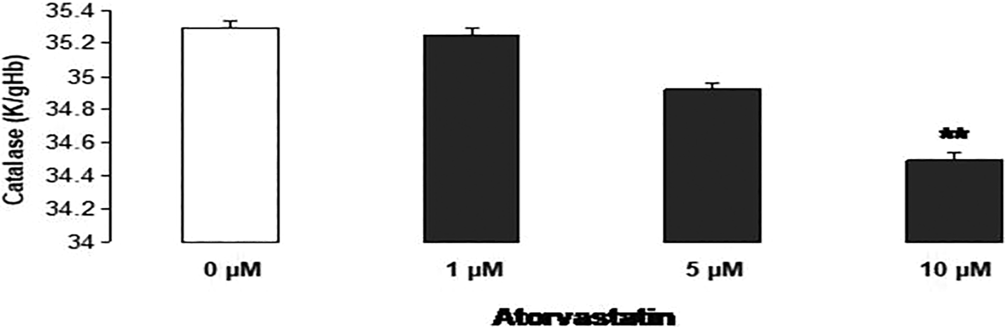
Effect of atorvastatin on catalase activities (K/gHb) in erythrocytes. Arithmetic means ± SEM (n = 20) of the catalase in treated erythrocytes exposed for 48 hours to Ringer solution without (white bar) or with (black bars; 1-10) atorvastatin. Y-axis bars show the SEM. **P < .01 indicates significant difference from the absence of atorvastatin (ANOVA). ANOVA indicates analysis of variance; SEM, standard error of the mean.

Effect of atorvastatin on glutathione peroxidase activities (UlgHb) in erythrocytes. Arithmetic means ± SEM (n = 20) of the glutathione peroxidase in treated erythrocytes exposed for 48 hours to Ringer solution without (white bar) or with (black bars; 1-10 µM) atorvastatin. Y-axis bars show the SEM. *P < .05 indicates significant difference from the absence of atorvastatin (ANOVA). ANOVA indicates analysis of variance; SEM, standard error of the mean.
Figure 4 illustrated that 48-hour exposure of erythrocytes with atorvastatin (1-10 µM) resulted in apparent increase in human MCV, which may be due to erythrocytes membrane blebbing. RBC membrane blebbing, such as swelling or protrusions, is a marker of eryptosis. 28

Effect of atorvastatin on erythrocytes mean cell volume (fL). Arithmetic means ± SEM (n = 10) of erythrocytes exposed for 48 hours to Ringer solution without (white bar) or with (black bars; 1-10 μM) atorvastatin. Y-axis bars show the SEM. ***P < .001 indicates extremely significant difference from the absence of atorvastatin (ANOVA). ANOVA indicates analysis of variance; SEM, standard error of the mean.
Disposing of defective erythrocytes before hemolysis is an important physiological mechanism known as eryptosis. 29 Hemoglobin is released through hemolyzed erythrocytes that may be filtered through kidney or may precipitate in acidic lumen of renal tubules. 30 Figure 5 illustrates that 48-hour exposure of erythrocytes with atorvastatin showed no significant increase in hemolysis percentage as compared to control.

Effect of atorvastatin on hemolysis (%) in erythrocytes. Arithmetic means ± SEM (n = 6) of the hemolysis in erythrocytes exposed for 48 hours to Ringer solution without (white bar) or with (black bars) 0 pM (without) and 10 pM (with) atorvastatin. Y-axis bars show the SEM. ***P > .001 indicates highly significant difference from the absence of atorvastatin (ANOVA). ANOVA indicates analysis of variance; SEM, standard error of the mean.
Increased calcium activity ([Ca2+]i) also resulted in triggering of eryptosis. 10 For the confirmation of Ca+2 role in the stimulation of eryptosis by atorvastatin, calcium channel blocker amlodipine (10 µM) was used. Figure 6 shows the cell size measurement of erythrocytes after 48-hour exposure to atorvastatin (1-10 µM) and amlodipine that resulted in significant decrease in cell size and blebbing, which may be due to calcium entry inhibition. Amlodipine nonselectively inhibits cation channels and stops the Ca+2 entry in cell. 31 Nonselective cation channels are triggered by oxidative stress. 10 In this experiment, no membrane blebbing is observed, which is a characteristic of eryptosis. 12 By removing intracellular and extracellular Ca+2, similar effects would be observed. 32

Cell size measurement of atorvastatin-exposed erythrocytes after calcium channel inhibition. Arithmetic means ± SEM (n = 10) of erythrocytes exposed for 48 hours to Ringer solution without (white bar) and with (black bars) 10 pM atorvastatin. Y-axis bars show the SEM. ***P < .001 shows the significant effect of erythrocytes size measurement activity between 0 and 10 μM atorvastatin-treated erythrocytes. ### P < .001 indicates significant difference in cell size in comparison to untreated cells (ANOVA). ANOVA indicates analysis of variance; SEM, standard error of the mean.
The present study concluded that atorvastatin (10 µM) may enhance the rate of erythrocytes death by eryptosis with possible mechanism of oxidative stress.
Footnotes
Authors’ Note
Rumaisa Bashir Rana and Kashif Jilani contributed equally and thus shares first authorship.
Declaration of Conflicting Interests
The author(s) declared no potential conflicts of interest with respect to the research, authorship, and/or publication of this article.
Funding
The author(s) received no financial support for the research, authorship, and/or publication of this article.