
Abstract
The purpose of this study is to investigate the potentiality of Gafchromic external beam therapy 3 (EBT3) film to measure low dosage of solar ultraviolet (SUV; 0-10 600 mJ/cm2) and x-ray (0–750 mGy) radiation. In this experiment, 2 groups of EBT3 films were prepared with size 2 cm × 1 cm. The first group of films was exposed by incremental SUV dose in the middle of the day. The other group was irradiated by x-ray at 100 kVp, 100 mA, and 2 S of tube voltage, tube current, and exposure time, respectively. The measured SUV consists of 90% ultraviolet A (UVA) and 10% ultraviolet B. The film discoloration was represented by visible absorbance spectroscopy technique using Jaz spectrometer from Ocean Optics Inc. Simple linear regression produced high accuracy with coefficients of determination, r 2 of 0.9804 and root mean square error (RMSE) of 434.88 mJ/cm2 for the measurement of SUV dose. On the other hand, r 2 of 0.98 and RMSE of 31 mGy was produced for the measurement of x-ray dose. The application of multiple linear regression enhanced the measurement accuracy with R 2 of 99% and 99.7% and RMSE of 327.06 mJ/cm2 and 15.045 mGy for SUV and x-ray dose, respectively. The spectral analysis shows a promising measurement at selected wavelengths for SUV and x-ray dose.
Introduction
Radiation is production and emission of energy in the form of electromagnetic waves or particles. Electromagnetic radiation includes γ ray, x-ray, ultraviolet (UV), visible light, infrared, microwaves, and radio waves. Radiation can be categorized into ionizing and nonionizing radiation based on the radiation ability to ionize matters. Practically, the threshold between ionizing and nonionizing radiation is around 13.6 eV, which is the energy required to ionize hydrogen atom.
Knowledge of UV doses deposited in a matter is essential for an assessment of associated hazards. 1,2 Solar ultraviolet (SUV) is the primary natural source for UV radiation (UVR). A small amount of SUV is essential to produce vitamin D, but on the contrary, overexposure can cause various dangerous implications to human skin, immune system, and eyes. 3 The biological effects, however, depend on UV wavelength, location, and duration of exposure. 4 Solar ultraviolet spectrum is divided into 3 regions, ultraviolet A (UVA; 315-400 nm), ultraviolet B (UVB; 280-315 nm), and ultraviolet C (UVC; 100-280 nm) depending to their impact on human. 3 Solar ultraviolet radiation that reaches to earth comprises 95% UVA, and the other is UVB. 4
Ultraviolet radiation is a known carcinogen because the skin cancer increases due to the exposure to UVR. Over 90% of melanomas are caused due to SUV exposure in North America and Australia. The other risk of UV exposure is the cortical cataract. By 2050, it is expected that the incidence of cortical cataract due to UV exposure will increase up to 1.3% to 6.9% and the incidents cases will be 83 000 to 167 000 cases. 5,6
The implication of UV toward human is limited to the eyes and skin. Based on the global burden of disease data from the World Health Organization, excessive solar UV exposure caused the loss of approximately 1.5 million disability-adjusted life years and 60 000 premature deaths in the year 2000. The highest burden of disease results from cortical cataracts, malignant melanoma, and sunburn. However, no risk management processes are available to manage underexposure to UVR and the resultant health impacts particularly due to vitamin D deficiency though the impact (death and disability) of underexposure to UV is potentially thousands times higher than for overexposure to UV. 7
The intensity of SUV increases with increasing altitude and decreasing latitude. This intensity depends on the zenith angle. Thus, the intensity changes throughout the day time and the annual seasons. In the day, UVB is most intense in the middle of the day, that is, from 11:00
Ultraviolet dose can be measured with many types of dosimeters. Electronic dosimeters are expensive, and they read instantaneous UV irradiance but not the exposure. 3 Gafchromic films are sensitive to irradiation. This sensitivity is represented as a permanent change in their visible color. 3,8 Film discoloration depends on UV irradiance, UV wavelength, and exposure time. The darkness of the films is directly proportional to the concentration of light absorbed by the film’s material. 9 Three different generation of Gafchromic films were produced, external beam therapy called EBT, EBT2, and EBT3. 3 Comparing to EBT2, EBT3 films offer 2 main improvements: symmetry of the film and elimination of Newton ring coating. 10
The understanding of x-ray radiation dose is necessary to avoid the adverse effects due to the high dose. High dose of x-ray can cause biological effects including tissue reactions deterministic impacts such as erythema or cataract and cancer effect stochastic such as neoplasms. In tissue reactions, the number of cells are affected before the response is taken place, that is, there is a threshold for biological response to be observed. On the other hand, in cancer effect, the induced changes by the radiation appear in a single cell. These changes can initiate the biological process, which leads to the effect. 11,12
There are different types of x-ray detectors that can measure the absorbed dose, including thermoluminscent detector, semiconductor detector, and ion chamber. The EBT3 films can be used as x-ray dosimeter and also known to have good sensitivity for nonionizing UV dose measurement. Gafchromic films were originally made to quantify clinical x-ray doses due to their sensitivity to ionizing radiation. 1,4 Radiochromic films in general have solved several problems related to conventional 2-dimensional radiation detectors with its high spatial resolution, low energy dependence, and near-tissue equivalence characteristic. This is also served as the advantages in SUV measurement compared to conventional UV meter that captures the radiation in the unit of power per unit area (mW/cm2), instead of accumulated power over time (mW/cm2). The use of film-based dosimeter allows the SUV measurement to be made simultaneously across an area, an approach that will be costly if to be conducted using electronics UV meter. 13 These films can measure dose in the range from 0.2 up to 100 Gy for x-ray and up to 30 J/cm2 for SUV. 4,14 Information on EBT films absorption spectrum is important in designing efficient optical densitometer and achieving an accurate sense of the dosimetric film system. 15
Furthermore, until today, the most established methods employed for characterizing the color changes of EBT3 films either for UV or x-ray measurement is through flatbed scanner where the best response was commonly retrieved through red component of the images. 1,10,16 -19
Many published works reported dose measurements using EBT3 for x-ray and UV 20 and established a procedure to use EBT3 for measuring SUV radiation using red LED. Aydarous et al 4 showed that EBT3 could respond to UVA and UVB. León-Marroquín et al 15 studied analysis of EBT3 spectrum that irradiated with x-rays with a nominal energy of 6 MV.
Though various papers on the application of EBT3 are available thus far, comparative analysis between the films’ response toward UV and x-ray dose is not available. Hence, the purpose of this study is to compare the discoloration of EBT3 films through absorbance spectroscopy analysis toward low doses of SUV and x-rays.
Materials and Methods
SUV Exposure
In this experiment, International Specialty Products Gafchromic EBT3 films (lot no. 03071601) were cut into small pieces with a dimensional size of 2 cm × 1 cm. Forty-eight pieces were prepared to be exposed simultaneously to solar UV. Every 30 seconds, one exposed film was collected in order to get levels of accumulated UV doses. The irradiance of UVA + UVB was measured in mW/cm2 using UVA and UVB radiometers, model 4.0 and 6.0, respectively. The total irradiance of UVA + UVB was multiplied by the duration of exposure to get the UV dose values of films in mJ/cm2. 20 During the experiment, the temperature and humidity near films were measured using temperature humidity meter, Fluke971 device. Relative humidity range was (55%-65%), and the temperature range was (30.3°C-34.2°C). The exposed films then were put inside black envelopes to prevent films from other exposure. The absorbance of exposed films was measured after 48 hours to allow for color stabilization. 4
External Beam Therapy 3 Film Under X-Ray Irradiation
Twelve Gafchromic EBT3 films with the same dimensional sizes, 2 cm × 1 cm, were directly irradiated by x-rays (in the range 0-750 mGy). The parameters of the x-ray machine were set to be 100 kVp, 100 mA, and 2 seconds as they refer to the radiation quality, tube current, and the radiation exposure conditions, respectively. The variation of doses was obtained by varying the irradiation exposure time. Then, the film dose was measured using a dosimeter Model Diadose PTW Diagnostic Dosimeter T11003. The field size was 10 cm × 10 cm at a source to surface distance of 1 m. Next, the exposed films were kept in an envelope.
Films were then taken to the laboratory for the absorbance measurement via visible absorbance spectroscopy technique. Absorbance measurement was performed using Jaz spectrometer. The light source, which connected to Jaz spectrometer was a tungsten halogen emits light from 360 to 2000 nm. To capture and analyze the spectral data, SpecrtraSuite software program was used. The spectral reference used in this setup was an unexposed EBT3 film. The spectral sensitivity parameters were chosen to cover visible spectrum range between 400 and 700 nm.
Results and Discussion
Figure 1 shows the net visible light absorption spectrum (optical density) in the region between 500 and 700 nm for exposed EBT3 films when irradiated with incremental solar UV and x-ray dose. From these graphs, the net absorption spectra of the films show 2 absorption bands. The highest absorption value centered around 636 nm, while the lower one centered around 585 nm. Aydarous et al recorded absorbance peak for EBT3 at 633 and 582 nm for UV dose measurement up to approximately 60 J/cm2. On the other hand, León-Marroquín et al observed the highest absorbance peak at 636 nm and less intense peak at around 585 nm in the measurement of x-ray dose up to 50 Gy. 4,16 The absorption peaks change their positions toward shorter or longer wavelength depending on the dose. In this experiment, since the measurement of x-ray was only conducted for maximum dose of 884 mGy or correspond to 0.4 in absorbance value, the peak absorbance wavelength was identified to be located at 634 nm (for doses less than 589 mGy) and slightly shifted to lower wavelength of 632 nm for dose measurement at 884 mGy. On the other hand, since SUV dose measured in this experiment leads to much larger absorbance value for the films, the shift in peak absorbance wavelength is more significant. The peak absorbance for dose measurement of 423 mJ/cm2 is recorded at 631 nm (correspond to 0.2 in absorbance value), while the peak absorbance wavelength for SUV dose measurement of 10628.1 mJ/cm2 is recorded at 625 nm (correspond to 1.457 in absorbance value). As a result, the absorbance graph for the SUV dose measurement appears to have shoulder-like response at peak absorbance for higher dose measurement.

Visible absorbance spectra for EBT3 films irradiated by (A) x-rays and (B) SUV. EBT3 indicates external beam therapy 3; SUV, solar ultraviolet.
Higher doses of SUV and x-ray turned the EBT3 films to darker color and, as a result, raised the absorbance of the films. Although this research is specifically designed to target low level of doses, the gradual color changes may not be visually significance but can be adequately quantified through spectroscopy method.
The linear analysis was specifically conducted in the range from 540 up to 675 nm. This range shows a highly correlated in color changes of EBT3 films, which indirectly indicate UV or x-ray dose. Figure 2A shows the relationship of absorbance versus dose for both solar UV and x-ray at λ = 636 nm. Around this wavelength, the graphs present the highest value of absorbance. The absorbance values were multiplied by 1000 to produce a meaningful value of response slope. Figure 2B and C shows the graphs of a relationship between the coefficient of determination, r 2 value, and standard error versus wavelength. The r 2 was produced from the linear regression between dose and absorption of the films. From UV response as shown in Figure 2B, at λ = 605 nm, the graph can produce the highest linearity index with r 2 = 0.9804 and root mean square error (RMSE) = 434.88 mJ/cm2. On the other hand, for x-ray at λ = 636 nm, r 2 = 0.98, and RMSE = 30 mGy.
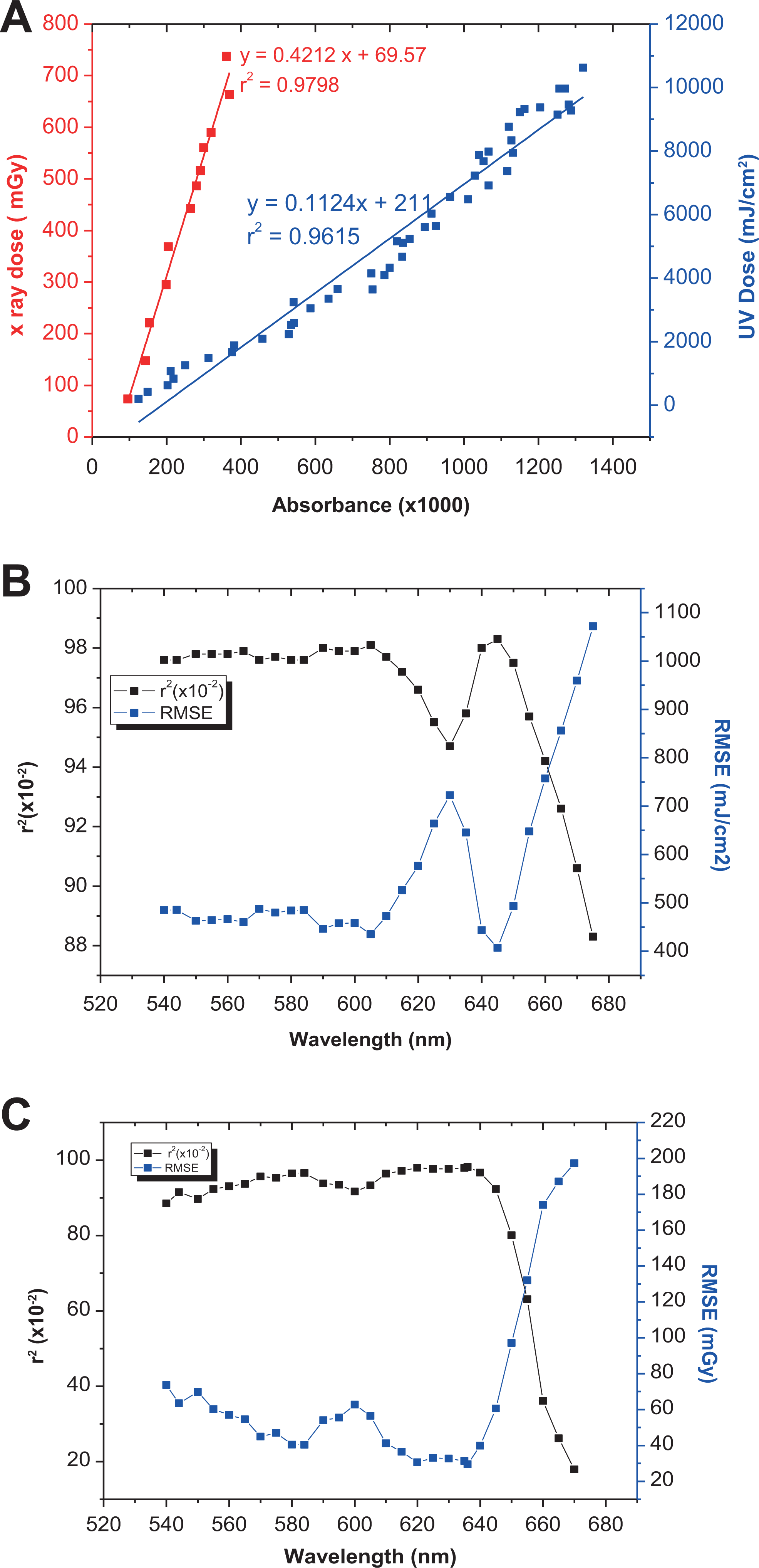
(A) Linear relationship between absorbance with SUV and x-ray doses at λ = 636 nm. Values of r 2 and RMSE generated for wavelengths between 520 and 670 nm in the measurement of (B) solar ultraviolet and (C) x-ray doses. RMSE indicates root mean square error; SUV, solar ultraviolet.
The line slope indicates the response or sensitivity of EBT3 color toward radiation exposure (ie, either SUV or x-ray) where higher slope indicates higher responsivity. This responsivity varies from one wavelength to the other depending on the transition range of EBT3 colors. Figure 3 shows the relationship between slopes, m, with wavelength for both SUV and x-ray, respectively. From Figure 3, the highest responsivity for SUV was recorded at λ = 620 nm with slope, m = 0.127, and for x-ray, the maximum value of slope, m = 0.485 at λ = 636 nm.

Relationship between slopes versus wavelength for both SUV and x-ray. SUV indicates solar ultraviolet.
Table 1 summarizes the maximum value of both coefficient of determination, r 2, and slope, m. In this work, the coefficient of determination r 2 will be chosen since the responsivity (m) can be amplified by multiplying the values by a factor.
Summary of Maximum Values of r 2 and m.

Abbreviation: SUV, solar ultraviolet.
Multiple linear regression (MLR) is used to determine the correlation between 2 or more predicted variables with one response variable and to make predictions for the response using the relation. Tables 2 and 3 present the best wavelength subsets using Minitab software version 18. The selected subset is that has the lowest value of RMSE and the highest coefficient of determination, R 2. From Table 2, the best combination of SUV dose in mJ/cm2 that satisfies previous condition is 544.5, 584.45, 605.12, 620.4, and 646.31 nm combination where RMSE = 327.06 mJ/cm2 and R 2 = 99%, and the developed algorithm can be represented using equation 1.

Results From Multiple Linear Regression Technique Using Selected Wavelengths in the Development of EBT3-Based Spectroscopic Algorithm in Measuring Solar Ultraviolet Doses.

Abbreviations: EBT3, external beam therapy 3; RMSE, root mean square error.
Results From Multiple Linear Regression Technique Using Selected Wavelengths in the Development of EBT3-Based Spectroscopic Algorithm in Measuring X-Ray Doses.
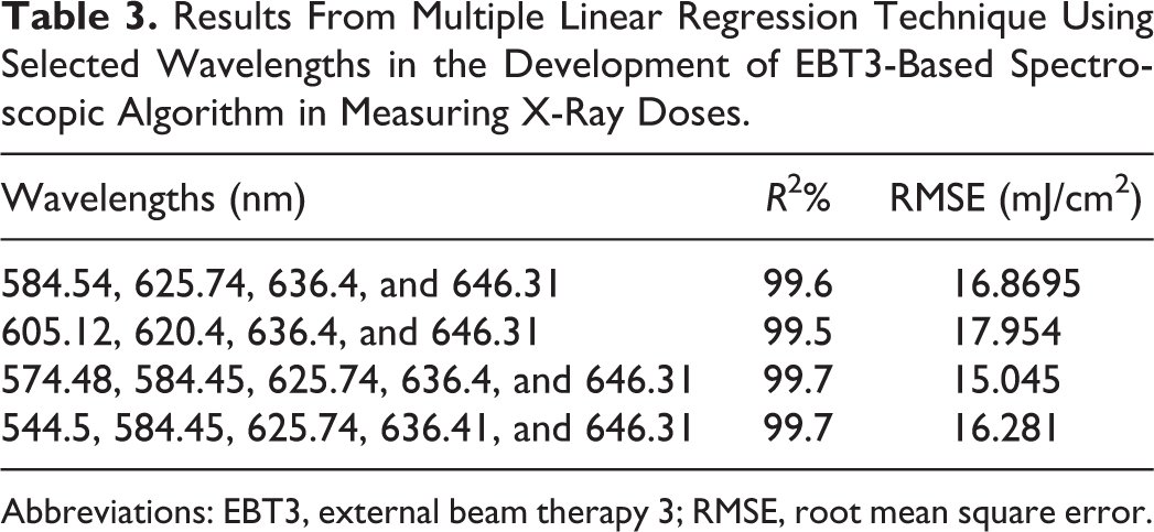
Abbreviations: EBT3, external beam therapy 3; RMSE, root mean square error.
where

Conclusion
This article shows that the EBT3 films are suitable to be used as SUV and x-ray dosimetry with high accuracy. Using visible absorbance spectroscopy technique, the film’s absorption spectra show 2 distinguishable absorption peaks around 580 and 630 nm. The linear analysis of the film’s absorbance presents the highly correlated variation in discoloration with r 2 = 0.9804 with RMSE = 434.88 mJ/cm2 at λ = 605 nm and r 2 = 0.98 with RMSE = 30 mGy at λ = 633 nm for SUV and x-ray, respectively. The MLR has produced high measurement accuracies with R 2 = 99% with RMSE = 327.06 mJ/cm2 and R 2 = 99.7% with RMSE = 15.045 mGy for SUV and x-ray, respectively. The high-accuracy spectroscopy–based algorithms computed in measuring the color changes of EBT3 films in relation to the absorbed SUV and x-ray dose may lead to the development of a single film–based dosimeter that is calibrated for various types of ionizing and nonionizing radiation measurement. Since this article focused on lower range of SUV and x-ray dose measurement, further exploration is, therefore, required to quantitatively determine the radiation doses from other UV and x-ray radiation sources through the development of spectroscopic algorithms for doses up to the films detection limit using common visible absorbance spectroscopy setup.
Footnotes
Acknowledgments
The authors thank the School of Physics at USM University for supporting this research and providing the appropriate research environment.
Declaration of Conflicting Interests
The author(s) declared no potential conflicts of interest with respect to the research, authorship, and/or publication of this article.
Funding
The author(s) disclosed receipt of the following financial support for the research, authorship, and/or publication of this article: This study was financially supported by the Malaysian Ministry of Higher Education Fundamental Research Grant Scheme (grant no. 203/PFIZIK/6711491). A sincere appreciation to the Deanship of Scientific Research at King Saud University for its funding of this research through the Research Group Project no. RGP-246. Our gratitude also goes to the RCMO USM, for supporting us with the Bridging grant (304.PFIZIK.6316276).