
Abstract
Purpose:
Radiochromic EBT3 film is gaining acceptance as a valuable dosimetry system for high-energy photon beams. The advantages of these films over other dosimetry systems are low spectral sensitivity and high spatial resolution. The aim of this study was to validate EBT3 film as a dosimeter for machine and treatment quality assurance (QA) of a 50-kV radiotherapy unit.
Methods and Materials:
Absolute and relative doses were acquired using EBT3 GafChromic films and compared to a parallel-plate ionization chamber (IC), the standard IC for low-energy X-rays. EBT3 was also used to evaluate beam profiles and output factors. Two films above each other, mimicking the clinical situation of a dosimeter on top of the skin, were simultaneously irradiated to evaluate EBT3 as in vivo dosimeter. All films were irradiated for 3 minutes, which corresponds with a surface dose of 3.25 ± 0.07 Gy.
Results:
A fifth-order polynomial function was found to be the best fit for the calibration curves. Good agreement between IC and EBT3 was found for absolute (0.92% for green and red color channels) and relative (1.2% and 1.0% for green and red color channels, respectively) dosimetry. Output factors for IC and EBT3 were comparable within 2.04% and 1.02% for the green and red color channels, respectively. Flatness and symmetry at the surface were within 2%. By applying film as in vivo dosimeter, an absorption of 4.70% needs to be taken into account with respect to the surface dose.
Conclusion:
EBT3 GafChromic film is a feasible and valuable QA and dosimetry tool for a 50-kV radiotherapy unit. EBT3 can be used for absolute and relative dosimetry, measurement of output factors and beam profiles. In vivo patient-specific QA can also be performed if one corrects for the dose absorption of the film.
Keywords
Purpose
The application of low-energy X-rays is being revisited in radiotherapy, particularly for the treatment of skin, rectal, and endometrial tumors and for the intraoperative irradiation after breast conserving surgery. 1 The original approach made use of the Philips RT50 machine, which had a short source to skin distance (SSD) of 3 to 5 cm. 2 After a period where no contact X-ray therapy machines were available, several new devices were introduced such as the Intrabeam (Carl Zeiss Meditec, Jena, Germany) and the Papillon50 (Ariane Medical Systems, Derby, England). When compared to a short SSD, the long SSD of 15 to 30 cm in those machines improves dose uniformity within the tumor. 3
In modern radiotherapy, machine- and patient-specific quality assurance (QA) is becoming essential. Film is often selected for various applications such as measurement of percentage depth dose (PDD) curves, QA of intensity-modulated radiation therapy, stereotactic radiosurgery, and brachytherapy. 4 -6 Moreover, there is a new trend going to patient-specific daily QA using online treatment verification systems highlighting the importance of in vivo dosimetry. 7 The use of radiochromic film has several advantages over other dosimetry systems, such as high spatial resolution and low spectral sensitivity. 8 Recently, EBT3 GafChromic film (Industrial Speciality Ingredients, Wayne) was released as a successor of the EBT2 film and has been gaining acceptance as a valuable dosimetry system. EBT3 film has the same active layer as its precursor EBT2 but has some new features such as the symmetrical structure and anti-Newton ring coatings, which reduce artifacts while scanning. 4 In the case of low-energy X-rays, originally BJR tabulated values 8 were used to obtain PDD curves. Later on, the AAPM TG-61 9 and the NCS 10 report 10 for low-energy dosimetry became available which recommended the use of a soft X-ray parallel-plate ionization chamber (IC). This type of chamber usually presents flatter energy dependence in the low-energy region than cylindrical chambers. 11 Furthermore, in a topical review, Hill et al 12 suggested that for the very low-energy X-ray beams, typically less than 50 kV, the gold standard detector is a low-energy X-ray parallel plate IC. Li et al 13 investigated 9 different detectors, including parallel plate ICs, for the use in the energy range of 50 to 300 kV. In comparison with Monte Carlo calculated data, they found that the parallel-plate IC is a reliable detector for depth dose measurements with an uncertainty of 3% without the need to apply correction factors.
With the introduction of film, many studies were performed to evaluate film as dosimetry tool. 4 -6,14,15 EBT and EBT2 GafChromic film showed strong energy dependence in the kilovoltage energy range, which makes these dosimeters not suitable for low-energy dosimetry due to beam hardening which influences the energy of the X-ray beam. 14 Brown et al found weak energy dependence over an energy range of 25 kV to 4 MV and suggested EBT3 to be a more suitable dosimeter for low- and high-energy X-rays. 15
In this feasibility study, we evaluated EBT3 as dosimeter over the entire treatment chain of a 50-kV radiotherapy unit and validated its use for absolute and relative dosimetry. Furthermore, we measured output factors and beam profiles (ie, symmetry and flatness) with EBT3. As a last step, we examined the use of EBT3 as in vivo dosimeter during contact radiotherapy.
Materials and Methods
Treatment Unit
A 50-kV radiotherapy unit, Dermopan 2 system (Siemens Industry, Berlin, Germany), operating with a 3 cm circular applicator and 1 mm inherent aluminum (Al) filtration was used to perform all measurements. The half-value layer of the beam was 0.92 mm Al, which was determined using high purity (99.99%) Al foils.
Irradiation Setup
A solid soft X-ray phantom type 2962 (PTW, Freiburg, Germany), designed for absolute and relative dosimetry in the range of 7.5 to 100 kV, was used to perform all measurements. It consists of polymethylmethacrylaat (PMMA) plates (13 × 13 cm2) of different thicknesses (1, 2, 5, and 10 mm) in which EBT3 films and the IC were inserted.
PTW 23342IC (PTW, Freiburg, Germany), which is the standard IC for dose measurements of low-energy photons, and EBT3 GafChromic film were assessed to perform the measurements. The IC has a vented sensitive volume of 0.02 cm3, a very thin entrance window of 0.03 mm polyethylene, and was calibrated for dose to water. The energy response is flat in the range of 10 to 100 kV. The IC readings were converted into dose to water at the water surface (Dw,Q) using the TRS-398 protocol. 16 EBT3 GafChromic film consists of a 28 μm thick active layer, which is in between 2 transparent polyester substrates of 120 μm 17,18 and changes color when exposed to ionizing radiation.
For the film calibration, EBT3 GafChromic films were cut into pieces of 5 × 5 cm2 and marked for orientation. Films were irradiated on the phantom’s surface and exposed to a dose ranging from 0 up to 5 Gy in steps of 0.5 Gy by placing the applicator in direct contact with the films, corresponding to an SSD of 30 cm. The dose response will vary with depth due to beam hardening, as suggested by Arjomandy et al. 19 For this reason, the calibration curves have been obtained at the surface and at 10 mm depth.
To obtain the calibration curves, optical densities (ODs) were plotted against absorbed doses measured with the IC. 17 Scan values for the nonirradiated and irradiated films were used to obtain OD. 20 Optical density was defined as −log(scan value/(216 − 1)). The scan values were acquired in Matlab R2012B (The Mathworks Inc, Massachusetts) for the green and red color channels using a circular region of interest (ROI) with a diameter of 1 cm in the center of the imported image. All calibration films and films from measurements were scanned 24 hours postirradiation, one by one in landscape orientation with an Epson 10000XL flatbed scanner and its associated software EPSON Scan version 3.49A (Seiko Epson Corporation, Nagano, Japan). Due to the symmetric structure of the EBT3 film, it is possible to scan the films with each side facing the scanner light source. 21 The scanner was warmed up by taking 5 preview scans to ensure reproducibility between measurements. To avoid rotational scanning artifacts and to ensure that a reproducible central location of the scanbed was used, a cardboard template was fixed on the scanbed in which the films fit. The scan software was used in professional mode with all image adjustment features turned off. Images of the films were registered in 48-bit color with a resolution of 75 dpi. Each film was scanned 3 consecutive times in order to compensate for changes in scanner lamp output, and images were stored in tagged image file format. The mean scan value was used for the OD calculation. More details about the film handling, calibration, and read-out procedure can be found in previous studies. 17,19,22
Absolute Dosimetry
Absolute dose measurements were performed by placing the IC or a piece of EBT3 film (5 × 5 cm2) at the surface of the phantom. The IC and the films were irradiated separately for 3 minutes (absorbed dose of 3.25 Gy) by direct contact with the applicator toward the IC and film. Scanned images were imported in Matlab, splitted in green and red color channels. Subsequently OD were calculated using the scan values of the ROI and converted into absolute doses using the calibration curves at 0 mm.
Depth Dose Curves
Depth dose curves were acquired by irradiating the IC and EBT3 films for 3 minutes (surface dose of 3.25 Gy) in the phantom. Films were placed perpendicular to the beam axis at depths from 0 to 30 mm (per 1 mm for 0-15 mm and per 2 mm for 15-30 mm). Optical densities, obtained in a centrally ROI of the image using Matlab, were converted into doses using the calibration curves. EBT3 films were also exposed parallel to the beam axis by placing the film at the border of the plates, which makes it possible to bring one side of the film in direct contact with the applicator. After placing the film in the right position, the PMMA plates were taped together to avoid an air gap. Doses for all depths were determined on a pixelline on the beam axis from 0 to 30 mm depth using Matlab and the calibration curves. The uncertainty of the measurements was given by the distribution obtained from 3 repeated measurements for each experiment.
Output Factors, Beam Profiles, Symmetry, and Flatness
Relative output factors for nonstandard applicator sizes (2 cm and 1 cm diameter) were determined by irradiating EBT3 film perpendicular to the beam axis for 3 minutes. These were calculated as the ratio of the dose rate for a given applicator to the dose rate of the standard applicator of 3 cm.
To obtain the beam profiles, the center of the circular irradiation field was automatically defined using an in-house developed script in Matlab. Absorbed doses were determined on a pixelline across the center. The absorbed dose in the center of the circular field was normalized to 100%.
In order to evaluate the symmetry and flatness of the beam, the beam profile (0 mm) was analyzed. For the symmetry, doses from 2 points at equivalent distance of the central axis were compared. The difference between these 2 points may not exceed 3%. The flatness was appreciated by looking at absorbed doses into the flat region of the beam profile. The optimal level of deviation should be within ±3% of the central axis dose as stated by Kouloulias. 23
In Vivo Measurements
In order to evaluate whether EBT3 film can also be used as in vivo dosimeter, 2 pieces of film above each other were irradiated for 3 minutes (surface dose of 3.25 Gy) on top of the phantom. The upper film represents the in vivo dosimeter and the lower film represents the skin of the patient, the latter to measure the actual delivered treatment dose. The dose for each film was obtained and compared in order to evaluate whether the upper film can reproduce the skin dose in in vivo situations while the lower film receives the prescribed dose.
Results
The images were obtained by scanning the EBT3 films when the polymerization was stable. In order to determine the postirradiation timepoint at which the films need to be scanned, OD of several films were investigated at 0, 1, 2, 3, 4, 6, 9, 12, 18, 24, 33, and 48 hours after irradiation. Stabilization of the polymerization process and OD was found 24 hours after irradiation. Furthermore, OD variation between 24 and 33 hours was less than 0.001 for all doses under study (results not shown). These findings are in accordance with Casanova Borca et al, 21 who reported variations in OD of less than 0.005 for a 6-MV X-ray beam between 24 and 72 hours after irradiation of EBT3 films in a dose range from 0.3 to 4 Gy. Several films were also scanned and analyzed at a resolution of 300 dpi, which showed a small difference of 0.13% in scan values (results not shown).
The calibration curves at 0 and 10 mm depth for EBT3 GafChromic film in terms of absorbed dose versus OD for the green and red color channels in a dose range from 0 to 5 Gy are displayed in Figure 1. A fifth-order polynomial function was found to be the best fit between dose and OD (R2 > .999) and used to plot the calibration curves. When comparing equal doses, the red color channel showed the highest response in OD. Due to beam hardening, the calibration curve at 0 mm was used to convert OD into dose for films irradiated between 0 and 9 mm depth. The calibration curve at 10 mm was used to convert OD into dose for films irradiated at 10 mm depth and deeper.

Fifth-order polynomial calibration curves at 0 and 10 mm depth.
Absolute Dosimetry
Absolute dose measurements for IC at the surface gave an absorbed dose of 3.25 ± 0.07 Gy. EBT3 gave absorbed doses of 3.28 ± 0.03 Gy for green and red color channels. A difference of 0.92% between the IC and EBT3 (both color channels) was found.
Depth Dose Curves
Depth dose curves obtained with IC and EBT3 film perpendicular to the beam axis are shown in Figure 2. Good agreement between both detectors over the whole range of depths was found. The average difference between IC and EBT3 film is 0.039 ± 0.036 Gy (1.21% ± 1.11%) and 0.033 ± 0.031 Gy (1.01% ± 0.95%) for the green and red color channels, respectively, confirming good concordance between both detectors. A maximal difference of 0.11 Gy (3.5%) for the green and 0.10 Gy (3.1%) for the red color channels was found. The mean uncertainty on the curves was found as 0.017 Gy (maximal 0.024 Gy), 0.064 Gy (maximal 0.125 Gy), and 0.050 Gy (maximal 0.102 Gy) for the IC, green, and red color channels, respectively.

Depth dose curves obtained from parallel-plate ionization chamber and EBT3 film measurements perpendicular to the beam axis (mean error of 0.017, 0.064, and 0.050 Gy for the ionization chamber, green color channel, and red color channel, respectively).
Figure 3 shows a depth dose curve acquired with EBT3 film exposed parallel to the beam axis and plotted against the IC. Good agreement in absolute doses was found between EBT3 film and IC starting at 3 mm depth. The average absolute difference between both detectors is 0.03 ± 0.03 and 0.04 ± 0.02 Gy for the green and red color channels, respectively. A maximal absolute difference of 0.08 Gy was found for both color channels. The mean uncertainty on the absolute depth dose curves was found as 0.017 Gy (maximal 0.024 Gy), 0.053 Gy (maximal 0.116 Gy), and 0.055 Gy (0.093 Gy) for the IC, green, and red color channels, respectively. Figure 4 combines the depth dose curves of the IC, perpendicular, and parallel film measurements.

Depth dose curves from ionization chamber and EBT3 film measurements parallel to the beam axis (mean error of 0.017, 0.053, and 0.055 Gy for the ionization chamber, green color channel, and red color channel, respectively).

Depth dose curves from ionization chamber and EBT3 film measurements perpendicular and parallel (starting at 3 mm depth) to the beam axis.
Output Factors, Beam Profiles, Symmetry, and Flatness
Output factors obtained for nonstandard applicator sizes are listed in Table 1. For the 1 cm applicator, a difference of 3.3% and 2.2% was found between IC and EBT3 for the green and red color channels, respectively. Compared to IC, EBT3 showed agreement within 2% for the green and 1% for the red color channels for the 2 cm applicator. Best agreement between film and IC was found for the red color channel.
Output Factors for Different Applicator Sizes.

Figure 5 shows the beam profiles obtained with EBT3 for the 3 cm circular applicator at 0 mm depth in the PMMA phantom. The flat region of the profile varied from 98% to 102% of the central axis dose, meeting the criteria for flatness of 3%. All points at equivalent distance from the central axis were within 2% deviation from each other, confirming good symmetry of the beam.
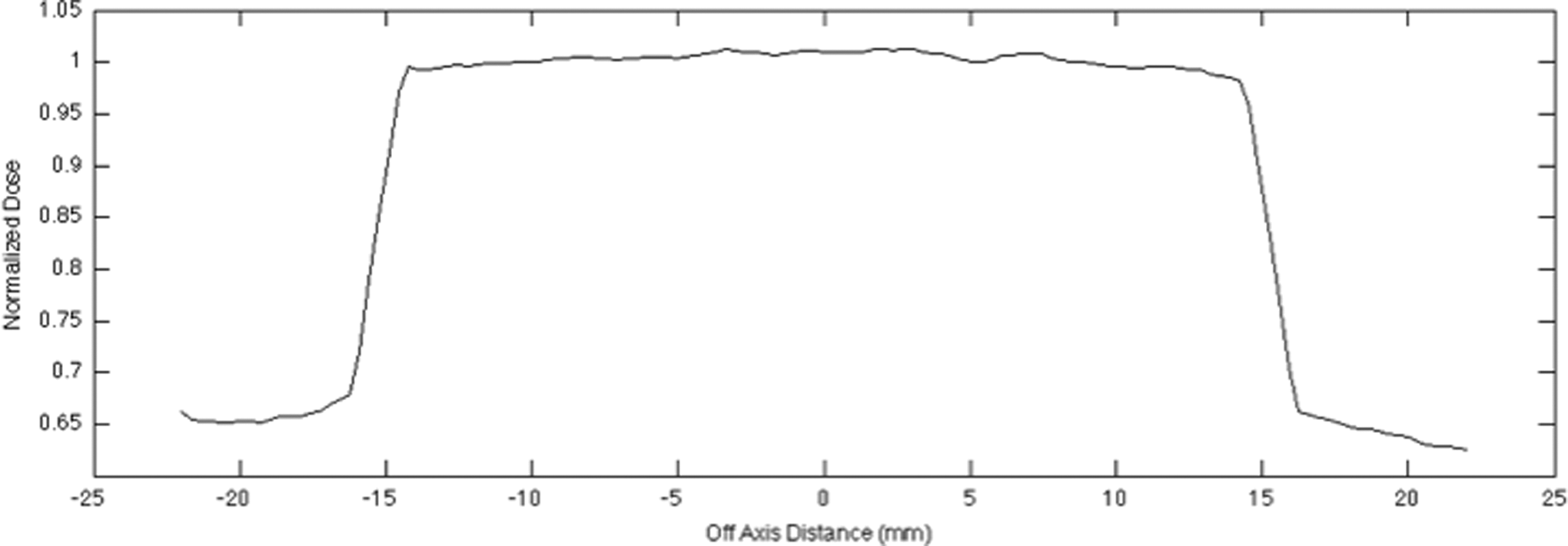
Beam profile for the 3 cm applicator at 0 mm, obtained with EBT3.
In Vivo Measurements
For the simulated in vivo evaluation, an average attenuation factor of 1.047 ±0.024 was found between the upper and lower film. The upper film absorbs 4.70% of the dose, which implies that if EBT3 is used as in vivo dosimeter, the lesion will be underdosed if not corrected for.
Discussion
Nowadays, machine and patient-specific QA is of crucial importance in modern radiotherapy. Recommendations and guidance on QA already exist for contact therapy (TG-61, 9 NCS report 10, 10 and TRS-398 16 ), but these are generally limited to specific tests of basic parameters. With the revival of contact radiotherapy for different treatment sites, QA procedures also need to be improved for kV systems. Such quality testing is required during the commissioning of a system for the introduction of new techniques or applicators and at regular scheduled testing to assure continued system performance. The challenges in low-energy dosimetry are the steep dose gradients, use of small field sizes, and surface measurements. Parallel-plate IC is recommended to perform all QA measurements. EBT3 was recently released as the successor of EBT2. As described in the literature, low-energy measurements (<100 kV) were problematic using EBT2 film due to high-energy dependence. Although, as stated by Brown et al, 15 GafChromic film has potential for dose measurement in contact radiotherapy due to its low-energy dependence, which is advantageous when beam hardening occurs. From a dosimetrical point of view, film can be used to measure absolute doses, depth dose curves, output factors, and beam profiles. Even in high-gradient regions and for small field sizes, where IC measurements are limited to the physical size of the sensitive volume, films can be used. Another interesting application is the use of EBT3 as in vivo dosimeter to verify the delivered dose and identify systematic and random errors as part of the QA procedure 24 and in radiobiological experiments, as it can determine the dose delivered to the cell samples and evaluate the dose homogeneity. 25,26 In this study, we evaluated and validated the feasibility of using EBT3 as reliable dosimeter for kV contact therapy.
Prior to performing EBT3 film measurements, a defined film marking and scanning protocol need to be taken into account to mitigate some uncertainties arising from scanner warm-up and film orientation and placement on the scanbed. Film orientation at scanning has been measured to be up to 8.4% when the film is rotated to 90°. 6 The scanned film side, being found to be within the general scan-to-scan variation of up to 0.5%, is not expected to affect the signal due to the symmetric construction of EBT3. 6 Scanner warm-up characteristics required the first 4 scans to be discarded to ensure 0.2% repeatability between scans. 27 Interfilm uniformity, defined as the difference in measured scan values between different films of 1 batch, and whole-film uniformity, defined as the difference in measured scan values between different parts of 1 single film, were already investigated by Sorriaux et al. 28 They reported an interfilm uniformity of 0.04% and 0.02% for the red and green color channels, respectively. Whole-film uniformity was found as 0.07% and 0.06% for the red and green color channels, respectively. These uncertainties gave confidence of the reproducibility between different parts of 1 film and different films of 1 batch in general. Nevertheless we avoided the use of different film batches and recommend the use of 1 calibration for 1 batch of films.
Avanzo et al 24 confirmed our statement that the red color channel has to be used for doses up to 10 Gy. Brown et al 15 found weak energy dependence for EBT3 and suggested that the energy response is dependent on the chemical composition of the film, which can differ between several lots. Therefore, we recommend calibrating each batch of film before use to eliminate errors arising from the energy dependence of the film.
Until now, EBT3 film was performed for relative dosimetry. We investigated the use of EBT3 film as absolute dosimeter and found a good agreement compared to IC. These results confirm and suggest the use of the EBT3 as a valid alternative for IC. On the other hand, EBT3 film was also used to measure depth dose curves, which were plotted against IC measurements with the dose maximum at the surface, where contact radiotherapy treatments are usually prescribed. Compared to conventional radiotherapy, which makes use of megavoltage X-rays, kilovoltage PDD curves have a steep dose fall-off with depth. This characteristic is advantageous for organs at risk in the environment of the lesion and allows radiotherapists to prescribe higher doses to the lesion. Good agreement was found between depth dose curves measured with IC and EBT3 GafChromic film. Important to note is that simultaneous measurements of IC and EBT3 GafChromic film gave significantly different results (P > .05; data not shown). If one prefers to measure absolute and relative doses or plot calibration curves with both detectors at the same time, which is less time consuming, extensive evaluation of all factors that can influence the reading of the detectors is needed in order to obtain good results. 24
For the depth dose measurements, films were exposed in 2 directions, either perpendicular or parallel to the beam axis. Both directions show good agreement between IC and EBT3 for both color channels. For films exposed parallel to the beam axis, the depth dose curve was underestimated when the first millimeters of the film were used and normalization was performed at 0 mm depth. Depth of 3 mm was found adequate to start the depth dose curve implying that the first 2 mm at the border of the film can’t be used when performing parallel film measurements due to cutting the film into pieces. This finding is in concordance with Mayers 29 who reported the damage to the film measured approximately 3 mm from the line of incision. Comparable uncertainties were found at the first centimeter of the borders of the original film. 30
Output factors, beam profiles, symmetry, and flatness, which cannot be measured with an IC, are also important in the QA procedure of contact therapy. EBT3 allows the user to obtain beam profiles and perform a quick analysis of the symmetry and flatness of the beam, which was not always possible with the EBT2 due to Newton ring formation while scanning. It was reported that these artifacts could lead to uncertainties up to 5%. 24 Due to the innovation of anti-Newton ring coatings, EBT3 shows better performances. Our experiments showed that beam profiles, symmetry, and flatness could easily be acquired with EBT3. For contact radiotherapy of the skin, where self-developed circular collimators (1-10 mm) were used, the measurement of output factors is of utmost importance to ensure adequate dose to the lesion. Output factors are difficult to assess with IC due to the limited resolution of the sensitive volume, while our measurements proved that the use of EBT3 film is suitable.
Our measurements confirm the usefulness of EBT3 GafChromic film as dosimetric tool for absolute QA and depth dose measurements of kV contact radiotherapy. Furthermore, Pidikiti et al suggested that GafChromic film is a reliable dosimeter for other kV X-ray applications such as a small animal irradiatior. 22
We believe that this specific machine QA using EBT3 film can be extended to patient-specific QA of contact therapy to ensure adequate dose delivery to the lesion. To prove this statement, EBT3 film was examined as in vivo dosimeter. A difference of 4.7% was observed in dose between the 2 films due to dose absorption of the film. Arjomandy et al 19 stated that the film has a near water-equivalent effective atomic number (Zeff [EBT3] = 6.84 compared to Zeff, [water] = 7.3) but exhibits an absorbed dose energy dependence significant below 100 kV. 14 If EBT3 is applied as in vivo dosimeter, one has to take into account this attenuation factor to correct the prescribed treatment dose. The strong point of this patient-specific QA procedure is that at the same time absolute dose, flatness, homogeneity, and symmetry of the irradiation toward the lesion can be reviewed. Homogeneity and flatness are also important, as lesions in direct contact of the beam will be treated, where homogeneous dose in the whole applicator opening is important.
Conclusion
This aim of this feasibility study was to assess the reliability of EBT3 GafChromic film as a dosimeter for machine and treatment QA of a 50-kV radiotherapy unit. EBT3 film can be used either parallel or perpendicular to the beam axis. Red color channel was preferred to green color channel to obtain comparable doses to IC in the therapeutic dose range. An advantage of EBT3 film is that flatness and symmetry of the beam profiles can be evaluated and output factors can be measured when necessary. Another potential is the application of EBT3 as in vivo dosimeter to perform patient-specific QA if one takes into account the absorption of the EBT3 film to correct the prescribed treatment dose.
Footnotes
Abbreviations
Declaration of Conflicting Interests
The author(s) declared no potential conflicts of interest with respect to the research, authorship, and/or publication of this article.
Funding
The author(s) received no financial support for the research, authorship, and/or publication of this article.