
Abstract
The aim of the present study is to assess the effects of low-dose occupational exposure on T helper response. One Hundred five employees working in Nuclear Power Plant, Kozloduy, Bulgaria and control group of 32 persons are included in this investigation. Flow cytometry measurements of T-cell populations and subpopulations and natural killer T cells are performed and levels of G, A, and M immunoglobulins and interleukin 2 (IL-2), IL-4, and interferon γ were determined. The data interpreted with regard to cumulative doses, length of service, and age. The results of the present study are not enough to outline a clear impact of occupational radiation exposure on T helper populations. Nevertheless, the observed even slight trends in some lymphocyte’s populations and in cytokines profile give us the reason to assume a possibility of a gradual polarization of T helper 1 to T helper 2 immune response at dose range 100 to 200 mSv. The results of the present study indicate the need to perform a more detailed epidemiological survey including potential confounding and misclassifying factors and possible selection bias that could influence the results.
Introduction
The radiation impact on the immune system as an integral part of the body determines the alterations in the homeostasis and/or in its components. Long-term epidemiologic studies demonstrate that ionizing radiation may induce a dose-dependent impartment of the immune system with a deregulation of cytokine production and persistent inflammation, which is supposed to increase the risk of both cancer and noncancerous diseases. 1 –5 The radiation-induced oxidative stress leads to higher expression of some markers of inflammation such as adhesion molecules and cytokines, which when interacting with cell’s surface receptors activate specific mechanisms and stimulate immune response. T helper 1 (Th1) lymphocytes producing pro-inflammatory cytokines are induced immediately after irradiations, while T helper 2 (Th2) lymphocytes producers of anti-inflammatory cytokines restore homeostasis. The balance between pro- and anti-inflammation tendencies could fluctuate long after a radiation impact and will persist until the elimination of the stress factor. The published results on the effect of ionizing radiation on the Th1/Th2 balance and its influence on human health are controversial. They suppose a pronounced inflammatory effect of a high-dose exposure 6 and an anti-inflammatory effect at low-dose irradiation (single doses ≤1.0 Gy). 7,8
The epidemiological studies of atomic bombing survivors and Chernobyl emergency and clean-up workers found long-term imbalance in Th1/Th2 responses shifted toward an inflammatory profile. 1 –4,9 –11 It is suggested 1 –5,9 that doses in the range of 10 to 100 mGy lead to the prevalence of T1 helper subpopulation, while doses above 200 mGy switch to prevalence of T2 helper immune response. Experimental studies reveal a decrease after low-dose exposure and an increase after high-dose exposure of expression in both Th1 and Th2 type cytokines. 12
The assessment of low-dose radiation on changes in immunological parameters and state of subclinical inflammation requires a careful examination of the immune status of occupationally exposed persons. Our previous data on lymphocyte populations’ profiles in Nuclear Power Plant (NPP) workers 13,14 raised the issue with respect to the prevalence of Th1 immune response at low-dose exposures and dominating of Th2 with increasing cumulative doses above 100 to 200 mSv. Therefore, the current study aims to extend the investigation on NPP workers with special focus on cytokine profiles—interleukin 2 (IL-2), interferon γ (IFNγ), IL-4, and T helper subpopulations—in order to determine some imbalance in Th1 and Th2 immune response.
Material and Methods
One Hundred five employees working in NPP, Kozloduy, Bulgaria, were included in the study. The group of men occupationally exposed to external γ radiation was selected from the service personnel of units 5 and 6 of NPP Kozloduy. A control group of 32 male participants was selected at similar age and with similar length of service who do not have any work related to ionizing radiation. The study was carried out thanks to a contract between the National Centre of Radiobiology and Radiation Protection of the Ministry of Health of Bulgaria and the NPP Kozloduy. An informed verbal consent was obtained from all participants. The study was performed within the framework of a bilateral contract between the National Centre of Radiobiology and Radiation Protection, part of the Ministry of Health and NPP Kozloduy. The data for individual monitoring exposure were submitted by NPP Kozloduy dosimetry service, approved by the regulatory body. The determination of the cumulative radiation doses is the part of NPP individual exposure monitoring program.
Workers were divided in groups according to cumulative dose received as follows: controls, up to 25 mSv, up to 100 mSv, up to 200 mSv, and above 200 mSv (Table 1). As low doses were defined those below 0.1 to 0.2 Sv, this was the main criteria in determination of the groups, namely with very low initial doses, low doses, and high doses above 0.2 Sv). Criteria such as working condition with predominantly gamma ray exposure and without having undergone medical exposure for the past 3 years have been taken into account in the study.
NPP “Kozloduy” Personnel Data According to Age, Length of Service, and Cumulative Dose.

Abbreviation: NPP, Nuclear Power Plant; SD, standard deviation.
All participants complete a questionnaire and undergo medical examinations and basic hematological studies to assess their health status. The individual variability in cytokine secretion and its dependence on age, morbidity, and preexisting diseases, which could confound the obtained results, were taken into consideration. Cardiovascular diseases (hypertension, ischemic heart disease) and obesities in some of responders were mainly identified.
Blood samples were collected from each participant by venipuncture into Vacutainer EDTA tubes (Greiner Bio-One GmbH, Kremsmunster, Austria). The total white blood cells (WBCs) and lymphocytes were counted using an automatic hematology analyzer ABX Pentra 60 C+ (HORIBA Group ABX Diagnostics, Montpellier, France) operated in cell blood count + 5 population differential count modes. Blood smears were prepared to observe WBC morphology.
The lymphocyte subpopulations were determined using a 4-parameter flow cytometry in whole blood after lysis of erythrocytes by the method of Jackson. 15 The staining of lymphocytes was carried out using the following kits: BD Multitest—CD3/CD16+CD56+/CD45/CD19/; BD Multitest—CD3/CD8/CD45/CD4/; BD Multitest—CD45RA/CD62 L/CD3/CD4/; BD Simultest—CD8/CD28 (Becton Dickinson Biosciences, San Jose, California). The samples were acquired and analyzed by FACSCalibur flow cytometer (Becton Dickinson Biosciences, Software V 2.1) and Cell Quest software. The following lymphocyte populations were examined: CD3+ (total T lymphocytes), CD19+ (B lymphocytes), CD3+ CD4+ (helper-inducer T lymphocytes), CD4+ CDRA+ CD62L+ T lymphocytes, CD4+ CDRA+ CD62L− T lymphocytes, CD4+ CDRO+ CD62L+ T lymphocytes, CD3+ CD8+ (cytotoxic suppressor T lymphocytes), CD8+ CD28+ cytotoxic T lymphocytes, CD3− CD16+ CD56+ (natural killer cells), CD3+ CD16+ CD56+ (natural killer T [NKT] cell).
Serum immunoglobulin (Ig) G, M, and A were determined by radial immunodiffusion using immunoplates (EASY RID, Roseto, Italy). Plasma levels of IL-2, IL-4, and IFNγ (Table 2) were assessed by commercial enzyme-linked immunosorbent assay (ELISA) kits (Gen-Probe Diaclone SAS, Besancon, France) according to the manufacturer’s instructions following the construction of standard curves for each ELISA system protocols. Since there are no accepted reference standards for ILs, we have prepared our own reference standards for the studied ILs, based on data obtained for control group and taking into account published data of measurement cytokines in the same conditions. 16,17 The generally low and no measurable level of the investigated cytokines in healthy persons, obtained using conventional ELISA methods, and their large dispersion required the application of frequency analysis to compare the percentage of individuals with values outside the accepted reference ranges by group and to outline a more clearly defined trend.
Reference Values of Studied Interleukins.
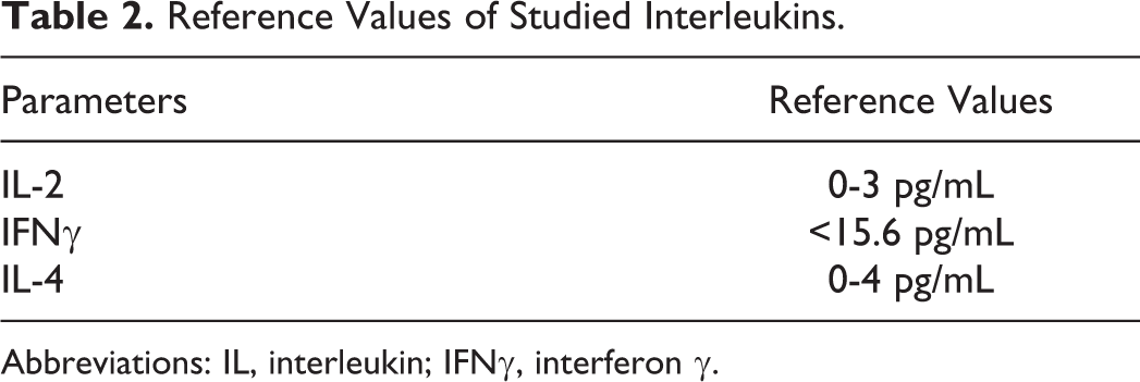
Abbreviations: IL, interleukin; IFNγ, interferon γ.
Statistical Methods
The following statistical methods were applied to process the results.
Parametric methods
One-way analysis of variance (ANOVA) was used to check the equality of more than 2 mean values in a normal distribution.
Nonparametric methods
Kolmogorov-Smirnov and Shapiro-Wilk tests were used to check the normality of distribution of quantitative variables; Mann-Whitney test was used for comparison of averages in 2 groups of 1 quantitative variable when the distribution is not normal; Kruskal-Wallis test was used for comparison of averages in more than 2 groups of quantitative variables when the distribution is not normal.
Variation analysis of quantitative variables
Mean, standard deviation, standard error of the mean, and 95% confidence interval of the mean were analyzed.
Correlation analysis
Coefficient of linear correlation included parametric (Pearson) and nonparametric (Spearman); partial correlation. The data are not presented in tables, but only correlation coefficients were shown, where necessary. Frequency analysis deals with the number of occurrences (frequency) and analyzes measures of central tendency, dispersion, percentages. SPSS version 11.0.1 for Windows was used for data processing. 18,19
Results
The relative and absolute values of cell parameters were analyzed. Data from the variation analysis of the studied main lymphocyte populations are presented in Table 3.
Variation Analysis of Main Lymphocyte Populations According to Cumulative Dose.

Abbreviations: SD, standard deviation; X, mean.
The parametric ANOVA test was applied to analyze CD3+ (percentage) and CD4+ (percentage) T lymphocytes. Mann-Whitney и Kruskal-Wallis tests were used to analyze the parameters that lack normal distribution. There was no significant difference for all parameters as shown in Table 3—between the control and exposed individuals and between the 4 exposed groups. The correlation analysis showed no dependence on age, dose, or length of service for total T cells, helper-inducer T cells, suppressor cytotoxic T cells, and B lymphocytes.
The variables were compared with nonparametric Kruskal-Wallis test because of the lack of normal distribution found by Kolmogorov-Smirnov test. No statistically significant difference was found between groups for cytotoxic CD8+ CD28+ T lymphocytes, but a tendency of decrease in their average was observed in groups with cumulative doses above 100 mSv, confirmed by weak negative correlations with cumulative dose (r = −0.249 at P = .003). These results are in accordance with the reports of Liu et al 20,21 who established similar trend in the values of cytotoxic T-cell subpopulation (CD8+ CD28+) with the increase of cumulative doses.
The observed increase of NKT cells in groups with cumulative doses up to 200 mSv, although insignificant (Table 4), also was confirmed by the weak correlations with cumulative dose (r = 0.171 at P = .045). The stimulatory effects of low radiation doses on the innate immunity probably determined the observed trend. 22 –24
Variation Analysis of CD8+ CD28+ T Lymphocytes, NK, and NKT Cells According to Cumulative Dose.
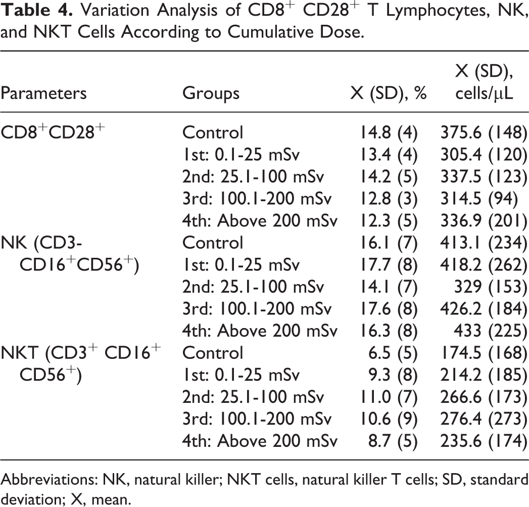
Abbreviations: NK, natural killer; NKT cells, natural killer T cells; SD, standard deviation; X, mean.
The tendency of dose-depended decrease of helper-inducer CD3+ CD4+ T lymphocytes observed in our previous studies supported further investigations of their subpopulations. Reported differentiation of memory CD45RO+ CD4+ cells in helper 1 and helper 2 lymphocytes based on the expression of L-selectin receptor (CD62 L) 25,26 also suggested continuation of research of CD4+ T lymphocytes subpopulations.
The results of variation analysis on T helper lymphocyte subpopulations are presented in Table 5. Mann-Whitney and Kruskal-Wallis tests were applied for analysis, as variables lacked Gauss distribution. The values of the coefficient of significance did not show statistical reliability either to control or between exposed groups. Exception was recorded for the absolute values of central memory CD4+ CDRO+ CD62L+ T lymphocytes between the groups with lowest and highest cumulative doses (P = .012). For the same subpopulation, a slightly significant positive dependence on age (0.245 at P = .007) and length of service (0.315 at P = .001) was found for percentage values and on the dose (0.216 at P = .015) for its absolute ones, by performing correlation analysis with Spearman test. Naive CD4+ CDRA+ CD62L+ T lymphocytes have shown a relative decrease, although no significant, in groups with cumulative doses higher than 25 mSv, which could be explained by the greater radio sensitivity of naive T cells, subjected to apoptosis in a dose-dependent manner. 27,28 Low positive correlation of this subpopulation with length of service (0.272 at P = .003) was found.
Variation Analysis of T Helper Lymphocyte Subpopulations According to Cumulative Doses.

Abbreviations: SD, standard deviation; X, mean.
a Statistical confidence between the first and fourth group for central memory T cells (P = .012). There is significant statistical difference between the groups in boldface. It is described within the text.
Table 6 present concentrations of serum immunoglobulins IgG, IgA, and IgM expressing the functional activity of B-lymphocytes. The variation analysis was done by nonparametric Kruskal-Wallis test, for lack of normal distribution of variables. There was no statistically significant difference for the 3 tested immunoglobulins within groups. A tendency of an increased average levels of IgA was observed by the established low positive correlations with age (r = 0.037 at P = .185) and length of service (r = 0.236 at P = .008). Contrarily, a trend of decreasing values of IgM in comparison to the controls was found, most pronounced in the group with doses above 200 mSv, confirmed by low but significant negative correlation of IgM with cumulative dose (r = −0.220 at P = .011).
Variation Analysis of Serum Levels of IgG, IgA, and IgM According to Cumulative Dose.

Abbreviations: Ig, immunoglobulins; SD, standard deviation; X, mean.
Table 6 presents the results of variation analysis of plasma levels of IL-2, IL-4, and IFNγ. Cytokines, as the most important mediators for immune cell communication, could be up- or downregulated by low-dose radiation. Interleukins IL-2, IL-4, and IFNγ are associated with T-cell growth and differentiation as well as with the distinction between Th1 and Th2 immune response. Inflammatory cytokines, such as IFNγ, IL-2, IL-6, and TNF-α, increased expression, providing evidence of persistent inflammatory responses, while IL-4 is an antioxidant, anti-inflammatory cytokine restoring homeostasis.
Interleukin 2 did not show any significant difference in control or between groups of exposed persons by Kruskal-Wallis test. Relatively higher plasma levels of IL-2 were observed in all exposed groups compared to control, more pronounced in the group with cumulative doses 25 to 100 mSv. Frequency analysis confirmed the highest percentage of persons with elevated values in the same group—23.3% as their number gradually decreased, reaching the lowest percentage of persons with deviations in the group with a dose over 200 mSv (6.82%; Table 7). No significant correlation for IL-2 was found with age, length of service, or cumulative dose as well as with increased frequency of chronic diseases.
Frequency Analysis of Interleukins.
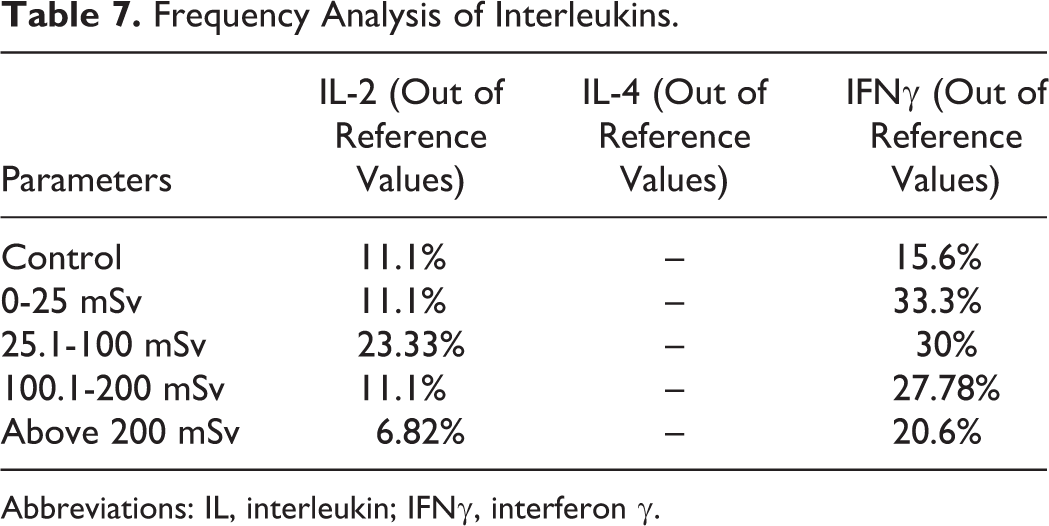
Abbreviations: IL, interleukin; IFNγ, interferon γ.
For the other pro-inflammatory cytokine, IFN-γ, crucial for immunity against intracellular pathogens and for tumor control, no significant difference, either between groups or to control, was established (Table 8). This cytokine also showed lack of dependence on age, dose, length of service, viral, and chronic diseases. However, the frequency analysis established the highest percentage (33.3%) above the reference in the group with cumulative doses 0.1 to 25 mSv, which gradually decline with increasing the cumulative dose, reaching 20.6% in the group with cumulative dose above 200 mSv (Table 7). The analysis of the anti-inflammatory cytokine IL-4 by Kruskal-Wallis test did not show any significant difference between groups of NPP workers and the control. However, comparison within exposed groups by Mann-Whitney test showed a significant difference with cumulative doses 100 to 200 mSv and above 200 mSv. The obtained results in all groups were within reference values, and there was no correlation of IL-4 with age, cumulative dose, and length of service, or chronic diseases.
Variation Analysis of Plasma Levels of Interleukins According to Cumulative Dose.

Abbreviations: IL, interleukin; IFNγ, interferon γ; SD, standard deviation; X, mean.
a Statistical confidence between the third and fourth group for IL-4 (P < .05).
Discussion
The present study on the effects of occupational radiation dose at the immune system did not register any statistical differences within the main lymphocyte populations and their subpopulations. The results varied widely and only a few of studied parameters showed weak correlation with cumulative dose, age, or length of service. Other surveys on such contingents 29 –31 reported similar results. However, the established slight deviation of some of studied subpopulations and their low correlation with cumulative radiation dose and age could be discussed.
The observed trend of increasing of NKT cells is similar to the one reported by Kuzmenok et al, 11 who found increased number of NKT cells as late effects in Chernobyl liquidators. According to Subleski et al, 32,33 NKT levels increase with increasing the total dose as they could rapidly express pro- and anti-inflammatory cytokines determining the type and the magnitude of the immune response.
Probably, the reduction of cytotoxic CD8+ CD28+ T cells with increasing of cumulative doses established in studied contingent reflected an activation of suppressor regulatory mechanisms. This presumption was based on studies of Liu et al 20 who supposed that while the low doses upregulate the expression of CD28 molecule, leading to immune enhancement, higher doses upregulate the CTLA-4 expression and lead to immunosuppression. In the present research on CD3+ CD4+ subpopulations, data for increasing of central memory CD4+ CDRO+ CD62L+ T lymphocytes, cells with anti-inflammatory cytokine production were obtained. This suggested some impact of low-dose radiation on CD3+ CD4+ subpopulation and it could be assumed that they reflect the gradual polarization of helper 1 to helper 2 balances. The trend of the decreasing naive CD4+ CDRA+ CD62L+ T lymphocytes with increasing cumulative dose could be in confirm of Kusunoki et al 10 report for similar variation in memory and naive CD4+ T-cell populations at atomic bombings survivors, interpreted as an expression of radiation-induced accelerated aging of the immune system. It could be suggested that the obtained discrete deviation in some of studied lymphocyte’s populations demonstrated a gradual tendency of polarization of helper 1 to helper 2 immune response, but the insufficient number of respondents did not allow us of making a general conclusion.
The results of the plasma immunoglobulins confirmed the data of our previous studies. 13,14 Epidemiology investigations 34,35 of radiology and X-ray department workers established reduction of IgM, which was also registered in our study. Contrarily, Japanese authors, 4,36 who performed a long-term study of atomic bomb survivors, reported elevated serum concentration of IgM due to compromised immune system. Similar to our results, an increasing of IgA were found by Rybkina et al 37 in Mayak workers chronically exposed to external radiation, which authors explained by switches of immunoglobulins biosynthesis to IgA isotype. Kluciñski et al, 38 on other hand, consider that smoking amplified the suppressive effect of occupational exposure on the production of IgA and IgG. It is obvious that no consistent position exists in concern to the effects of chronic low-dose radiation on the immunoglobulins concentration. Such discrepancies could be due both to confounding lifestyle/environmental risk factors and to epidemiological situation during the survey because of the short half-life of immunoglobulins.
In our survey, we observed higher percentage of persons with above the normal values of IL-2, in the group with cumulative dose 25 to 100 mSv, that gradually decline in groups with increasing cumulative dose. These results were in accordance with studies of Xu et al 39 and Zakeri et al 31 who also reported increased secretion of IL-2 at occupational radiation exposure in the same dose interval, namely in the range of 30.5 (24.3) mSv. The results for IFNγ, other representative of pro-inflammatory ILs demonstrated similar dynamic—a gradual reduction in percentage of persons with higher than normal values with increasing the cumulative dose. While the results of pro-inflammatory IL-2 and IFNγ cytokines showed a slight trend of decreasing with increasing of cumulative dose, those for anti-inflammatory cytokine IL-4 were very dispersed, with no clear dependency to cumulative doses. The established significant difference for IL-4 between the group with cumulative doses of 100 to 200 mSv and cumulative doses higher than 200 mSv raises the question of the cause for obtained results. As known, production of IL-4 is carried out by several cells: mast cells and basophils, γ/δ T cells, subpopulation of NKT cells, and naive CD4+ T cells, but Th2 cells are particularly the most important producer of IL-4, required for their own development. Although NKT cells could secrete large amounts of IL-4 immediately after T-cell receptor stimulation, there are reports 40 about their important role in the immune balance at late stage after radiation, when their removal is associated with enhanced production of IL-4 and IgE. That could be one of the reasons about the lower average of IL-4 in the group, with doses from 100 to 200 mSv, where the relative higher number of NKT cells was obtained. On the other hand, the increase of IL-4 in group with cumulative dose above 200 mSv could be explained with the significant increase in the number of central memory cells, representing characteristic of cells with anti-inflammatory cytokine production.
Based on the observed insignificant deviations in studied cytokines levels, more likely trends of decreasing of pro-inflammatory cytokines IL-2 and IFNγ, and the obtained results for IL-4, we could speculate for gradual switch from Th1 to Th2 immune response at dose interval of 100 to 200 mSv. Such an idea launched Attar et al 41 who found higher lymphocyte-induced IL-4 and IL-10 production and lower IL-2 and IFN-γ production in the exposed inhabitants of high background area of Ramsar. The authors suggested that the immune system of individuals exposed to high-dose ionizing radiation has adapted to its environment by shifting from a type 1 to a type 2 response to promote anti-inflammation. Based on obtained results for plasma concentration of these proteins, we could suggest that there is some prevalence of pro-inflammatory immune response to cumulative dose 200 mSv, which gradually switch to type 2 at doses above 200 mSv, confirmed in other studies.
The secretion and activity of cytokines is very individual depending on many confounding social, genetic, and epigenetic factors and especially health status of individuals, so that it is necessary to precise the impact of other factors on radiation effects.
Finally, we could summarize that the results of the present study are not able to outline clear conclusions about the impact of occupational radiation doses on Th1/Th2 immune balance. We consider that testing such parameters, whose normal values vary in a relatively wide range and which are too sensitive to environmental impact, require carrying out a number of studies to monitor any statistical reliability. The relatively small number of respondents within each group of our survey does not allow us to draw definite conclusions. Nevertheless, the observed even slight trends in some lymphocyte’s populations (NKT, CD8+28+, and CD4+ CDRO+ CD62L+ T lymphocytes) and in cytokines profile give us the reason to assume a possibility of a gradual polarization of Th1 to Th2 immune response at dose range of 100 to 200 mSv. The evidence presented in this study indicates a need to conduct more detailed epidemiological studies that are capable of addressing potential confounding and misclassifying factors and possible selection bias that could influence the results.
Footnotes
Declaration of Conflicting Interests
The author(s) declared no potential conflicts of interest with respect to the research, authorship, and/or publication of this article.
Funding
The author(s) received no financial support for the research, authorship, and/or publication of this article.