
Abstract
Polylactic acid matrix composites are widely used in packagings and biomaterials. The specific surface area, flexibility and degradation efficiency of the material are the key factors to determine its application in these fields. In this study, a series of poly(L-lactic acid) (PLLA)/graphene oxide (GO) composite nanofiber webs were prepared using electrospinning technique. The scanning electron microscope (SEM) image of PLLA/GO nanofibers showed a rougher surface and a smaller average diameter compared with that of pure PLLA nanofibers, and the nanofibers with 6 wt% GO in PLLA matrix looked like flatter ribbon. Accordingly, the tensile stress test of the electrospun webs with different GO contents showed high performance, 400% increment in the tensile stress at presence of 6 wt% GO. The hydrolytic degradation behavior of composite the nanofiber webs exhibited that the presence of GOs greatly improved the degradation rate, after 9 days, the degradation ratio of PLLA/GO can reach 16.83%. of the PLLA matrix, resulting from the better hydrophilic property and absorbability. Using GO to improve the preparation of new biocompatible materials from PLLA can provide a reference for problems in the field of packaging materials.
Keywords
Introduction
Poly(L-lactic acid) (PLLA) has attracted much attention in various applications such as packagings, 1 fibers, 2 tissue engineering scaffolds, 3 would dressings 4 and drug delivery systems 5 due to its renewability and degradability. However, the inherent brittleness, low heat resistance and slow degradation rate of PLLA have blocked its further development and extensive practical application. 6 PLLA nanocomposites are usually blended with nanofillers like carbon nanotubes (CNTs), cellulose nanofibers, hydroxyapatite, and graphene to overcome these disadvantages. 7
Graphene has many outstanding properties including superior mechanical strength, good electrical conductivity and large surface area. These properties make graphene extensively accepted as an effective nanofillers in PLLA to improve its mechanical and thermal properties. 8 Li et al. added 0.5 wt% graphene oxide (GO) into the PLLA matrix, tensile strength of PLLA/GO nanocomposites increased from 35 MPa of PLLA to 53 MPa (an increase of 51.4 %), and the elongation at break increased from 6.50% to 8.91% (an increase of 37.1%). 9 Papageorgiou GZ et al. reported that the crystallization and biodegradation rates of the PLLA nanocomposites increased with increasing the GO and organomodified-GO content. 10 Chen et al. prepared a series of of GO incorporated into PLLA matrix, the presence of the GOs accelerates the thermal degradation of the PLLA. 11
A variety of methods have been applied to fabricate graphene nanosheets reinforced in polymers, including solution (or melt) blending and in situ polymerization.12–14
Electrospinning is often used to fabricate flexible PLLA nanofiber webs. 15 And it is a versatile method of incorporation of nanofillers into electrospun nanofibers during electrospinning of blend solution.16,17 The nanocomposite membranes have the advantages of small fiber diameter, large specific surface area, controllable mechanical properties and porous network structure. 18 Therefore, nanofiber webs can be used in battery, supercapacitor electrode, air purification, wastewater treatment, tissue engineering, drug delivery, wound dressing bioactive materials, product packaging and other fields. In addition, “green materials” are needed to minimize the environmental and human health impacts of nanofiber webs, such as biodegradable and recyclable biogenic materials. There are many reports regarding the fabrication of carbon nanotube (CNT)/polymer nanocomposite fibers due to its tube like structure which can effectively incorporated into polymer matrix during electrospinning of blend solution.19,20 Mei et al. successfully fabricated an electrospun PLLA/MWNT/hydroxyapatite composite fibrous membrane with a weight ratio of MWNT/HA to PLLA of 1:9, and found the degradation characteristics were far improved. 21 However, there are very few articles about the GO sheets incorporated into PLLA matrix for its hard to incorporate GO sheets through polymer matrix during electrospinning due to their micro sized sheets and high degree of agglomeration.22,23 Soghra Ramazani et al. produced poly(e-caprolactone) (PCL) nanofibers by the electrospinning process were studied in the presence of different amounts of graphene oxide with different oxidation levels. 24 Zhang et al. fabricated an electrospun PLLA/GO nanofibrous mats with a weight ratio of GO to PLLA of 1:100, and found the hydrophilic performance were improved. 25 Therefore, more attention should be paid to eco-friendly PLLA to prepare nanofiber webs.
We noted that the addition of graphene nearly always not more than 5 wt% in the traditional electrospun nanofibers. In order to investigate the influence of amount of GO on the morphology, diameter, tensile strength and degradation of the electrospun nanofibers, composite nanofibers based on PLLA and GO are prepared by the electrospinning technology in this paper. In addition, we also explored the effect of graphene on the degradation of PLLA materials. The results could provide a basis for the design and development of a new type of packaging textiles based on PLLA.
Experimental
Materials
PLLA (trade name 6252D, molecular weight 60,000 g/mol) was obtained from Shanghai Tong-jie-liang Biomaterials Co. Ltd. Graphite powder (<30 μm) was purchased from the Sinopharm Chemical Reagent Co. Ltd., Shanghai. Sodium nitrate (NaNO3), potassium permanganate (KMnO4), sulphuric acid (H2SO4), chloroform and N,N-dimethylformamide (DMF) were analytical grade used as received.
Preparation of GO powder
Graphite oxide (GO) was synthesized by modification of the Hummers method. 26 The mixture of graphite flakes (2.0 g), NaNO3 (1.0 g) and KMnO4 (8.0 g) were added into 67 mL of H2SO4 solution at 0°C to avoid violent reaction. After the heat released, the reaction continued for 2 h at 50°C. After that it was diluted with water (300 mL), followed by addition of 30 wt% H2O2 until the solution turn to yellow. The system was washed with water on the centrifuge for several times until the supernatant fluid to neutral. After filtration and freeze-drying in a lyophilizer for 48 h, graphene oxide powder could be obtained.
Preparation of electrospinning solutions
GO powder was dispersed in DMF by stirring for 24 h followed by ultrasonic vibration for 4 h at room temperature. PLLA was dissolved in CHCl3 under continuous magnetic stirring overnight at room temperature to obtain a 9 wt% PLLA solution. The composite PLLA/GO suspensions were obtained by dropping GO suspensions (2, 4 and 6 wt% relative to PLLA, respectively) to PLLA solutions under continuous stirring, which were named PLLA/GO1, PLLA/GO2 and PLLA/GO3. 9 wt% PLLA/(CHCl3:DMF = 9:1 v/v) was prepared as a control blank (named PLLA). Before electrospinning, the PLLA/GO mixture was ultrasonicated for 30 min.
Electrospinning
The solution was loaded into 10-mL plastic syringes fitted with a stainless steel needles (Gauge20) mounted on a syringe pump (789100C, Cole-Pamer Instrument Co., Vernon Hills). The working voltage was adjusted between 13 kV and the flow rate was 1.2 mL/h. The nanofibrous membrane was collected on a thin sheet of aluminum foil which was varied between 15 cm away from the needle as shown in Figure 1.

Schematic illustration of the preparation of PLLA/GO nanofibers.
Characterization
The morphology of electrospun nanofibers was studied using a scanning electron microscope (SEM) (Hitachi S-4800, Japan) at an accelerating voltage of 1 kV. The diameter of the nanofibers was measured directly from the SEM micrographs using ImageJ software, with the average value being calculated from 100 measurements.
A transmission electron microscope (TEM) (Talos F200X, USA) was employed to investigate the internal structure and GO distribution of nanocomposite fiber. For this study, the nanofibers were directly electrospun onto a copper grids coated with a carbon support film (mesh number: 230, Beijing Zhongjingkeyi Technology Co., Ltd, China).
The changes in glass transition temperature and crystallization behavior due to graphene loading were examined by a differential scanning calorimeter (DSC) instrument (NETCSCH 204F1, German) in a nitrogen flow (40 mL/min). The samples (~5 mg) were heated from 40 to 200 oC with a heating rate of 10°C/min.
Thermogravimetric analysis (TGA) was done on a TGA Instrument (NETZSCH STA-409 PC, German) to determine the thermal stability of the nanofibrous webs. The samples (~5 mg) were heated from 25 to 800°C at a rate of 20°C/min in a nitrogen atmosphere. The degradation temperatures were calculated from the differential TGA curves.
The mechanical properties of the electrospun nanofibrous mats were tested using a universal testing machine for fabrics (Instron 3345, USA). Rectangular specimens of dimensions 5 × 50 mm were used for testing at a cross-head speed of 10 mm/min and a 10 N load cell at ambient conditions. The tensile stress and elongation at break were calculated from the obtained stress-strain curve.
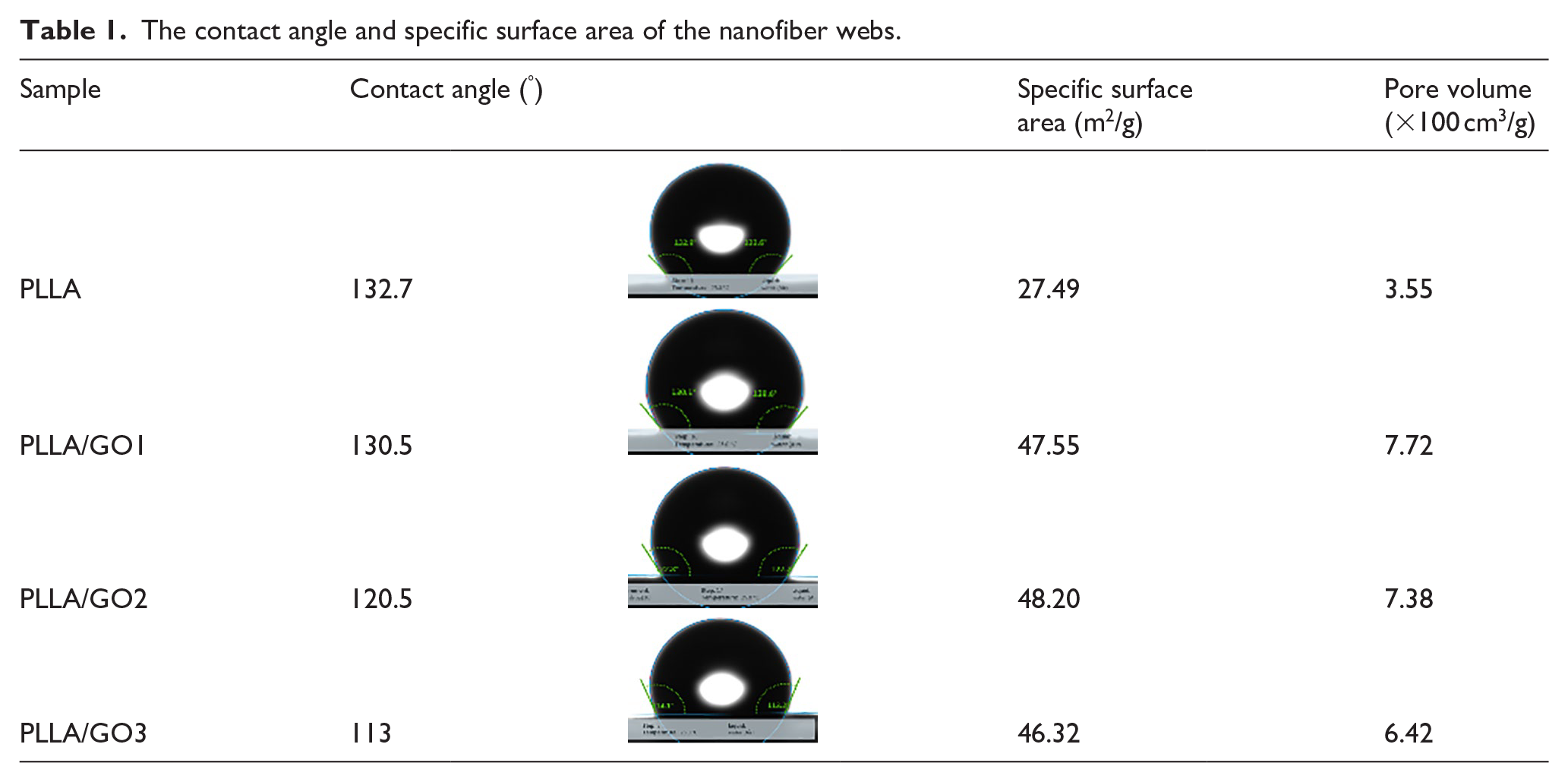
Contact angle was measured on a drop shape analysis system (KRÜSS, DSA 30) at the temperature of 25°C. A drop of 2 μL distilled water was dropped on the surface of a sample slice to analyze the contact angle.
The Brunauer-Emmett-Teller (BET) specific surface area and porosity were tested with Nova1000e instrument (Quantachrome, USA). The tube was weighed and recorded, then about100 mg sample was put into the tube and degassed for 4 hrs.
Specimen was cut into 4 cm × 4 cm rectangular shape and measured the initial weight for degradation study. Then the samples were immersed into the phosphate buffer solution (PBS) for 9 day period, with refresh the PBS solution every day. Considering that graphene is a good far-infrared absorber, a 100 W infrared lamp was placed above the solution (30 cm). The samples were weighted after dried at each time interval. The mass loss was calculated according to the following equation:

where m0 and mt represent the initial mass and the dry mass at time t, respectively.
Results and discussion
Morphology of composite nanofibers
Morphology of the PLLA/GO nanofiber webs with different composition of GO are shows in Figure 2. Compared with neat PLLA ( Figure 2(a) ), PLLA/GO ( Figure 2(b–d) ) nanofibers exhibit smaller diameters with the increasing amount of GO. PLLA shows a mean fiber diameter of 1652 ± 791 nm, the presence of GOs reduce the diameters from 1154 ± 343 nm of PLLA/GO1, to 721 ± 207 nm of PLLA/GO2, to 652 ± 202 nm of PLLA/GO3. It is well known that the diameter of electrospun fibers is affected by internal factors such as solution viscosity and conductivity. In this study, PLLA/GO suspensions showed a higher viscosity and a higher conductivity compared with neat PLLA solution (1.57 × 10⁻⁵ S/m and 7000 mPa·s). The values are increased to (3.39 × 10⁻⁵ S/m and 7050 mPa·s), (1.21 × 10⁻⁴ S/m and 7150 mPa·s) and (5.85 × 10⁻⁴ S/m and 7750 mPa·s), respectively, for the PLLA/GO1, PLLA/GO2 and PLLA/GO3 composite suspensions. In general, the fiber diameter increases with the increase of the viscosity of the electrospinning solution, and an increase in the composite solution conductivity tends to produce fibers with smaller average diameters. The increment in the conductivity of the composite suspension is higher than that in solution viscosity, hence the effect of conductivity on the diameter of electrospun composite fibers is more prominent than that of viscosity. 27 Therefore, the fiber diameter decreased significantly with the increase of GO content. SEM images also highlight that the PLLA/GO nanofibers have different morphologies. PLLA micron fibers looks cylindrical with porous surface due to the competition between phase separation and solvent evaporation. 28 No beadings were observed in the composite nanofibers (Figure 2(b–d)). As the GO content increased to 6 wt % (Figure 2(d)), the composite nanofibers surfaces were found to be oblate exhibiting flatter ribbon-like morphology. The GO nanosheets might be embedded in the nanofibers and aligned along the axial direction similar to the literature reported by Lee and his co-workers. 29
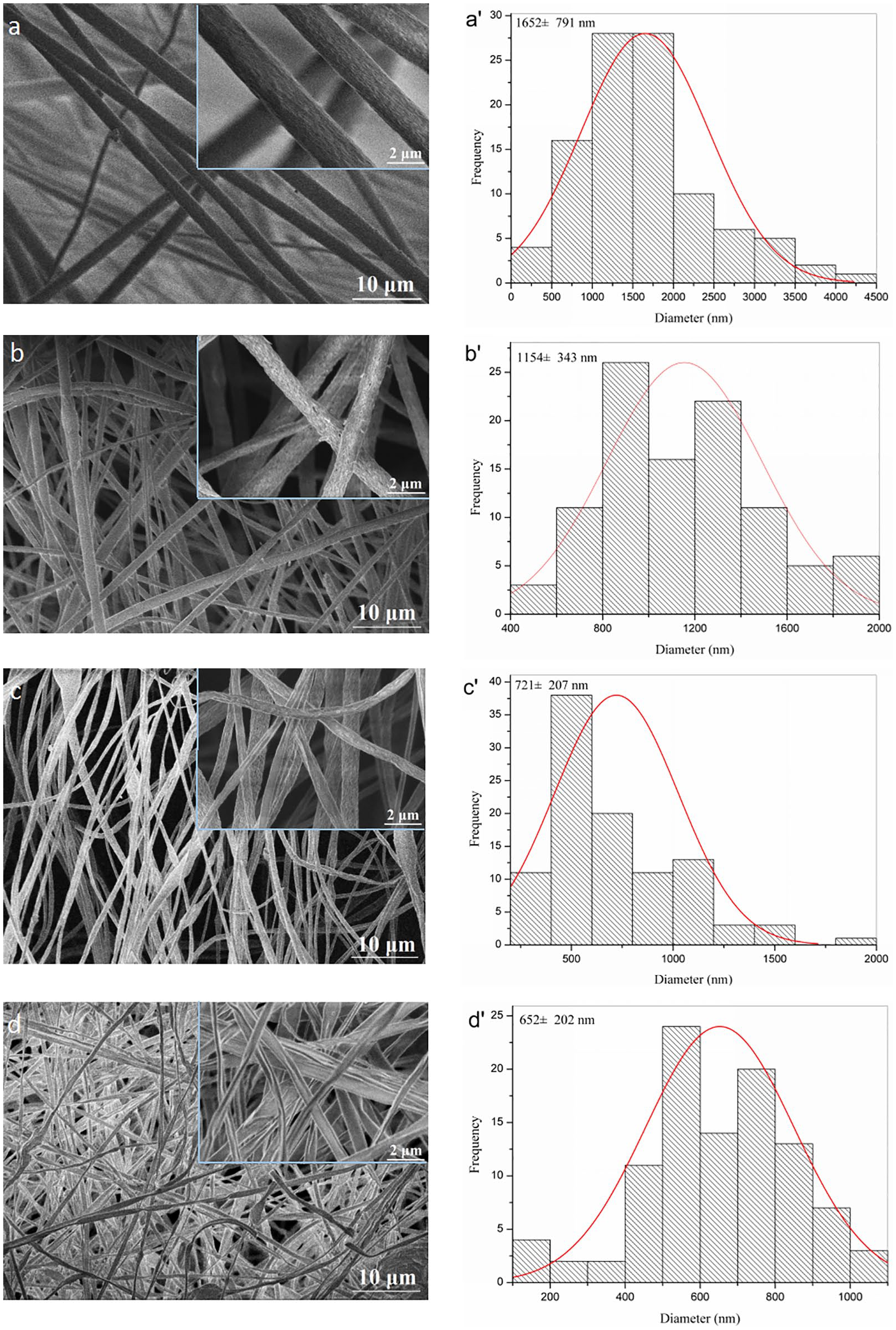
SEM images (left) and diameter distribution (right) of the fibrous mats of (a) PLLA, (b) PLLA/GO1, (c) PLLA/GO2 and (d)PLLA/GO3, respectively.
To further explore the dispersion of the GO sheets in the nanofibers, TEM is shown in Figure 3 . As a control, neat PLLA fibers had a relative smooth surface and uniform size in Figure 3a . The TEM image ( Figure 3b ) of PLLA/GO3 nanofibers exhibited a rough surface and uneven thickness, revealed that GO sheets dispersed PLLA nanofibers were successfully fabricated. To observe the structure of rough parts in more detail, the high-magnification TEM images of PLLA/GO3 nanofiber compared with GO sheets were shown in Figure 3(c) and (d). GO sheets showed a natural tendency to unfold and a flake-like and wrinkled morphology (Figure 3(c)). 30 While the GO sheets in nanofibers were well-oriented in the fiber axis direction, which was attributed to the higher draw ratio. During electrospinning, the stress on the fiber was larger as it was being formed due to the higher draw ratio, and the two-dimensional GO nanosheets were properly aligned in the fiber axis.

TEM micrographs of (a) PLLA, (b) PLLA/GO3, (c) GO and (d) high-magnification PLLA/GO3 respectively.
Thermal analysis
The crystallization behaviors, glass transition temperature (Tg) and melting temperature (Tm) of the samples was investigated by DSC (Figure 4(a)). In the curve of neat PLLA, it showed that the Tg and Tm of PLLA were about 54.2°C and 166.9°C, respectively. 31 With the addition of GO, Tg increased to 57.7°C for PLLA/GO1, 58.1°C for PLLA/GO2 and 56.7°C for PLLA/GO3, while Tm increased to 168.4°C, 168.4°C and 167.6°C, respectively. The increases of the Tg and Tm values in PLLA/GO composite nanofibers may be due to the constrained chain mobility of PLLA by hydrogen bonding and electrostatic attraction with GO sheets. 32

DSC thermograms (a), TG (b) and DTG (c) profile of the fibrous mats.
The TG curves and corresponding differential thermogravimetric analysis (DTG) curves of PLLA/GO composite nanofibers are shown in Figure 4(b) and (c), respectively. All the composites decompose in a one-step process. Compared with neat PLLA, the TGA curves shift to higher temperatures with the incorporation of GO. The peak temperature of DTG curve (Figure 4(c)) represents the maximum rate of weight losing. This temperature for PLLA/GO1 is 335.6°C, for PLLA/GO2 is 338.6°C and PLLA/GO3 is 347.1°C, higher than that of neat PLLA for 330.6°C. The improved thermal stability for composites prepared electrospinning may be also attributed to the good thermal stability of graphene sheets and their well dispersion.
Mechanical property
The typical tensile stress-strain curves of the PLLA/GO composite nanofiber webs are presented in Figure 5(a) . The variations of modulus as a function of GO content are plotted in Figure 5(b) . The tensile strength of PLLA/GO composite nanofiber webs are significantly increased as compared to neat PLLA fiber web (0.12 MPa), and reach to a maximum of 0.61 MPa at GO loading of 6 wt%. This result shows that PLLA is highly reinforced by GO which due to the well dispersion of the GO nanosheets oriented in the nanofiber axis direction. 33 Wan have shown a significant enhancement of 95% in the tensile stress of PCL fibrous webs electrospun from solution 10 wt% PCL and 0.3 wt% GO due to strong interfacial interactions between the two phases, the good dispersion of GO in the PCL matrix and the intrinsic properties of GO nanosheets. 34 Other researchers have also shown increment in the mechanical properties of electrospun mats at presence of GO and RGO nanosheets owing to the similar reasons. 35

Typical stress-strain curves of neat PLLA and PLLA/GO composite nanofiber webs (a); tensile strength (b) as a function of GO content.
Wettability
Hydrolytic degradation property is related to the hydrophobicity, specific surface area and pore volume of nanofiber webs. Therefore, before degradation study, we first studied the contact angle and specific surface area of the nanofiber webs, as shown in Table 1. The contact angle of the pure PLLA fibrous web was 132.7° due to its large number of hydrophobic ester bonds. The graphene oxide contains a large number of hydrophilic groups such as carboxyl and hydroxyl groups. Therefore, with the increase of the content of graphene oxide, the contact angle of PLLA/GO nanofiber webs decreased gradually. Usually, the nanofibers obtained by electrospinning have a larger specific surface area. Compared with the pure PLLA matrix, the specific surface area and pore volume of the composite nanofibers are significantly increased by GO modification. With the decrease of fiber diameter, the pore volume and specific surface area of the web increased, in accordance with the results of BET. Compared with the pure PLLA matrix, the specific surface area and pore volume of the composite nanofibers are significantly increased by GO modification. The specific surface area increased from 27.49 m2/g of PLLA to 48.20 m2/g of PLLA/GO2, which increased by 75.3%. The average pore volume increased from 3.55 × 10⁻² cm3/g of PLLA to 7.72 × 10⁻² cm3/g of PLLA/GO1, and increased by 117.5%. The increasing specific surface area and pore volume of PLLA/GO nanofiber webs may be caused by the Graphene oxide is intercalated with PLLA, and the surface of graphene oxide is wrinkled, resulting in changes in the pore structure of the sample, By GO addition, the pore structure of the PLLA/GO samples are changed. The graphene oxide lamellar is intercalated by the molecules of PLLA, and the surface of the graphene oxide is wrinkled, thus increasing the specific surface area and pore volume of the polylactic acid.
The contact angle and specific surface area of the nanofiber webs.
Degradation behavior
A large number of PLLA packaging wastes are produced in people’s daily life, which will cause great damage to the ecological environment of the earth. It is expected that PLLA with rapid degradation to relieve environmental pressure. The flatter ribbon-like PLLA/GO nanofiber webs would be easy to degrade due to its specific surface area, wettability, far-infrared absorber. Figure 6(a) shows the degradation profiles of pure PLLA and PLLA/GO nanofiber webs. The neat PLLA without any additives exhibited a slow weight loss rate and the weight loss reached 3.45% during the 9 days of study. For the PLLA composites with GO, a significant difference in degradation behavior compared with the neat PLLA was observed. The PLLA with GO exhibited a slower weight loss rate during the first 3 days, and after that the weight loss rate appeared gradual increase. As the content of GO increased from 2 wt% to 6 wt%, the trend of degradation increased. After 9 days degradation, the weight loss of PLLA/GO nanofiber webs reached 10.71 wt% for PLLA/GO1, 13.45 wt% for PLLA/GO2 and 16.83 wt% for PLLA/GO3, respectively. This result suggested that the degradability of PLLA/GO composite nanofiber webs was higher than that of neat PLLA. It is likely because the presence of the graphene in the matrix has an obvious accelerating effect on the degradation of PLLA. 36 The presence of GO can be likely associated to the pro-degradative effect 37 induced by the oxygenated functional groups of GO which interact with the PLLA chains activating their hydrolytic scission. The better hydrophilic property and absorbability, which can accelerate the hydrolysis of PLLA, since the hydrophilic fillers promote the penetration of water inside the polymer matrix. A similar enhanced effect on PLLA hydrolytic degradation was also reported from functionalized MWCNTs. 38 Besides, the addition of GO breaks the crystal of PLLA, thereby raising the risk of structure destroying. Therefore, the PLLA/GO composite nanofibers are easier to be degraded and suitable to be used as tissues scaffold.

Weight loss as a function of time during degradation tests of the PLLA/GO nanofiber webs (a), and optical images of the PLLA/GO nanofiber webs.
SEM was also used to further study the morphology changes of the samples after degraded at different times as shown in Figure 7 . Before degradation, the surface of the nanofibers was smooth and there did not exist obvious junctions among fibers (Figure 2(b)). However, after three days immersion, the surface of electrspun fibers became a little rough and conglutinated with the adjacent fibers (Figure 7(a)–(d)). With the degradation time being prolonged to nine days, the fibers become thinner due to degradation, and the surfaces of the fibers became more rougher (Figure 6(a’)–(d’)). The rougher surfaces of the samples mean more serious hydrolytic degradation. 39 These observations support the degradation curves in Figure 6.
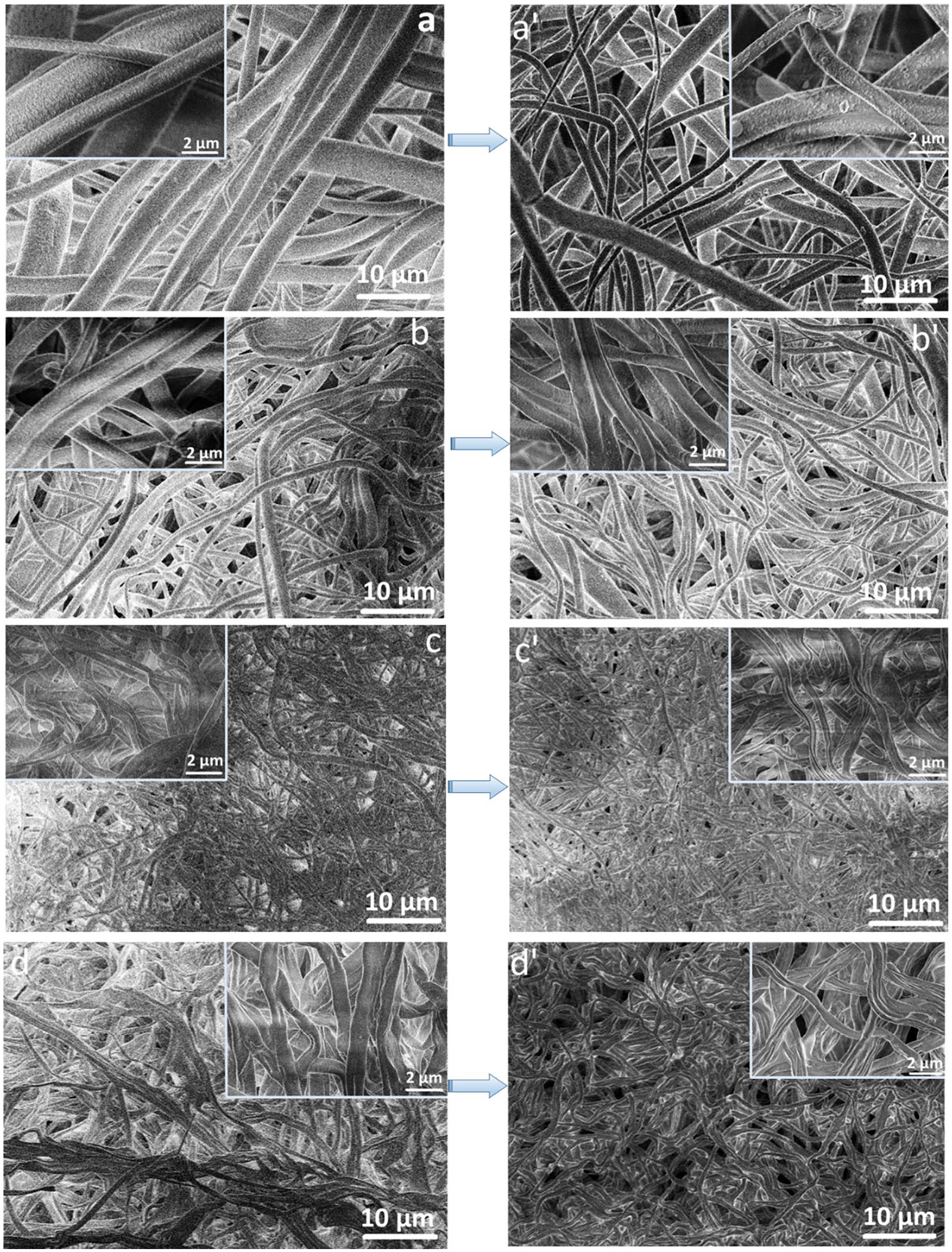
SEM images showing the morphology changes of the nanofiber webs during different degradation periods: 3 day ((a) PLLA, (b) PLLA/GO1, (c) PLLA/GO2 and (d) PLLA/GO3) and 9 days ((a’) PLLA, (b’) PLLA/GO1, (c’) PLLA/GO2, (d’) PLLA/GO3).
Conculsions
In conclusion, nanofiber webs based on PLLA embedded with GO nanosheets were prepared by electrospinning technique. The morphology of the PLLA nanofibers exhibited round with pores distribution throughout the fibers, while it was flatter ribbon-like surfaces of the composite nanofibers as a result of high GO concentration. Compared with neat PLLA, and their average diameter was decreased with the increasing incorporation of GO sheets. Furthermore, remarkable promotion in mechanical properties of PLLA/GO composite nanofibers was obtained with the content of GO. The simultaneous enhancement in thermal and mechanical properties could be due to the homogeneous dispersion of GO in PLLA matrix, improved interfacial interaction. With the incorporation of GO, the degradability of PLLA/GO composite nanofiber webs was higher than that of neat PLLA. As the content of GO increased from 2 wt% to 6 wt%, the trend of degradation increased. After 9 days degradation, the weight loss of PLLA/GO nanofiber webs reached 10.71 wt% for PLLA/GO1, 13.45 wt% for PLLA/GO2 and 16.83 wt% for PLLA/GO3, respectively. This result suggested that the graphene is very effective in improvement of the PLLA composites and may promise multipurpose of PLLA applications.
Footnotes
Declaration of conflicting interests
The author(s) declared no potential conflicts of interest with respect to the research, authorship, and/or publication of this article.
Funding
The author(s) disclosed receipt of the following financial support for the research, authorship, and/or publication of this article: This work was supported by the Jiaxing Project of Science and Technology (2019AD32011).