
Abstract
The main objective of this study was to develop antibacterial materials based on polyacrylonitrile for potential application in protective face masks to combat airborne pathogens. To achieve biocidal properties, 1-chloro-2, 2, 5, 5-tetramethyl-4-imidazolidinone as a kind of N-halamine was introduced into the polyacrylonitrile nanofibers by an electrospinning technique to form nanofibers by an electrospinning technique to form polyacrylonitrile/1-chloro-2, 2, 5, 5-tetramethyl-4-imidazolidinone-5% nanofibers. Scanning electron microscopy and Fourier transformed infrared spectroscopy were employed to characterize the structure of nanofibers. The antimicrobial efficacies of electrospinning nanofibers with 1-chloro-2, 2, 5, 5-tetramethyl-4-imidazolidinone against both Staphylococcus aureus and Escherichia coli O157:H7 were evaluated at different contact times. The antimicrobial efficacies against bioaerosol of S. aureus were also performed. The polyacrylonitrile/1-chloro-2, 2, 5, 5-tetramethyl-4-imidazolidinone-5% nanofibers possess excellent antimicrobial efficacies against bacteria bioaersol, and it has good air permeability.
Introduction
Recently, polymeric nanofibers have attracted increasing attention. Nano-sized fibers have good advantages in diameter, porosity, and especially in surface properties, such as high surface area.1,2 The small pore sizes of the nanofibrous mats were confirmed to be essential for air permeability. In contrast to other materials, nanofibers have a wide range of applications including battery, 3 filtration,4–7 drug delivery8,9 and so on.
Electrospinning is one of the common methods to prepare nanofibrous materials and has aroused much attention due to its efficiency and simplicity.10,11 Many researchers have made great efforts to develop and modify the electrospinning technology. Polyacrylonitrile (PAN) is one of the common materials used in electrospinning for various applications. 12 The effects of temperature, amount of PAN, voltage on structure, and thermal properties have been studied.13–15 To make the nanofibers antibacterial, antibacterial agents are introduced to PAN nanofibers creating a potential use in the area of biomedical materials.
There are many literatures about the antibacterial agents, 16 which have been used in microbial infection control including metal,17–19 quaternary ammonium salts, 20 N-halamine,21–24 and so on. Among these, N-halamines are the most effective antimicrobial materials against a broad spectrum of microorganisms,25–27 such as bacteria, viruses, fungi, and yeasts, 28 and with a great research interest due to their long-term stability and low environmental impact. In recent years, several methods were used to produce antibacterial fibers such as blending, composite spinning, and finishing process. However, few studies using nanofibers for protective face masks have been reported. Ren et al. 7 added N-halamine precursors into PAN solutions to produce nanofibers by electrospinning and acquired antimicrobial activity through chlorination. However, the chlorination process might affect the structure of nanofibers. Antibacterial agent 1-chloro-2, 2, 5, 5-tetramethyl-4-imidazolidinone (MC) was used widely without a chlorination process, as it helped reduce the waste of water and other resources. MC has been applied to water disinfections due to its high efficacy and is inexpensive to synthesize. 29 It provided a rapid and effective antibacterial material. It showed good stability in deionized water. 6 In recent report, Huang et al. prepared a new kind of packaging materials with MC loaded with absorbent pads in meat packaging. 30 In this study, we used the electrospinning to produce PAN nanofibers with an addition of the N-halamine (MC) in the spinning solution without a need for the chlorination process. The biocidal efficacies against Staphylococcus aureus and Escherichia coli O157:H7 in solution and bioaerosol of S. aureus were evaluated.
Experimental setup
Materials
PAN with a molecular weight of 150,000 Dalton was purchased from Aldrich Chemical Co., Ltd., Shanghai, China. MC was prepared according to the procedure described by Tsao. 29 All chemicals were used without further purification.
Electrospinning
The PAN spinning solutions at 5, 8, and 10 wt% were prepared by adding PAN into dimethylformamide (DMF) and stirring at ambient temperature until homogeneous solutions were formed. A 20-mL syringe was filled with the prepared solution, equipped with a flat-tipped stainless steel 22-gauge needle and placed in a syringe pump. The voltage supply was connected to the needle tip, 15 kV was applied, and the flow rate was set to 1.5 mL/h. Fibers were collected on a grounded target covered in aluminum foil, and the perpendicular distance from the needle tip to the collector was 15 cm.
Preparation of PAN/MC-5% nanofibers
A known amount of wt% of PAN was immersed into DMF and stirred at ambient temperature to produce a homogeneous solution. Then, 5 wt% MC was dissolved in the PAN solution to obtain antimicrobial properties. The PAN/MC-5% nanofibers were produced using the electrospinning technique.
Instruments
Syringe pump (JZB-1800, 20 mL) was purchased from Fresenius Kabi Jian Yuan (Changsha) Medical Technology Co., Ltd. Thermal analysis was conducted in a thermo-gravimetric analyzer (TA Instruments, the United States). About 1 to 3 mg samples were scanned from 30oC to 600oC at a heating rate of 10oC/min under nitrogen atmosphere. Fourier-transformed infrared (FT-IR) spectral analysis was carried out with a NICOLET iS10 spectrometer (the United States). These IR spectra were collected over a range of 4000 to 500 cm−1. The surface morphology of the nanofibers was examined by a scanning electron microscope (TM3030, Japan) operated at 25 kV with different magnifications, and SEM micrographs were taken for analysis. Different sample was taken once on SEM. Fabric air permeability instrument (YG (B) 461E, China) was used to measure the air permeability of samples.
Iodometric titration
An iodometric/thiosulfate titration procedure was applied to measure the oxidative chlorine content on the nanofibers. Triplicate samples were performed, and the average chlorine contents were reported. It was calculated using the following equation

where, N and V are the normality (equiv/L) and volume (L) of sodium thiosulfate, respectively, and W is the weight (g) of the nanofibers.
Chlorine stability test
In order to test the stability of the chlorine in the swatch, a series of nanofibrous mats were immersed into 10-mL deionized water with shaking (40 rpm) at ambient temperature. 31 After samples were removed at 3, 6, 24, 48, 72, and 96 h of shaking, the water samples were titrated to measure the quantity of the released chlorine in the deionized water following the National Environmental Methods Index (NEMI) test method 4500-ClB. Triplicate samples were performed, and the average chlorine contents were reported. The active chlorine content was calculated by the following equation

where, N and V are the normality (equiv/L) and volume (L) of sodium thiosulfate, respectively, and V′ is the volume of deionized water.
Shelf-life stability of PAN/MC-5% films
Nanofibrous mats were stored separately in a dark environment at ambient temperatures or under UVA light at 60oC. After designated storage times (0, 2, 3, 5, 8, 12 weeks), the samples were removed and the active chlorine contents were measured. The stability of the PAN/MC-5% was assessed by the remaining chlorine content in the samples. Triplicate samples were performed, and the average chlorine contents were selected.
Biocidal efficacy analysis
Staphylococcus aureus (ATCC 6538) and E. coli O157:H7 (ATCC 43895) were used to test the biocidal efficacy of the PAN nanofibrous mats and PAN/MC-5% nanofibrous mats. The inoculum was about 106 CFU bacteria per sample. AATCC Test Method 100-2004 with modification was used. In brief, an aliquot of 25 µL of bacterial suspension was added to the center of a 2.54-cm square swatch, and another same swatch was placed on the top. After a certain contact time, the samples were quenched with 5.0 mL of sterile 0.02 N sodium thiosulfate solution to neutralized chlorine residuals and vortexed vigorously to remove survived inoculum from mats to solution. Then 10-fold serial dilutions were made using pH 7, and 100 μM phosphate buffer; and each dilution was plated on trypticase soy agar plates in triplicate. After the plates were inoculated at 37°C for 24 h, the average bacterial colony number was counted for antibacterial effectiveness analysis.
Biocidal efficacy using an aerosol test
Biocidal efficacy was tested using the standard test method—ASTM Method F 2101.01. During the test, S. aureus was used as a biological aerosol to test the bacterial filtration efficiency of the nanofibrous mats. All materials were sterilized before use. Briefly, PAN/MC-5% nanofibrous mats with PAN mats as a control were installed on the experimental setup. The bacteria were aerosolized using a jet nebulizer through the chamber to contact samples. In this setup, the streaming air pressure and airflow were set at 20 psi and 259 mL/min, respectively. The duration of treatment was 3 h. After the experiment, samples were aseptically removed and placed into 5 mL 0.02 N sodium thiosulfate solution to isolate survived bacteria following the procedures mentioned in the previous section. Then, the biocidal efficacy was analyzed.
Results and discussions
Characterization of PAN and PAN/MC-5% nanofibers
SEM micrographs of various PAN contents are shown in Figure 1. It was found that the average fiber diameter of 8 wt% PAN nanofibers was around 350 nm. Compared to 5 wt% PAN and 10 wt% PAN, the 8 wt% PAN was chosen for the experiment due to the presence of knots in the 5 wt% PAN and the large diameter in the 10 wt% PAN. Also, the nanofibers mats became thicker with increasing concentration of PAN. The increase in viscosity of PAN spinning solution led to the increase of surface tension, which made it harder to prepare thinner nanofibers mats.

SEM micrographs of (a) 10 wt% PAN, (b) 8 wt% PAN, (c) 5 wt% PAN, and their diameter distributions.
The PAN/MC-5% nanofibers were synthesized for using as the antibacterial material. By contrast, the surface morphology of nanofibers was not influenced by the addition of the MC antibacterial compound (Figure 2).
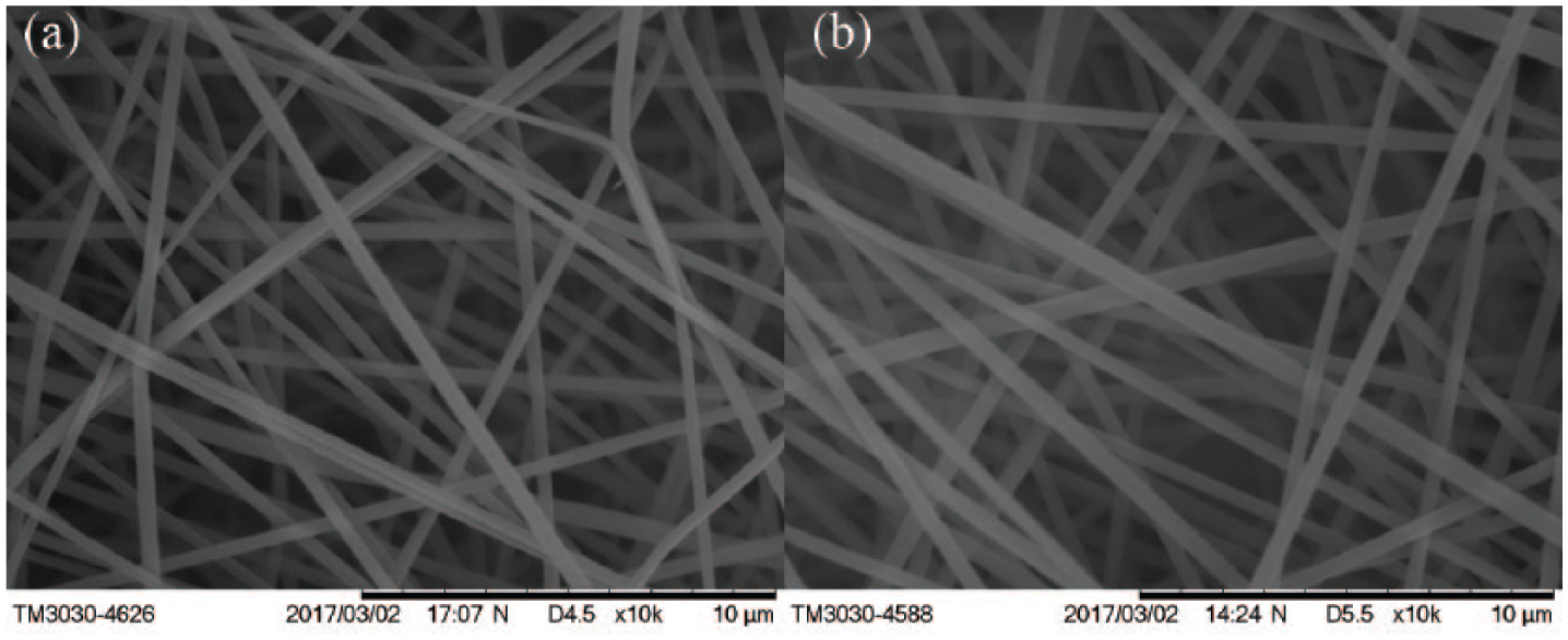
SEM micrographs of (a) 8% PAN, (b) 8% PAN incorporated with 5% MC.
The FT-IR spectra of MC, PAN, and PAN/MC-5% are shown in Figure 3. The strong bonds at 2247 cm−1 and 1080 cm−1were attributed to the nitrile (C≡N) functionality of PAN (Figure 3(b)).32–34 The appearance of a new functional group on PAN/MC-5% was shown by the presence of new peak at 1715 cm−1, which was assigned to C=O stretching vibration of MC (Figure 3(a)). The results indicated that the antibacterial agent MC existed in the nanofibers.

The FT-IR spectra of (a) MC powder, (b) PAN, (c) PAN/MC-5%.
The decomposition temperatures (°C) of PAN and PAN/MC-5% nanofibers against weight loss (%) are shown in Figure 4. It was found that there were two clear weight losses in the TGA (thermogravimetric analysis) curve of PAN nanofibers. The onset of weight loss of PAN nanofibers was at 100°C due to the water evaporation. The second loss was found to begin at 283°C. The temperature of the maximum decomposition rate was at 294°C. When the temperature reached 330°C, the PAN nanofibers showed 40% weight loss. In the TGA curve of PAN/MC-5% nanofibers, there were three weight losses. The weight of PAN/MC-5% nanofibers slightly declined at about 157°C, which was related to the melting point of MC. 6 It is believed that the antibacterial agent MC has no effect on the thermal stability of PAN nanofibers.

TGA and DTG curves of (a) PAN nanofibers and (b) PAN/MC-5% nanofibers.
Stability of PAN/MC-5%
The stability of chlorine in PAN/MC-5% was tested by dipping these samples into deionized water in a state of oscillation for designated time intervals. The oxidative chlorine content in the sample was 0.30%. As shown in Figure 5, the active chlorine released into the solution increased gradually with time. When it reached 3 h, the active chlorine in the solution was about 0.3 ppm and increased to 1.69 ppm after 96 h. The chlorine content was lower than the EPA maximum residual disinfectant level (4 ppm) for drinking water. 35 The excellent stability of the fixed chlorines in nanofibers indicated the great potential applications in air and water filtration.

Released chlorine contents for PAN/MC-5% nanofibers.
The storage stability and UVA light stability of PAN/MC-5% nanofibers are shown in Table 1. After 12-week storage, the chlorine loading of PAN/MC-5% nanofibers had no significant decrease under dark storage. The chlorine loadings dramatically decreased under UVA light, and this was similar to the result reported by Demir et al. 6 The chlorine loss was related to the N-Cl bond photo dissociation. 36
Storage Stability of PAN/MC-5%. a

PAN: polyacrylonitrile; MC: 1-chloro-2, 2, 5, 5-tetramethyl-4-imidazolidinone.
The error in the measured Cl + loading was about ± 0.01%.
Chlorine loadings are reported in wt% Cl +.
Air permeability
Figure 6 shows the air permeability of the nanofibrous mats with thickness of 0.46 mm. According the ASTM D 737 criterion, 34 the air permeable property was tested under a differential pressure of 100 Pa, and the materials were measured at least 3 times on the condition that the diameter of the circular samples was 70 mm. The average air permeability of the PAN and PAN/MC-5% are 33.3 mm s−1 and 27.3 mm s−1, respectively. The air permeability decreased by 20% between PAN and PAN/MC-5% nanofibers. For the commercial nano-surgical mask and Nano-N95, it was reported that the air permeability was below 100 Pa (around 20 mm s−1 and 12 mm s−1). 37 Therefore, the membranes are more breathable.

Air permeability of PAN and PAN/MC-5% nanofibers.
Antimicrobial efficacy
Bacterial log reduction is commonly used to express the antimicrobial ability of biocides. The biocidal activity results are presented in Table 2. Pure PAN nanofibers showed 0.768 log reduction for S. aureus and 0.110 log reduction for E.coli O157:H7 after contact time of 30 min. PAN/MC-5% nanofibers with chlorine loading of about 0.29% showed a robust reduction of inoculated bacteria. It inactivated all inoculated S. aureus (6.04-log reduction) within 1 min. The PAN/MC-5% nanofibers caused 1.63 logs and 6.60 logs (100%) reduction of E. coli O157:H7 in 5 min and 10 min, respectively. The results showed that the excellent biocidal function of the PAN/MC-5% was from MC.
Biocidal efficacy against bacteria.

PAN: polyacrylonitrile; MC: 1-chloro-2, 2, 5, 5-tetramethyl-4-imidazolidinone.
The inoculum was 1.10 × 10⁶ CFU/sample.
The inoculum was 3.97 × 10⁶ CFU/sample.
The PAN and PAN/MC-5% samples were also tested against S. aureus by an aerosol test. The results are presented in Table 3. After the test, the PAN/MC-5% had 2.58 × 10⁶ (about 6.41-log reductions) and 1.47 × 10⁶ (about 6.17-log reductions) for S. aureus compared with PAN control, in the Experiment 1 and the Experiment 2, respectively. The filters behind control and sample had no bacteria recovered, which indicated the PAN and PAN/MC-5% samples had good performance in filtration against bacteria. The PAN/MC-5% nanofibrous materials showed significant reduction against S. aureus aerosols and could inactivate most bacteria in the aerosols collected on the materials. Furthermore, a real use in food package made it clear that MC could be applied to an internal melt-blown layer, which would not contact the skin of the user. Therefore, it was concluded that there are no issues of biocompatibility or toxicity since MC did not evaporate readily at normal temperature and pressure, and it did not send out any chlorine gas. Considering the air permeability of PAN/MC-5% nanofibrous used as protective materials, this typical experiment suggested that PAN/MC-5% materials could have potential applications in filter materials, medical products, and protective masks.
Biocidal efficacy of fabrics against Staphylococcus aureus bioaerosols.

PAN: polyacrylonitrile; MC: 1-chloro-2, 2, 5, 5-tetramethyl-4-imidazolidinone.
Conclusion
PAN/MC-5% antibacterial nanofibers were synthesized successfully from PAN and MC by electrospinning. These nanofibers have smooth surface as viewed under SEM. The PAN/MC-5% exhibited excellent storage stability and strong antimicrobial efficacies against both S. aureus and E. coli O157:HH7. Within the contact time of 1 and 10 min, it was able to inactivate all inoculated S. aureus and E. coli O157:H7 with 6.04 and 6.60 log reductions, respectively. PAN/MC-5% materials can also inactivate S. aureus bioaerosols with more than 6 log reductions after 3 h exposure.
Footnotes
Declaration of conflicting interests
The author(s) declared no potential conflicts of interest with respect to the research, authorship, and/or publication of this article.
Funding
The author(s) disclosed receipt of the following financial support for the research, authorship, and/or publication of this article: The authors thank for the financial supports from the Fundamental Research Funds for the Central Universities (JUSRP51722B and JUSRP11702) and the Project for Jiangsu Scientific and Technological Innovation Team.