
Abstract
KLF2, a member of the Kruppel-like factor (KLF) family, is thought to be a tumor suppressor in many kinds of malignant tumors. Its functions in prostate cancer (PCa) are unknown. This study aimed to explore the role of KLF2 in the migration and invasion of PCa cells. The expression of KLF2 was measured by immunohistochemistry in PCa tissues and in paired non-tumor tissues. KLF2 and MMP2 expression in cells was measured by Western blot and RT-qPCR. Adenoviruses and siRNAs were used in cell function tests to investigate the role of KLF2 in regulating MMP2. Interactions between KLF2 and MMP2 were analyzed by a luciferase activity assay. The present study, for the first time, identified that KLF2 was downregulated both in PCa clinical tissue samples and in cancer cell lines. The overexpression of KLF2 inhibited the migration and invasion of PCa cells via the suppression of MMP2.This study demonstrates that KLF2 might act as a tumor suppressor gene in PCa and that the pharmaceutical upregulation of KLF2 may be a potential approach for treatment.
Prostate cancer (PCa) is one of the most common solid malignancies, and its mortality rate ranks second among malignancies in males (Siegel, Naishadham, & Jemal, 2012). In the United States and Europe, the incidence of PCa ranked first (Siegel, Miller, & Jemal, 2016). In China, its incidence is also rising yearly; it is estimated that there were over 60,300 new cases of PCa in China, accounting for approximately 26,600 deaths in 2015 (Chen, Zheng, et al., 2016).
Kruppel-like factors (KLFs) have Cys-2/His-2 zinc-finger domains that bind to GC boxes in DNA sequences, acting as transcriptional activators or repressors by binding to target promoters (Bialkowska, Yang, & Mallipattu, 2017). KLFs have also been reported for transcriptional regulation in cell differentiation and proliferation (Suske, Bruford, & Philipsen, 2005). KLF2, a principle member of the family, is particularly interesting in terms of cancer prevention. Studies suggested that KLF2 was downregulated in breast (Zhang, Levi, Banerjee, Jain, & Noy, 2015), colorectal (Wang, Cao, et al., 2017), and lung cancers (Li et al., 2016). Another study reported that KLF2 was abundantly expressed in normal epithelial cells but was remarkably downregulated in several cancers of various grades and stages (Wang et al., 2005). Many studies demonstrated that KLF2 was an inhibitor of cell proliferation, migration, and metastasis and a promoter of cell apoptosis (Nie et al., 2016; Xie et al., 2011; Zhang et al., 2016).
Matrix metalloproteinases (MMPs) belong to a multifunctional family of zinc dependent endopeptidases, the main function of which is lysis of the extracellular matrix (ECM), a collection of extracellular molecules secreted by cells that provide structural and biochemical links to normal physiological cells. MMP2 is one member of the MMP family. The major activity of MMP2 is the hydrolysis of collagens (Types I, IV, V, VII, X, and XI), which are the most abundant proteins in the ECM, including fibronectin, laminin, aggrecan, and elastin (Banday et al., 2016). MMP2 plays a key role in cell migration because it can dissolve collagens, allowing malignant tumors to penetrate basement membranes. Numerous studies have reported that MMP2 was associated with tumor invasion (Chen, Yang, et al., 2016; Huang et al., 2014; Lu et al., 2014).
This research hypothesized that KLF2 might be an important gene involved in the development of cancer, particularly PCa. In the present study, we investigated the expression of KLF2 in PCa clinical samples and explored the biological functions of KLF2 in the progression of PCa.
Materials and Methods
Cell Culture
Human PCa cell lines (PC-3 and 22Rv1) and a normal epithelial cell line (RWPE-1) were purchased from the Institute of Biochemistry and Cell Biology of the Chinese Academy of Sciences (Shanghai, China). Media for cell culture and fetal bovine serum (FBS) were obtained from GIBCO. The PC-3, 22Rv1, and RWPE-1 cell lines were cultured in Ham’s F12 nutrient medium (Invitrogen). RPMI 1640 medium (Invitrogen) were supplemented with 10% FBS (Sigma) and keratinocyte serum-free medium (Invitrogen), respectively. All the cells were cultured at 37 °C in an atmosphere of 5% CO2.
Cell Transfection
KLF2 adenovirus was a recombination product, encoding human KLF2 that was inserted into the gene vector pCMV5-Nter-FLAG. A total of 1 μl of KLF2 adenovirus and KLF2-null adenovirus was added to the PCa cell lines. KLF2 siRNA was designed according to the KLF2 messenger RNA (mRNA) sequence. After the efficacy test, the most efficient one was chosen as the target siRNA. KLF2 siRNA and negative control siRNA were synthesized by Invitrogen (Table 1). The cells were cultured in six-well plates for 24 hr before transfection. The siRNA was transfected by Lipofectamine RNAiMIX (Invitrogen) in FBS-free RPMI 1640 medium for 6 hr, followed by addition of 10% FBS. After 48 hr, the cells were harvested for RT-qPCR and Western blot.
Primer List.
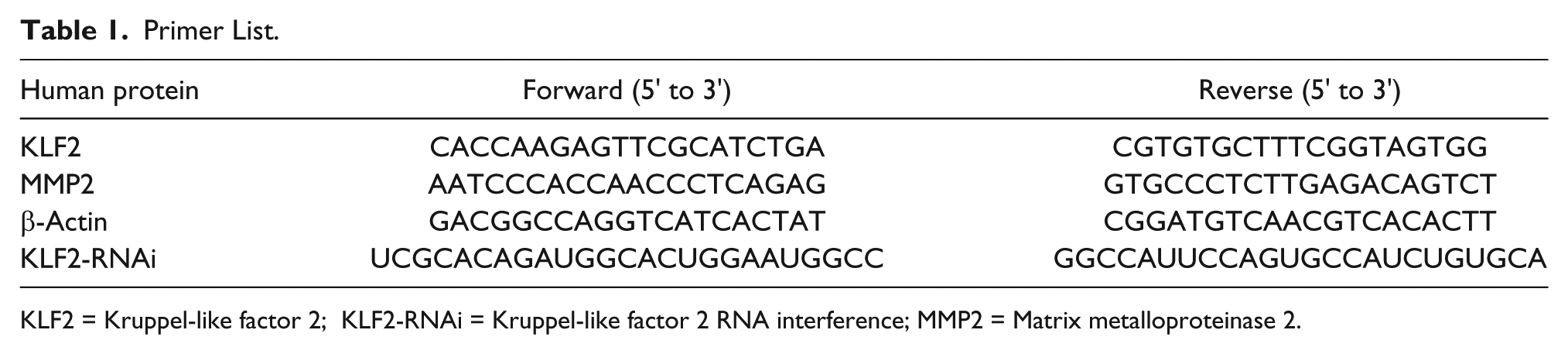
KLF2 = Kruppel-like factor 2; KLF2-RNAi = Kruppel-like factor 2 RNA interference; MMP2 = Matrix metalloproteinase 2.
Clinical Tissue Samples and Immunohistochemistry
The study was approved by the Research Ethics Committee of Peking University Third Hospital (Beijing, China). Radical prostatectomy PCa tissues samples had been obtained after surgery. Written consent was obtained from patients. Out of 30 PCa patients, 3 cases were T1c, 4 cases were T2a, 7 cases were T2b, 11 cases were T2c, and 5 cases were T3a. The expression of KLF2 protein was measured by immunohistochemistry in both PCa tissues and adjacent normal tissues. The samples were fixed in formalin, embedded in paraffin, and cut into 4-μm thick sections. After de-paraffin and antigen recovery, the sections were washed three times in 0.01 mol/L phosphate buffered saline (PBS) for 5 min each, blocked for 1 hr in 0.01 mol/L PBS contained 0.3% Triton X-100 and 5% BSA, followed by addition of anti-KLF2 (1:50) (Abcam) antibody at 4 °C overnight. After washes with PBS, sections were incubated with 0.01 mol/L PBS containing horseradish peroxidase conjugated rabbit anti-goat IgG (1:500) (Zhongshan) for 2 hr, followed by incubation with 0.003% H2O2 and 0.03% 3,30-diaminobenzidine in 0.05 mol/L Tris–HCl (pH 7.6).
Western Blotting
Western blot analysis was performed according to standard protocols. The cells were lysed by RIPA Lysis Buffer (Genstar) containing protease inhibitors and phosphatase inhibitors. The lysate proteins were separated by 10% of sodium dodecyl sulfate-polyacrylamide gels, which were then transferred to polyvinylidene fluoride membranes (Millipore). The membrane was incubated overnight with primary antibodies to KLF2 (1:1,000), MMP2 (1:600), and β-actin (1:1,000) (Abcam). After the secondary antibody of anti-mouse IgG and anti-rabbit IgG (Zhongshan Golden Bridge) probed for 2 hr, the blots were visualized using luminal reagent kit (Immuno Cruz) via X-ray films (Kerui).
Quantitative RT-PCR
The total RNA was extracted from the cells by TRIzol reagent (Invitrogen) according to the manufacturer’s instructions. A total of 3 μg RNA was reverse-transcribed in a volume of 20 μl by using random primers Reverse Transcription Kit (Takara). Real-time PCR analyses were carried out with SYBR Green (Takara), following the manufacturer’s instructions. β-actin mRNA expression was considered as the internal control. The RT-qPCR and the data collection were performed on a Quantstudio 5 Real-time-PCR instrument (Applied Biosystems, Thermo Fisher). Relative expression was calculated and normalized using the 2−ΔΔCt method relative to β-actin. The primer sequences are listed in Table 1. The experiments were repeated three times.
Wound-Healing Assay
KLF2 and KLF2-null adenoviruses were transfected into cell lines. Once the cell density reached 80%, a “wound” was created by scratching the culture surface with a sterile 20 µl tip and cultures were washed with PBS to remove the detached cells. Medium with 1% FBS was used for cell culturing over the following 24 hr. At three time points (0, 6, 12 hr) images of the plates were obtained using the microscope camera system. ImageJ program was used to calculate the migration area in order to indicate the migration trends. We applied the area of 0 hr as the frame of reference, so the relative change of the area at 6 hr or 12 hr was normalized to the area at 0 hr. The experiments were repeated three times in each cell line.
Cell Invasion Assays
The Transwell assay was performed using a Boyden chamber (Corning) covered in Matrigel. At 48 hr post-transfection of KLF2 adenovirus and KLF2-null adenovirus, 1 × 10⁵ cells with serum-free medium were placed into the upper chamber. Medium containing 10% FBS was added to the lower chamber. After 12 hr incubation, the cells remaining in the upper chamber were removed with a cotton swab. The cells invading through the Matrigel membrane were fixed with methanol and were stained with 0.1% crystal violet. The invaded cells were counted via an inverted microscope (Leica). The number of cells were counted in four different fields in every well and images were captured under a light microscope. Each experiment was repeated three times.
Reporter Vector Construction and Luciferase Activity Assay
MMP2 promoter and KLF2 sequence were searched through the National Center for Biotechnology Information (NCBI) database. The KLF2 and MMP2-promoter genes were synthesized from Genechem and were inserted into the expression vector GV230 (CMV-MCS-EGFP-SV40-Neomycin) (Genechem) and GV238 (MCS-firefly luciferase) (Genechem) with cloning sites KpnI and XhoI, respectively. Transfection for the luciferase activity assay was performed in 24-well plates using X-tremegene HP (Roche). Two cloning vectors of 0.5 μg each were co-cultured in 293T cells for 48 hr. A Renilla luciferase-containing plasmid (Genechem) was used to control for transfection efficiency. Luciferase activity was determined by a dual-luciferase reporter assay system (Promega). MMP2 promoter-driven luciferase activity was normalized to thymidine kinase Renilla activity. The experiments were repeated three times. All data were presented as the means ± standard deviations (SD). Student’s t-test and two-way analysis of variance were used for the comparison of paired observations.
Results
The Expression of KLF2 Is Downregulated in PCa Tissues and PCa Cell Lines
Pathological results showed all 30 patients were prostate adenocarcinoma. Pathological Gleason score: There were 3 cases that less than 6 points, and 9 cases that have a score of 7 points, 18 cases were higher than 8 points. Immunohistochemistry results reported that the intracellular localization of KLF2 were cytoplasm and nucleus in epithelial cell (Figure 1A). The quantity of KLF2 expression in cancer tissues and normal tissues with different groups are summarized in Table 2. KLF2 expression in tumor tissues was lower than that in normal tissues (p = .005; Figure 1B). In addition, the result of western blot revealed that KLF2 expression in RWPE-1 was higher than PC3 and 22Rv1 (Figure 1C).

KLF2 Expression in Different Patients.

The Overexpression of KLF2 in PCa Cells Inhibits Cell Migration and Invasion
To evaluate the biological function of KLF2 in PCa cells, the role of KLF2 overexpression in the regulation of PC3 and 22Rv1 cells migration was investigated. The wound healing assay proved that the migration ability at different time points was reduced in KLF2 transfected cells. Cells transfected with KLF2 would get a slower closing towards scratch wounds compared with the control cells (Figure 2 A and B). Furthermore, the invasion ability (penetrating Matrigel) of cancer cell was evaluated by Transwell assay. The results identified that the invasion of PC3 and 22Rv1 cell lines were reduced by the overexpression of KLF2 in vitro (Figure 2C).

MMP2 Is a Potential Downstream Target of KLF2
Western blot and qPCR results demonstrated that the overexpression of KLF2 was associated with the suppression of MMP2. The overexpression of KLF2 reduced expression of MMP2 at both the protein and mRNA levels (Figure 3A and B). Conversely, decreased KLF2 expression induced higher MMP2 expression (Figure 3C and D). The luciferase activity assay reported that the promoter activity was regulated by transcription factors which indicated MMP2 was regulated by KLF2 at the transcription level (Figure 4).


Discussion
Recent publications have reported that several KLF family members, including KLF4, KLF5, and KLF9, served as tumor suppressor genes in PCa (Ci et al., 2015; Shen et al., 2014; Wang et al., 2010). Wang et al. have reported that KLF2’s expression level were substantially lower in both gastric cancer cell lines and human tumor specimens and negative correlated with patient survival (Wang, Li, Duan, Wang, & Chen, 2017). Previous studies have shown that KLF2 expression is downregulated in many human cancers, such as ovarian and lung cancer (Wang et al., 2005; Yin et al., 2015). Unexpectedly, KLF2 was found to be upregulated in hepatocellular carcinoma and appeared to be an oncogene (Zou et al., 2016). These studies suggested that the role of KLF2 may vary from different types of cancers. However, whether KLF2 acts as an oncogene or tumor suppressor in PCa remains unknown.
In the present investigation, experimental data strongly supported an association between KLF2 and PCa. First, KLF2 expression was significantly decreased in PCa tissues compared with normal prostate tissues. Second, this research proved that KLF2 expression downregulated the expression of MMP2 in vitro. Moreover, this phenomenon was attenuated by transfection of siRNAs of KLF2. MMP2 secretion increased in many types of cancer diseases, and its abnormal expression is associated with a poor prognosis. MMP2 is one of the most important factors in tumor metastasis and is often abundantly expressed in tumors, degrading ECM and promoting the penetration of cancer cells (Jablonska-Trypuc, Matejczyk, & Rosochacki, 2016). MMP2 was proved to be associated with the increased migration and invasion ability of PCa cells (Kowalska, Habrowska-Gorczynska, Urbanek, Dominska, & Piastowska-Ciesielska, 2018; Peng & Kopecek, 2015; Tsui et al., 2018). As increased levels of MMP2 is one of the hot spots correlating with increased metastatic potential, the mechanisms regulating the production of MMP2 have been an active area of research (Jacob et al., 2013).
As an important hallmark of cancer, activating invasion and metastasis make cancers hard to treat and have been the main cause of patient death. Deaths due to metastasis constitute the major threat of PCa. It is critical to prevent metastasis in the progression of PCa for effective therapeutic intervention (Chen et al., 2017). In the present study, elevating KLF2 by adenovirus transfection repressed the migration ability of two types of PCa cells in a time-dependent scratch assay. In addition, the invasiveness of the PCa cells was impeded by KLF2 transfection.
It is well known that MMP2 is a classic tumor activator and its aberrant expression has often been found in various cancers. KLF2 could regulate the expression of multiple genes. By searching the Universal Protein Database, it was predicted that KLF2 binds to the CACCC box, and CACCC box was part of the promoter of MMP2. Therefore, we hypothesized MMP2 would be regulated by KLF2 through an unknown pathway. To test whether MMP2 could be regulated by KLF2, RT-PCR and luciferase activity test were performed. The result indicated that KLF2 can regulate MMP2 expression by interacting with the MMP2 promoter region. Specific regulatory mechanisms between KLF2 and MMP2 require further research. Taken together, these results strongly indicate that KLF2 might act as a tumor suppressor gene by significantly decreasing the ability of invasion and the immigration of PCa cells, possibly shedding light on the pathway to a novel pharmaceutical target. Further in vivo insights into the functions and interactions of KLF2 are required to elaborate the appraisal of novel diagnostic or therapeutic biomarkers for prostate tumors.
Supplemental Material
supplemental_data_MMP2 – Supplemental material for KLF2 Inhibits the Migration and Invasion of Prostate Cancer Cells by Downregulating MMP2
Supplemental material, supplemental_data_MMP2 for KLF2 Inhibits the Migration and Invasion of Prostate Cancer Cells by Downregulating MMP2 by Binshuai Wang, Mingyuan Liu, Yimeng Song, Changying Li, Shudong Zhang and Lulin Ma in American Journal of Men’s Health
Supplemental Material
supplement_table-KLF2 – Supplemental material for KLF2 Inhibits the Migration and Invasion of Prostate Cancer Cells by Downregulating MMP2
Supplemental material, supplement_table-KLF2 for KLF2 Inhibits the Migration and Invasion of Prostate Cancer Cells by Downregulating MMP2 by Binshuai Wang, Mingyuan Liu, Yimeng Song, Changying Li, Shudong Zhang and Lulin Ma in American Journal of Men’s Health
Footnotes
Declaration of Conflicting Interests
The author(s) declared no potential conflicts of interest with respect to the research, authorship, and/or publication of this article.
Funding
The author(s) disclosed receipt of the following financial support for the research, authorship, and/or publication of this article: This study was supported by the Beijing Municipal Science & Technology Commission (Z161100000116037) and the Tianjin Natural Science Foundation (16JCYBJC26500).
Supplemental Material
Supplemental material for this article is available online.
References
Supplementary Material
Please find the following supplemental material available below.
For Open Access articles published under a Creative Commons License, all supplemental material carries the same license as the article it is associated with.
For non-Open Access articles published, all supplemental material carries a non-exclusive license, and permission requests for re-use of supplemental material or any part of supplemental material shall be sent directly to the copyright owner as specified in the copyright notice associated with the article.