
Abstract
Although, superparamagnetic iron oxide nanoparticles (SPIONs) have extensively been used as a contrasting agent for magnetic resonance imaging (MRI), the lack of intrinsic fluorescence restricted their application as a multimodal probe, especially in combination with light microscopy. In Addition, the bigger size of the particle renders them incompetent for bioimaging of small organelles. Herein, we report, not only the synthesis of ultrasmall carbon containing magneto-fluorescent SPIONs with size ∼5 nm, but also demonstrate its capability as a multicolor imaging probe. Using MCF-7 and HeLa cell lines, we show that the SPIONs can provide high contrast mulicolor images of the cytoplasm from blue to red region. Further, single particle level photon count data revealed that the SPIONs could efficaciously be utilized in localization based super resolution microscopy in future.
Keywords
Introduction
Over the past few years, superparamagnetic iron oxide nanoparticles (SPIONs), due to their strong magnetic properties and magnetic susceptibility, have been extensively used in magnetic resonance imaging (MRI), 1 -3 terabit magnetic storage devices, 4 magnetically guided cancer diagnosis, 5 theranostic, 6,7 drug delivery, 8 -10 hyperthermia, 11 photoacoustics 12 and cell separation. 13 Despite such vast applications utilizing their paramagnetic behavior, the exploration of SPIONs as a multimodal probe for various techniques, especially in combination with optical microscopy for high resolution bioimaging is rarely reported. This is attributed to the lack of inherent fluorescence in SPIONs. Henceforth, engineering SPIONs by decorating the fluorescence properties and to get magneto-fluorescent SPIONs is highly desirable to function as a multimodal probe. The magneto-fluorescent behavior of SPIONs will be extremely beneficial as the drawback of 1 technique could easily be rectified by others, e.g. deep tissue cellular imaging, which is not possible in optical microscopy can be achieved by MRI. On the other hand, optical bioimaging is capable of attaining a high spatial resolution which is not possible by MRI imaging.
Nevertheless, few reports on the magneto-fluorescent SPIONs are available in the literature. The common synthesis approach includes the co-assembly of 2 types of nanocrystals having distinct properties into larger colloidal particles at the mesoscopic scale or by surface functionalization of the SPIONs with fluorescent materials such as quantum dots (QDs) or organic dyes. 14,15 Strategies involving the template based heterostructure crystal growth have also been reported. 16,17 However, the conjugated magneto-fluorescent SPIONs, synthesized by the above methods, have a few limitations. For example, the attached fluorescent molecules may suffer quenching in their fluorescence when attached to the nanoparticles and hence compromise with the effective fluorescence quantum yield (QY) and therefore restrict the long term bioimaging. In addition, the tedious multistep synthesis processes are challenging in terms of reproducibility and colloidal stability.
On the other hand, the synthesized magneto-fluorescent, reported so far are majorly very large in size, which obstructs their utilization in viewing smaller organelles inside the cells. 18 The size was found to be a critical criterion that decides the resolving power of the optical bioimaging, the internalization mechanism and kinetics of the cellular uptake efficiency of the nanomaterials into the cell. Moreover, the renal clearance of these bigger size particles is also an issue. 19 Few reports have discussed the small size fluorescent SPIONs. Riju et al. described the synthesis of fluorescein labeled small iron oxide nanoparticles as a T1 MRI contrasting agent and as a viable tool for cell labeling. 20 Jing et al. reported ultrasmall superparamagnetic iron oxide nanoparticles bound with NIR dyes as a theranostic agent. 21 Jabadurai et al. developed flavin mononucleotide coated fluorescent iron oxide nanoparticles. 22 Unfortunately, multistep tedious processes were involved in all these reports, where the fluorescent molecules were conjugated to the SPIONs. While synthesizing small size SPIONs is a challenge in itself, incorporating fluorescence properties adds another hurdle in the process to achieve homogeneous particles without losing both the magnetic and optical properties. Here, we were not only successful in synthesizing ultrasmall magneto-fluorescent carbon composite SPIONs with greater ease via a single step 1 pot procedure but also it demonstrated as a magneto-fluorescent probe for multicolour bioimaging from blue to red region. Using 2 different cell lines (MCF-7 and HeLa cell lines), we reveal the fast cell internalization of the SPIONs with bright multicolor confocal image of the cytoplasm. Additionally, the fluorescence properties at single particle level have also been studied. These studies reveal that the developed material can be efficiently used as a probe for super resolution microscopy in future.
Materials and Methods
All glasswares were washed with aqua regia, followed by rinsing several times with double distilled water. Ferrocene, Oleic acid, Hexadecene and H2O2 were purchased from Merck Chemicals. All chemicals were used without further purification. Double-distilled (18.3 mΩ) deionized water (ELGA PURELAB Ultra) was used throughout the entire process.
The particle morphology was observed by transmission electron microscopy (TEM) using FEI Tecnai TEM equipped with a LaB6 source operating at 200 kV. Powder X-ray diffraction (PXRD) pattern was recorded on a Rigaku Smart Lab diffractometer, using CuKα radiation from 5° to 80° with a scanning rate of 2°/min. Fourier transformed infrared (FTIR) spectra were recorded with Agilent Technologies Cary 6000 series FTIR spectrometer at wavenumber from 400 to 4000 cm-1. X-ray photoelectron spectroscopy (XPS) measurements were carried out on a NEXSA surface analysis model by Thermo Fisher Scientific using Al-Kα (1486.6 eV) X-ray radiation. Energy dispersive X-ray analysis (EDX) and elemental analysis of SPIONs was characterized by using a field emission scanning electron microscope (FESEM) FEI Nova SEM-450. The XPS data were acquired with a spot size 400 µm having a standard lens mode. Magnetic properties were analyzed through vibrating sample magnetometer-superconducting quantum interference device (VSM-SQUID) under the vibrating magnetic field of 4 T at 300 K. UV-Vis absorption spectra were measured with Shimadzu U-2450 UV-Vis spectrophotometer from wavelength range of 200-800 nm. Fluorescence spectra and absolute quantum yield were recorded on the Horiba spectrophotometer.
Confocal Imaging of Cells Labeled With SPIONs
Coverslip Preparation
The glass slides and coverslips were cleaned by incubating in freshly prepared Piranha solution for 30 min and finally washing by MiliQ water in bath sonication then dried under nitrogen.
Cell Culture, Fixation and Staining
Cells were grown in Dulbecco’s Modified Eagle Medium (DMEM) with 10% fetal bovine serum. The cells were grown in 6-well plate on the coverslips with a density of 104 cells per 100 µl. Each well was filled with 2 ml of cell suspension in growth medium and the cells were allowed to grow overnight for the proper adherence and growth. The growth and the attachment of the cells to the coverslips were examined by an optical microscope. Once the cells reached proper attachment and growth stage, they were stained with SPIONs for 4 h to achieve enough labeling density for confocal microscopy. Finally, the cells were fixed by incubating with 4% paraformaldehyde solution in 1x PBS buffer for 15 min. The fixed cells were then washed 4-6 times by PBS buffer to remove extra agents. The coverslips were fixed on a glass slide before imaging.
Confocal Microscopy
Nikon Eclipse Ti inverted microscope was used for the confocal microscopy and images were acquired using Nikon Nis-Element software. The cell samples were excited by the 4 lasers 401, 488, 561 and 639 nm. Finally, the images were collected by choosing a proper filter set.
Single Molecule Time Trace and Photon Counts
SPIONs were spin coated on a clean glass slide to collect single molecule time traces. The diffraction limited spots produced due to single molecule blinking were observed using 100x Nikon TIRF objective. A 532 nm diode laser with 50 mW maximum power was used. An oil immersion Nikon TIRF objective (100x magnification and 1.49 NA) was mounted on a custom build inverted optical microscope. A 532 nm high pass Dichroic (AHF Analysentechnik) was used to separate the excitation and emission light. Andor EMCCD iXon Ultra was used to record the single molecule photon events at the frame rate of 17 MHz and exposure time of 50 ms. The time trajectories were recorded and analyzed using the Andor Solis Software. The incident photons were converted to electrons and subsequent to digital counts by the EMCCD. The Andor Solis and Matlab ware used to extract the total photon counts. The movies recorded under the kinetic mode of EMCCD, time/frame trajectories of the counts/intensity at a given pixel, were obtained for the provided exposure time. These time trajectories were saved for further analysis.
Result and Discussion
The carbon composite SPIONs were synthesized following the thermal decomposition method. In brief, 0.49 g Ferrocene, 2 ml oleic acid and 20 ml hexadecene were taken in a 3 neck round bottom flask and stirred for 5 min at 25°C under vacuum, to thoroughly mix all the components. Then, the temperature of the mixture was increased to 270°C and the mixture was stirred for 1 h after which it was again cooled to room temperature. Thereafter, 10 ml H2O2 (30% w/w) was added to the mixture and the temperature was slowly increased to 130°C and stirred for 1 h. The entire solution was finally cooled down to room temperature. The precipitates from the reaction mixture were extracted in ethanol solution and centrifuged at 1400 rpm. In order to remove any remaining oleic acid and hexadecene the precipitates were washed with hexane and thereafter the magnetic particles were separated with the help of magnet. Finally, the particles were dried in hot air oven at 80°C and dried powder was collected for various characterizations.
Transmission electron microscopy (TEM) images revealed the formation of homogeneous particles. Further, it also depicted a nearly spherical morphology SPIONs (Figure 1A) with an average size of ∼5 nm in diameter. The size was obtained by calculating the average distribution of approximately 50 particles as shown in (Figure 1A, inset). A 2D lattice fringes from high resolution (HR-TEM) image (Figure 1B, inset) ∼0.20 nm confirms the existence of (102) lattice plane of graphitic (sp 2 ) carbon. 23,24 Powder X-ray diffraction (PXRD) analysis was performed which further verified the composition of material as an iron carbon composite. The diffraction pattern showed sharp reflections at the 2θ value for magnetite phase of iron. Peaks corresponding to (111), (220), (311), (400), (422), (511), and (440) plane belong to Fe3O4 with inverse spinel structure. 25 A diffraction peak around 24° represents the (002) plane of graphitic carbon 26 thereby validating the presence of graphitized carbogenic nature to the SPIONs (Figure 1D). 27

(A) TEM and (B) HRTEM image of SPIONs, (C) PXRD spectra showing the diffraction planes (111), (220), (311), (400), (422), (511), and (440) observed for Fe3O4 and (002) plane for graphitic carbon, (D) fourier-transform infrared (FTIR) spectra shows absorption peak for different functional groups, (E) full survey XPS spectra depicting the presence of C1 s, O1 s and Fe2p, respectively in scan profile, (F) high resolution deconvoluted Fe2p XPS spectra, (G) high resolution deconvoluted O1 s XPS spectra and (H) high resolution deconvoluted C1 s XPS spectra of SPIONs.
X-ray photoelectron spectroscopy (XPS), a surface sensitive technique also corroborated the presence of magnetite phase of iron. Full survey scan spectra showed the presence of carbon, oxygen and iron (Figure 1E). Deconvolution of the Fe2p spectrum (Figure 1F
VSM (vibrating sample magnetometer) measurement was carried out to find out the magnetic properties of the material. A material is said to be superparamagnetic if it magnetized up to its saturation magnetization during the application of external magnetic field and on removal of magnetic field it should not exhibit any residual magnetic interaction. 29 A magnetic hysteresis (M–H) curve (Figure 2A) shows saturation magnetization (Ms) at 1.94 emu/g, which seems to be in the little lower range. This could be attributed to the ultrasmall size of the SPIONs, which might show the canting effect. 30 It was previously shown that on decreasing the size of magnetic core the magnetic saturation value decreases. 31 Moreover, once the applied magnetic field was removed, the magnetic particles did not retain any residual magnetism at room temperature. Fe3O4 crystals with diameters of 20 nm or less are superparamagnetic as there is no hysteresis after applying an external magnetic field because each crystal acts as a single magnetic domain. 32 Remanent magnetization (Mr) 0.35 emu/g, is very low which further verified superparamagnetic behavior. Little broadening in M-H hysteresis loop reveals a weak ferromagnetic nature of SPIONs at room temperature as coercivity (Hc) comes to be 134. 33,34 Temperature dependent zero field cooled (ZFC) and field cooled (FC) also suggest about the superparamagnetic nature, to achieve the superparamagnetic nature blocking temperature (TB) should be much lower than room temperature. 35 ZFC and FC in (Figure 2B) therefore confirmed the superparamagnetic behavior of material as TB was found to be ∼50 K. Also, the TB value is related to the particle volume (V) and effective anisotropy (Keff) by the following equation; V = 25kBTB/Keff, where kB is the Boltzmann constant. 36 According to this, lower the TB value lower will be the volume, TB ∼50 K again confirms the smaller size of the material. The shape of the ZFC and FC curves were indicative of the smaller particle size and narrower size distribution for SPIONs 37 which is in accordance with TEM data. Hence, it could be concluded that the material has enough magnetic properties to be used as MRI contrasting agent.

(A) M–H hysteresis curve for SPIONs recorded at room temperature and (B) temperature dependent ZFC and FC curves of SPIONs under a magnetic field of 100 gauss.
Next, we measured the optical properties of the magneto fluorescent SPIONs. A broad absorption spectrum ranging from 200 to 800 nm was observed. This could be attributed due to the carbon dots like behavior of carbon coated on SPIONs (Figure 3A). 38 The observed emission spectra also support the typical signature of carbogenic nanomaterials. A broad emission spectrum with an emission maximum at 423 nm under 360 nm excitation was observed (Figure 3A). Further, the carbon coated SPIONs shows the excitation dependent multicolor emission (Figure 3B) as normally obtained in carbon dots. 39 The observed emission properties of SPIONs may be attributed to surface energy traps emission from rich carboxyl and hydroxyl groups, as reported earlier for carbogenic nanomaterials. The multicolor emission was also observed on the spin coated drop dried sample when observed in a confocal microscope using different laser excitation systems, along with proper optical filters. The sample was excited with 4 different lasers 401, 488, 532, and 639 nm and the different color emission (blue, green, yellow and red respectively) are presented in Figure 3C-F. The data clearly show that the SPIONs could be used as a multicolor imaging probe in real cellular systems.

(A) The absorbance (blue) and PL (green) spectra of SPIONs, (B) emission spectra of SPIONs at different excitation wavelengths ranging from 280-700 nm, and (C-F) confocal microscopy images of SPIONs spin coated on glass coverslip showing multicolor emission under different excitation laser wavelengths 401 (blue), 488 (green), 561 (yellow), and 639 (red) nm, respectively.
The above results motivated us to examine the bioimaging of SPIONs in 2 different cancer cell line i.e. MCF-7 and HeLa cells. The cells were grown in Dulbecco’s Modified Eagle Medium (DMEM) with 10% fetal bovine serum and further seeded on a glass coverslip with density of 104 cells per 100 µl. Once the cells were properly adhered, they were incubated with SPIONs for 4 h in order to achieve enough labeling density for imaging in confocal microscope. The cells were then washed with PBS buffer several times to remove any unbound SPIONs. Finally, the stained cell lines were examined through the confocal microscope. Surprisingly, Figure 4 shows that the SPOINs have the capability to show the multicolor bioimaging with greater efficiency, but with a little decrease in intensity in the red region in both the cell lines. This could be justified by the emission intensity of the bulk measurement. It could be seen in Figure 3B that the emission intensity is maximum in the blue and green region, while it decreases substantially in the red region. As a result, in the confocal image, the number of excited particles is also less and hence the emission intensity decreases in the cells.
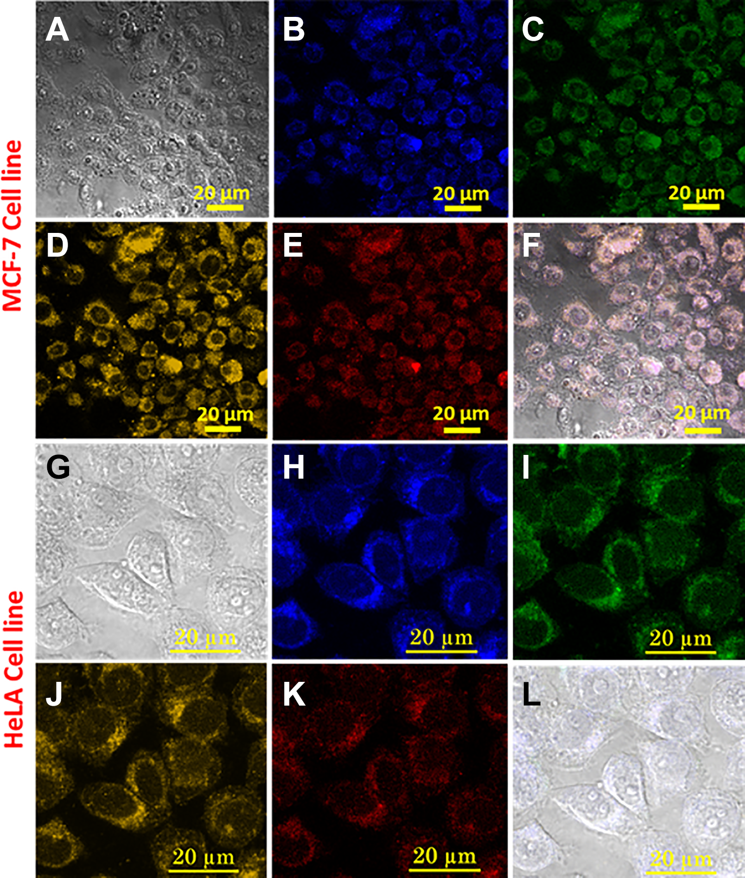
(A) TD image, (B-E) confocal microscopy image of MCF-7 cell line stained with SPIONs under different excitation laser wavelengths 401, 488, 561, and 639 nm, respectively, (F) overlay of Figure 4 (B-E), (G) TD image, (H-K) confocal microscopy image of HeLa cell line stained with SPIONs under different excitation laser wavelengths 401, 488, 561, and 639 nm, respectively and (L) overlay of Figure 4 (H-K).
Finally, the fluorescence emission behavior of SPIONs was performed at the single particle level. The dispersed spincoated particles on a coverslip were illuminated by a 532 nm laser and corresponding fluorescence was collected by set of appropriate dichorics and filters. (Figure 5A and B

(A, B) Single molecule fluorescence time traces of SPIONs, both single step photobleaching and blinking were observed, (C) total numbers of photos found were 11474 and (D) photons per cycle were found to be 1994.
Conclusion
In summary, we have successfully synthesized carbon coated magneto-fluorescent ultrasmall superparamagnetic iron oxide nanoparticles with a size ∼5 nm, with a greater ease in a single step 1 pot method. Along with their magnetic properties, the SPIONs have the capability to be used as a multicolor imaging probe. Using MCF-7 and HeLa cell lines, we have shown that the SPIONs efficiently stained the cytoplasm of the cells. Further, single particle level photon count study reveals that the SPIONs could efficiently be used in localization based super resolution microscopy in future.
Supplemental Material
Supplemental Material, Supporting_information_8 - Magnetofluorescent Nanoprobe for Multimodal and Multicolor Bioimaging
Supplemental Material, Supporting_information_8 for Magnetofluorescent Nanoprobe for Multimodal and Multicolor Bioimaging by Aditya Yadav, Chethana Rao, Navneet Chandra Verma, Pushpendra Mani Mishra and Chayan Kanti Nandi in Molecular Imaging
Footnotes
Author Contributions
A.Y. designed all the experiments. C.R. performed the TEM measurement. N.C.V. helped in analyzing the optical data at single particle level. P.M.M. completed the cell culture experiments. C.K.N guided the complete project and wrote the manuscript with the help of A.Y.
Acknowledgments
We are thankful Advanced Material Research Centre (AMRC) and BioX center of IIT Mandi, India for providing the facilities and the sophisticated instruments. A.Y. thanks to the Council of Scientific and Industrial Research (CSIR JRF:09/1058(0014)/2019-EMR-I), N.C.V. thanks the Council of Scientific and Industrial Research (CSIR SRF:9/1058(07)/2017-EMR-I), P.M.M. thanks the Council of Scientific and Industrial Research (CSIR JRF:09/1058(0013)/2019-EMR-I) and C.K.N. acknowledges IIT Mandi for the financial support.
Declaration of Conflicting Interests
The author(s) declared no potential conflicts of interest with respect to the research, authorship, and/or publication of this article.
Funding
The author(s) received no financial support for the research, authorship, and/or publication of this article.
Supplemental Material
Supplemental material for this article is available online.
References
Supplementary Material
Please find the following supplemental material available below.
For Open Access articles published under a Creative Commons License, all supplemental material carries the same license as the article it is associated with.
For non-Open Access articles published, all supplemental material carries a non-exclusive license, and permission requests for re-use of supplemental material or any part of supplemental material shall be sent directly to the copyright owner as specified in the copyright notice associated with the article.