
Abstract
Leclercq K, Liefferinge JV, Albertini G, et al. Epilepsia. 2019. doi:10.1111/epi.16055. Epub ahead of print. PMID: 31179549 The cystine/glutamate antiporter system xc- could represent a new target for antiepileptogenic treatments due to its crucial roles in glutamate homeostasis and neuroinflammation. To demonstrate this, we compared epilepsy development and seizure susceptibility in xCT knockout mice (xCT−/−) and in littermate controls (xCT+/+) in different chronic models of epilepsy. Mice were surgically implanted with electrodes in the basolateral amygdala and chronically stimulated to develop self-sustained status epilepticus (SSSE); continuous video-electroencephalography monitoring was performed for 28 days after SE and hippocampal histopathology was assessed. Corneal kindling was induced by twice daily electrical stimulation at 6 Hz and maintenance of the fully kindled state was evaluated. Next, messenger RNA (mRNA) and protein levels of xCT and of the proteins involved in the phosphoinositide 3-kinase (PI3K)/Akt/glycogen synthase kinase 3β (GSK-3β)/eukaryotic initiation factor 2α (eIF2α)/activating transcription factor 4 (ATF4) signaling pathway were measured at different time points during epileptogenesis in Naval Medical Research Institute mice treated with pilocarpine. Finally, the anticonvulsant effect of sulfasalazine (SAS), a nonselective system xc-inhibitor, was assessed against 6 Hz-evoked seizures in pilocarpine-treated mice. In the SSSE model, xCT−/− mice displayed a significant delayed epileptogenesis, a reduced number of spontaneous recurrent seizures, and less pronounced astrocytic and microglial activation. Moreover, xCT−/− mice showed reduced seizure severity during 6 Hz kindling development and a lower incidence of generalized seizures during the maintenance of the fully kindled state. In pilocarpine-treated mice, protein levels of the PI3K/Akt/GSK-3β/eIF2α/ATF4 pathway were increased during the chronic phase of the model, consistent with previous findings in the hippocampus of patients with epilepsy. Finally, repeated administration of SAS protected pilocarpine-treated mice against acute 6 Hz seizure induction, in contrast to sham controls, in which system xc- is not activated. Inhibition of system xc- could be an attractive target for the development of new therapies with a potential for disease modification in epilepsy.
Sears SMS, Hewett JA, Hewett SJ. Epilepsia Open. 2019;4(1):133-143. doi:10.1002/epi4.12307. eCollection 2019 March. PMID: 30868123. Although the cystine/glutamate antiporter system xc—(Sxc-) plays a permissive role in glioma-associated seizures, its contribution to other acquired epilepsies has not been determined. As such, the present study investigates whether and how Sxc—contributes to the pentylenetetrazole (PTZ) chemical kindling model of epileptogenesis. Male Sxc—null (sut/sut) mice and their wild-type littermates were administered PTZ (intraperitoneal) daily for up to 21 days (kindling paradigm). Seizure severity was scored on a 5-point behavioral scale. Mossy fiber sprouting, cellular degeneration, and Sxc—light chain (xCT) messenger RNA (mRNA) were explored using Timm staining, thionin staining, and real-time quantitative polymerase chain reaction, respectively. Levels of reduced and oxidized glutathione and cysteine were determined via high-performance liquid chromatography. Plasma membrane protein levels of glutamate and γ-aminobutyric acid (GABA) receptor subunits as well as the K+/Cl− co-transporter KCC2 were quantified via Western blot analysis. Repeated administration of PTZ produced chemical kindling in only 50% of Sxc—null mice as compared to 82% of wild-type littermate control mice. Kindling did not result in any changes in xCT mRNA levels assessed in wild-type mice. No cellular degeneration or mossy fiber sprouting was discernible in either genotype. Except for a small, but significant, decrease in oxidized cysteine in the hippocampus, no other change in measured redox couples was determined in Sxc - null mice. Cortical levels of the α-amino-3-hydroxy-5-methyl-4-isoxazolepropionic acid (AMPA) receptor subunit GluA1 were decreased in Sxc - null mice as compared to wild-type littermates, whereas all other proteins tested showed no difference between genotypes. This study provides the first evidence that Sxc-signaling contributes to epileptogenesis in the PTZ kindling model of acquired epilepsy. Further data indicate that a reduction in AMPA receptor signaling could underlie the resistance to PTZ kindling uncovered in Sxc-null mice.Objective:
Methods:
Results:
Significance:
Objective:
Methods:
Results:
Significance:
Commentary
Our current antiseizure drugs treat the symptoms of epilepsy but do not alter the course of the disease. Ideally, an antiepileptogenic therapy could be administered either soon after an acute brain insult to prevent the development of epilepsy, or after an epilepsy diagnosis to slow or reverse the naturally progressive worsening of the disease. If an ideal treatment were truly antiepileptogenic and did not simply suppress seizures, it could eventually be discontinued and the patient would remain seizure-free. Although there are no antiepileptogenic therapies in clinical use, preclinical studies have identified several potential antiepileptogenic compounds that act on a diverse set of molecular targets. 1 In these 2 papers, the authors investigated the antiepileptogenic effects of targeting a different type of protein, the amino acid transporter, system xc- (Sxc).
System xc is a heterodimeric complex composed of xCT and 4F2 chains. It is located on the plasma membrane of astrocytes and, possibly, on other glial cells and neurons. System xc imports cystine and exports the excitatory neurotransmitter, glutamate, and thus differs from excitatory amino acid transporters (EAATs) that import glutamate in conjunction with sodium. The export of glutamate into the interstitium likely activates a variety of neuronal ionotropic and metabotropic glutamate receptors and Sxc-mediated glutamate export has been directly shown to activate extrasynaptic N-methyl-D-aspartate receptors. 2 In addition to its role in excitatory neurotransmission, the importation of cystine, a precursor of the antioxidant, glutathione, suggests that Sxc may be involved in regulating responses to oxidative stress. However, xCT deletion mice (xCT–/–) that do not have another superimposed neurological condition do not have increased expression of oxidative stress proteins, reduced hippocampal glutathione concentrations, or histological evidence of neuronal death. 3 Nevertheless, because altered Sxc-mediated cystine may affect oxidative stress differently in the presence of an additional neurological condition, Sxc function is being actively studied in the context of several neurological and psychiatric disorders including epilepsy. 4 The link between Sxc and epilepsy was strengthened by the findings of increased xCT protein expression in surgically resected hippocampi of patients with mesial temporal lobe epilepsy 5 and increased chemoconvulsant seizure thresholds in xCT–/– mice. 3
While a previous study examined the effect of xCT deletion on seizure thresholds with acute chemoconvulsant administration, 3 the current 2 articles tested the effects of xCT deletion on epileptogenesis. The xCT deletion mouse used by Leclercq and colleagues (xCT–/–) was a targeted disruption of xCT exon 1, 6 while Sears et al studied xCT deletion in the subtle gray mouse (xCTsut/sut, Jackson 001310) which harbors a large spontaneous deletion in the xCT gene (Slc7a11sut) extending from intron 11 through exon 12. Importantly, these 2 papers evaluated the effect of xCT deletion in 3 separate models of epileptogenesis (Table 1). In one model, self-sustained status epilepticus (SSSE), the proepileptogenic insult was prolonged convulsive status epilepticus (SE), a severe brain insult, but one known to produce epilepsy in human patients. The other 2 models used kindling paradigms in which repetitive administration of low-intensity electrical stimuli or low-dose chemoconvulsants initially produced small seizures but ultimately produced large ones. Leclercq et al used a 6 Hz corneal stimulation kindling paradigm in which low-intensity, brief, twice-daily, electrical stimulations of the cornea first elicit focal seizures but eventually produce focal to bilateral tonic-clonic seizures (FBTCS). Shears and colleagues used a chemical kindling protocol in which low doses of the γ-aminobutyric acid type A antagonist, pentylenetetrazol, first produce mild generalized seizures (myoclonic- and absence-like seizures) but, after repeated administration, reliably evoke generalized tonic-clonic seizures (GTCS). In addition to testing the effects of xCT deletion, Leclercq et al also determined the effects of another epileptogenic model, pilocarpine-evoked SE on biochemical activation of a putative Sxc signaling pathway in wild-type NMRI (Naval Medical Research Institute) mice as well as the effects of acute pharmacological Sxc inhibition on acute seizures evoked after post-SE epileptogenesis.
System xc- in 4 Models of Epileptogenesis.
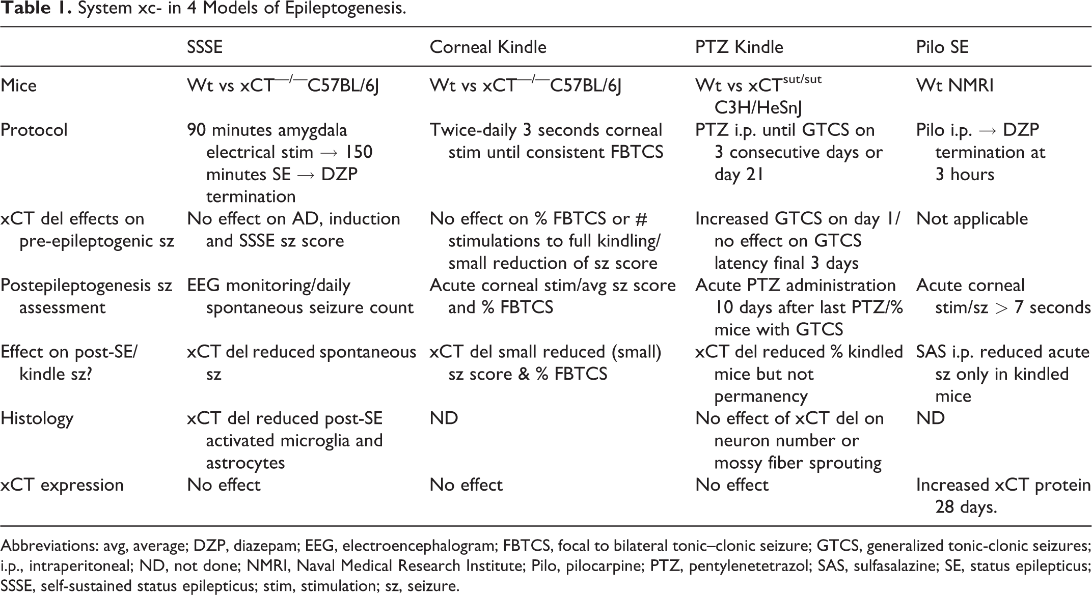
Abbreviations: avg, average; DZP, diazepam; EEG, electroencephalogram; FBTCS, focal to bilateral tonic–clonic seizure; GTCS, generalized tonic-clonic seizures; i.p., intraperitoneal; ND, not done; NMRI, Naval Medical Research Institute; Pilo, pilocarpine; PTZ, pentylenetetrazol; SAS, sulfasalazine; SE, status epilepticus; SSSE, self-sustained status epilepticus; stim, stimulation; sz, seizure.
It is possible that a genetic modification such as xCT deletion could reduce acute seizures associated with SE or kindling and thus appear to reduce epileptogenesis simply by blunting the initial proepileptogenic brain insult and not by modifying postinsult plasticity of the seizure network. As a clinical analogy, promptly terminating SE will reduce the probability that a patient will develop epilepsy, but acute SE treatments (eg, benzodiazepines) are not considered antiepileptogenic therapies. Because xCT–/– mice had increased seizure thresholds in response to acute chemoconvulsant administration 3 it was important for both groups to assess the effects of xCT deletion on seizures before or during the epileptogenesis period (Table 1). In the SSSE model, xCT deletion did not affect afterdischarge threshold or seizure score during SE induction or during the SSSE. For the corneal kindling model, there was no effect of xCT deletion on the percentage of mice experiencing a FBTCS or the number of stimulations required to reach a fully kindled state; however, xCT–/– mice did have reduced mean initial seizure scores. In the PTZ kindling model, a greater percentage of xCT–/– mice exhibited a GTCS on the first day of PTZ administration, a result opposite to what would be expected if xCT deletion was simply acting by reducing acute seizures. In addition, there were no differences in PTZ-evoked GTCS latency between wild-type and xCT–/– mice.
In all 3 models, xCT deletion reduced seizures/seizure severity after the epileptogenic period (Table 1). After SSEE, xCT–/– mice had a significantly longer latency before the onset of the first spontaneous seizure as well as fewer spontaneous seizures. In the corneal kindling model, xCT–/– mice had significantly fewer FBTCS as well as a lower mean seizure score; however, the magnitudes of both these effects were rather modest. In the PTZ kindling paradigm, a significantly smaller percentage (50%) of xCTsut/sut mice were kindled relative to wild-type mice (81.5%). There was no difference in the permanency of the kindled state.
In addition to evaluating the effects of xCT deletion on epileptogenesis, Leclercq et al determined the effects of systemic administration of sulfasalazine (SAS), a Sxc inhibitor, on acute seizures after pilocarpine kindling. Sulfasalazine administered for 3 days before the corneal stimulation challenge reduced seizures by greater than 50%. Interestingly, there was no effect of SAS on corneal-evoked seizures in mice that did not undergo pilocarpine SE, a result suggesting that Sxc was involved in modulating the seizure circuit resulting from epileptogenesis but not the native seizure circuit.
The effects of xCT deletion on postinjury histology were determined in the SSSE and PTZ kindling models (Table 1). After SSSE, hippocampi from wild-type mice, but not xCT–/– mice exhibited significantly increased staining for both ionized calcium binding adaptor molecule-1 protein and glial fibrillary acidic protein, markers of microglia and astrocytic activation, respectively. After PTZ-kindling, neither wild-type nor xCTsut/sut mice demonstrated hippocampal neuron loss or mossy fiber sprouting.
Resected hippocampi from human mesial temporal lobe epilepsy patients expressed increased xCT protein relative to autopsy controls. 5 However, in wild-type mice, PTZ kindling did not increase xCT mRNA and neither SSSE nor corneal kindling increased xCT mRNA or protein expression (Table 1). In contrast, pilocarpine SE caused a significant increase in hippocampal xCT protein expression 28 days after SE and a time-dependent, albeit nonstatistically significant increase in xCT mRNA expression. This elevated postpilocarpine SE xCT expression was accompanied by increases in phospho-Akt, phosphorylated GSK-3β, phosphorylated eIF2α, and ATF4, components of the PI3K growth factor signaling pathway which may play a role in xCT expression.
These 2 studies are important because they provide preliminary evidence that Sxc may be a novel target for antiepileptogenic therapy. While EAATs are also being investigated for antiepileptogenic therapy, 7 Sxc may be better adapted for drug targeting. Unlike Sxc which exports glutamate, EAATs import it and thus their activity must be enhanced in order to reduce extracellular glutamate concentration, a pharmacologically more difficult problem than inhibition. In addition, because investigators are testing the effects of Sxc inhibition in several neurological and psychiatric disorders, 4 it is more likely that Sxc antagonists will be clinically available in contrast to niche compounds that are only useful for preventing epileptogenesis.
It is also significant that these investigators found xCT deletion significantly reduced seizures in three very different models of epileptogenesis including SE and focal and generalized kindling. The use of multiple models provides confidence that the effect would be observed in a variety of proepileptogenic insults and thus ultimately applicable to a greater spectrum of patients.
Finally, given the possible role of Sxc in oxidative stress responses, it was notable that the investigators found that xCT deletion reduced markers of activated microglia and astrocytes in the SSEE model and did not cause neuron loss or mossy fiber sprouting in the PTZ kindling model. This finding provides reassurance that Sxc inhibition would not likely worsen neurological injury.
While these studies provide solid preliminary evidence of antiepileptogenic effects, more study is required. Are the observed effects truly antiepileptogenic? Clearly, unconditional genetic deletion models cannot fulfill the requirements of an antiepileptogenic therapy; namely, one that is administered at the time of neurological insult and eventually withdrawn. In contrast to the acute chemoconvulsant models studied previously, 3 the authors did find that xCT deletion did not alter the initial seizures prior to the epileptogenesis period, evidence that xCT loss did not act by merely ameliorating the initial neurological insult. However, it is possible that unconditional xCT deletion may alter neural plasticity mechanisms prior to the insult and that these preexisting changes may be needed prior to the proepileptogenic insult. Similarly, it is possible that xCT deletion is providing an anti-seizure effect after the epileptogenic period and, if it were possible to restore xCT expression, seizures would worsen. Future experiments using an inducible genetic knockdown strategy 8 employing specific Scx inhibitors are needed to answer these questions.
In conclusion, these 2 studies are notable for identifying Sxc as new potential target for antiepileptogenic therapy. Future studies that elucidate the time course and mechanisms of action will reveal if this potentially “exciting” development translates to useful pharmacological intervention.