
Abstract
Liu J, Baraban SC. eNeuro. 2019;6(1):ENEURO.0041-19.2019. doi:10.1523/ENEURO.0041-19.2019. eCollection 2019 Jan-Feb. PMID: 30895220. Seizures are characterized by hypersynchronization of neuronal networks. Understanding these networks could provide a critical window for therapeutic control of recurrent seizure activity, that is, epilepsy. However, imaging seizure networks have largely been limited to microcircuits in vitro or small “windows” in vivo. Here, we combine fast confocal imaging of genetically encoded calcium indicator–expressing larval zebrafish with local field potential recordings to study epileptiform events at whole-brain and single-neuron levels in vivo. Using an acute seizure model (pentylenetetrazole, PTZ), we reliably observed recurrent electrographic ictal-like events associated with generalized activation of all major brain regions and uncovered a well-preserved anterior to posterior seizure propagation pattern. We also examined brain-wide network synchronization and spatiotemporal patterns of neuronal activity in the optic tectum microcircuit. Brain-wide and single-neuronal level analysis of PTZ-exposed and 4-aminopyridine-exposed zebrafish revealed distinct network dynamics associated with seizure and nonseizure hyperexcitable states, respectively. Neuronal ensembles, comprised of coactive neurons, were also uncovered during interictal-like periods. Taken together, these results demonstrate that macro- and micro-network calcium motifs in zebrafish may provide a greater understanding of epilepsy.
Epileptic seizures result when large populations of neurons fire hypersynchronously. 1 Indeed, the unequivocal diagnosis of epilepsy often requires the identification of large amplitude, electrical waveforms on electroencephalography recordings that reflect the synchronized waves of synaptic currents in the superficial layers of the cortex. 2 However, the mechanisms that drive neuronal populations to develop and propagate such hypersynchrony during seizures remain largely unknown. At the whole-brain level, where does this synchronization start and how does it spread to other regions of the brain? At the local population level, how are synchronous neuronal subpopulations distributed across space and what percentage of the total neuronal population participates in seizures?
To convincingly address these questions, activity patterns across many neurons must be simultaneously monitored in vivo. Modern imaging approaches using genetically encoded activity sensors (eg, genetically encoded calcium indicator [GCaMP]) make such recordings possible. 3 However, imaging in live mammals is often complicated by an opaque skull, animal mobility, and limited scope of view. To overcome these limitations, Liu and Baraban recently employed large-scale imaging approaches in the larval zebrafish (Danio rerio), a vertebrate, to visualize the progression of seizures. To achieve image stability, the fish can be fixed in agar and immobilized with a muscle relaxant without compromising viability. Additionally, the existence of large neurons in the fish midbrain, coupled with the mosaic expression of the neurod1 promoter used to drive neuronal GCaMP expression, enabled the resolution of single neuron activity before and during seizures. Simultaneous electrophysiological measurements confirmed that GCaMP is sensitive enough to identify seizure-related electrical events. Indeed, the authors found a close approximation of the start times of calcium events and electrographic events, as well as a positive correlation (R2 = 0.839) between the corresponding durations, confirming the utility of calcium imaging for monitoring seizure-like activity.
With optical recording strategies in place, Liu and Baraban next employed well-established protocols to model the epileptic state. To generate seizures, the authors exposed fish to pentylenetetrazole (PTZ), a GABAA receptor blocker that blocks fast synaptic inhibition, thereby potentially facilitating excitatory loops that drive neuronal hypersynchrony. Interestingly, in addition to comparing PTZ-treated animals to control animals, the authors also evaluated neural activity patterns in fish treated with 4-aminopyridine (4-AP), a potassium channel blocker that depolarizes the resting membrane potential of neurons. By depolarizing neurons, 4-AP increased general neuronal excitability but did not precipitate ictal-like events.
Comparing whole-brain calcium activity across control, PTZ-treated and 4-AP-treated larvae revealed intriguing differences. Although 4-AP-treated larvae produced enhanced neuronal activity in the cerebellum and hindbrain relative to control larvae, PTZ-treated larvae reliably produced large-amplitude calcium events that were highly synchronized across all brain regions. By calculating a synchronization index based on calcium event phase differences between pairs of brain regions, the authors revealed highly synchronized activity patterns among virtually all brain regions in PTZ-treated larvae, but mostly weakly synchronized activity patterns between brain regions in 4-AP-treated and control fish. Furthermore, the midbrain neurons of PTZ-treated fish displayed a high degree of local synchrony. Taken together, the presence or absence of neuronal synchrony seems to be the defining feature distinguishing an epileptic brain state from a nonepileptic, albeit hyperexcitable brain state.
Next, the authors further investigated the initiation site and propagation of PTZ-induced seizures. To this end, the authors measured the relative time lag of ictal events across brain regions. They discovered that PTZ-induced seizures in zebrafish larvae almost always begin in the forebrain and then travel along the anterior–posterior axis to terminate in either the cerebellum or the hindbrain. In mammals, generalized seizures are also found to originate in the forebrain rather than in deeper structures. For instance, simultaneous intracranial recordings of absence seizures in inbred epileptic rats have suggested that the hypersynchronous electrical activity originates in the perioral region of the somatosensory cortex. 4,5 Network properties that lead to increased seizure susceptibility in the forebrain over other brain structures could thus be probed in the zebrafish model.
After characterizing structure-wide activity changes in control, 4-AP-treated and PTZ-treated fish, the authors increased the magnification on their microscope to evaluate mesoscopic activity patterns produced by individual midbrain neurons. In doing so, the authors unsurprisingly showed that most midbrain neurons in control larvae are quiet. However, provocative differences between PTZ- and 4-AP-treated fish were observed. Although elevated neuronal firing activity was observed during both treatments, neurons with the highest levels of activity were recorded in 4-AP treated, nonepileptic fish. Intriguingly, neuronal firing rates in PTZ-treated larvae had a much narrower and lower dispersion than in 4-AP-treated larvae. Hypersynchronous events seem to not only recruit normally silent neurons but also mitigate normally hyperactive neurons.
Finally, the authors analyzed the size and spatial extent of synchronous neuronal subpopulations. Such “ensemble events” were detected by comparing the number of coactive neurons in each 10 ms time bin of a recording session against 2000 surrogate data sets in which the activity of each neuron was randomly shuffled. Both the 4-AP and PTZ-treated larvae contained many more ensemble events relative to control larvae. However, the spatial extent and pattern of neuronal ensembles were dramatically different between the 4-AP and PTZ-treated fish. 4-Aminopyridine-treated ensembles contained fewer neurons and were spatially dispersed. In contrast, PTZ-treated ensembles contained more neurons that were clustered closely together in space (Figure 1). As PTZ-treated ensembles did not always coincide with seizures, the authors’ observations also suggest that anatomically proximal neurons are more likely to fire together even during interictal periods. In fact, intracranial microelectrode array recordings of humans with partial epilepsy revealed interictal epileptiform activity, termed “microseizures,” from spatially restricted neuronal populations. 6 Thus, spatial differences in neuronal coactivation may be yet another feature that distinguishes an epileptic brain state from a nonepileptic hyperexcitable brain state.
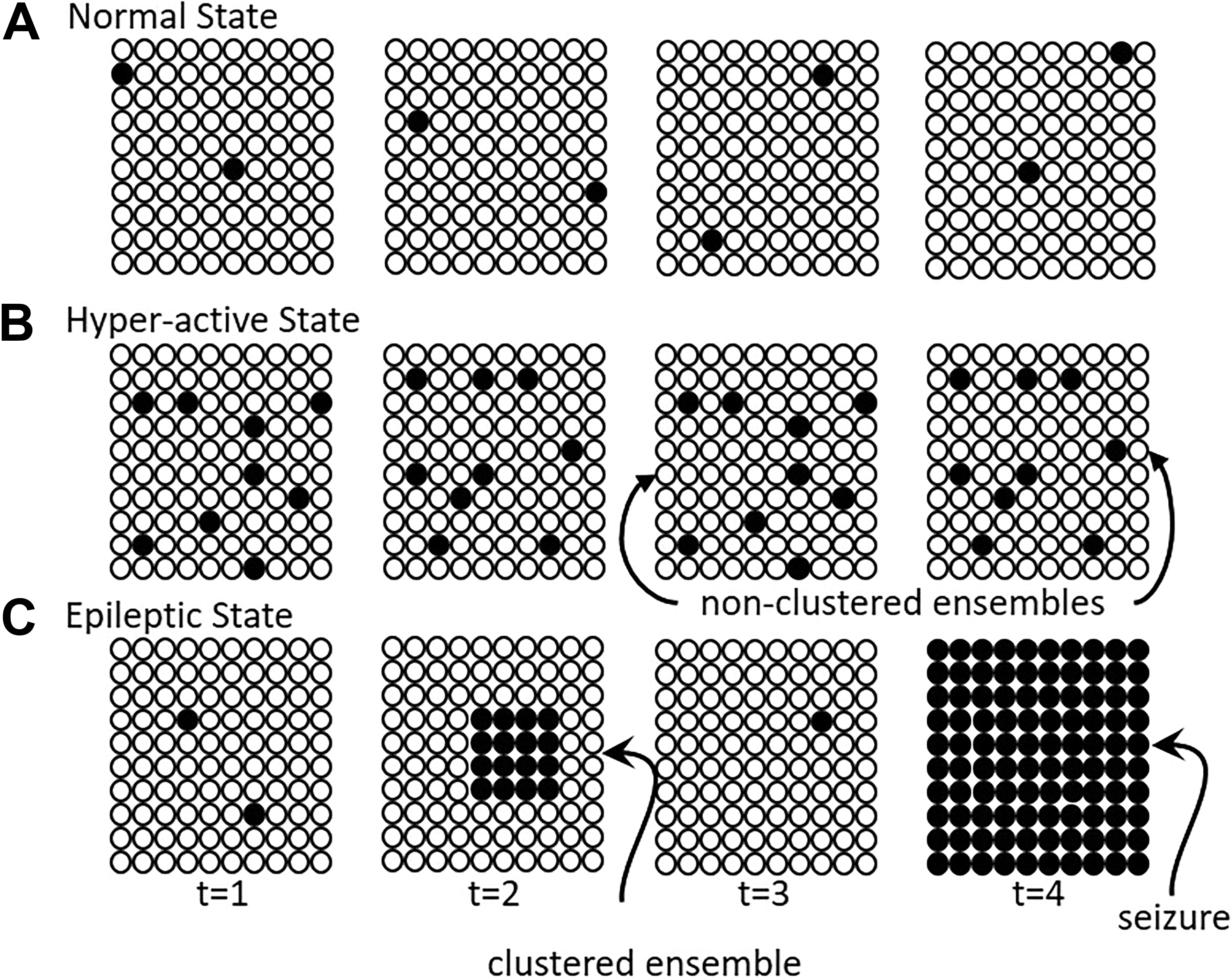
Patterns of neural activity in normal, hyperactive, and epileptic brains. A, In healthy brains, neural activity is generally sparse and not excessive. Each circle represents a neuron, and each grid represents an evolving moment in time. Open circles represent silent neurons while closed circles represent active neurons. At each moment, only a few neurons are active. B, In the hyperactive state (eg, following administration of 4-aminopyridine [4-AP]), many neurons are active at each time point, but coactive neuronal ensembles are spatially disparate. C, In the epileptic state, coactive neuronal ensembles are spatially clustered during the interictal period and evolve to brain-wide synchrony during a seizure.
In summary, the PTZ-treated larval zebrafish represents another useful model organism to better understand epilepsy and serves as an indispensable tool for the understanding of how the dynamics of small populations of neurons may lead to brain-wide synchronization. Arguably, there is no better alternative to date to catch all the players in action during seizures.