
Abstract
Whole body imaging technologies, coupled with the generation of stable light emitting strains of infectious agents, make it possible to monitor the location and kinetics of agent growth inside an animal by noninvasive nonterminal methodologies throughout the course of the infection. However, imaging of viable hazardous pathogens, such as Yersinia pestis, requires specialized containment facilities to prevent contamination of both personnel and the wider environment. Here we describe the successful establishment of an in vivo bioimaging capability specifically addressing the requirement for safe and secure work with hazard group 3 pathogens and demonstrate its utility for imaging mice infected with a bioluminescence producing strain of Y. pestis.
Introduction
In vivo biophotonic imagery refers to the detection of light sources within living animals. Particularly, animals, cells, microbes, and other entities may be tagged or labeled with a light emitting reporter molecule that can be detected by sensitive light detecting hardware. Since the process is akin to taking a photograph, it is noninvasive and nonharmful to animals and can be repeated over time to build up a dynamic picture of changes associated with the labeled moiety. Labeling a pathogen such as Yersinia pestis, the causative agent of plague, allows the pathogen to be monitored throughout the progression of infection in a living animal without the need for traditional terminal sampling methodologies. This method has been elegantly demonstrated in several studies detailing the production of bioluminescent strains of Y. pestis bacteria and in vivo imaging of the course of infection.1 –4 The technology has potential to contribute significantly to the refinement of animal models and to a reduction in the overall numbers of animals used, in line with the 3R’s aspirations for animal use in science. 5
When considering the issue of imaging animals infected with highly infectious pathogens, the overarching considerations are the safety of the operator, the welfare of the animals, prevention of pathogen release to the environment, and the practicalities and utility of the instrumentation under these conditions. At the most basic level of interpretation, the requirements for handling Health and Safety Executive (HSE) Advisory Committee on Dangerous Pathogens (ACDP) Hazard Group 3 (HG3) pathogens (equivalent to the US Department of Health and Human Services Biosafety Level 3 [BSL-3]/Animal Biosafety Level 3 [ABSL-3] pathogens) 7 calls for them to be handled under conditions that permit the isolation of the pathogen from the worker and the environment. 8 In the UK laboratory environment, this would normally mean that pathogen handling and manipulation is carried out within a microbiological safety cabinet 6 and that infected animals are housed within an isolator chamber. 9 Furthermore, the general laboratory space requires specific adaptation for working under these high containment conditions. For work with HG3 pathogens, there are structural considerations, specialized air handling systems generating directed air flow along negative pressure gradients, and self-contained effluent treatment systems that reduce the risk of viable agent escaping to the environment. Management practices, general laboratory codes of practice, training regimens, access control, and process-specific risk assessments and procedures also contribute to the overall containment of these pathogens within the laboratory environment.7,10,11
The combination of imaging technology, animal handling procedures, and containment of HG3 pathogens is a specialized requirement; thus, commercial, off-the-shelf solutions are not available. This article describes the successful establishment of an in vivo imaging capability in an HG3 containment facility and demonstrates the utility of the facility for imaging HG3 pathogens by imaging mice infected with a bioluminescence producing strain of Y. pestis.
Design of a Contained Imaging Facility
Internationally, different laboratories have approached the task of how to image infected animals safely in different ways. Published examples include the imaging suite at the Regional Biocontainment Laboratory (RBL), University of Pittsburgh, USA, which contains radiological, PET/CT, and optical imaging equipment all housed within a BSL-3 containment facility. 12 Mice are housed in biocontainment caging and prior to optical imaging are anesthetized and then placed directly within the optical imaging system, which is subsequently decontaminated after each use.
A second option is to isolate the infected animals within an isolation chamber for imaging, such as the AIIC isolation box produced by Caliper. This isolation box has been validated for use at the RBL, Duke University Medical Center, North Carolina, USA, for use under their BSL-3 containment regulations 13 ; here, personnel are required to wear specific personal protective equipment, including a powered air purifying respirator, to prevent exposure to pathogens while in the facility. Our preferred approach was to place the imaging device in full physical containment in-line with UK Control of Substances Hazardous to Health (COSHH) Regulations 2002 (as amended) and, in particular, to incorporate prescribed containment measures detailed in Schedule 3 of COSHH. 11 In conjunction with imaging specialists from CaliperLS UK, specialist isolator manufacturers Howorth, Ltd., UK (Bolton, UK), and decontamination specialists, Bioquell (Andover, UK), a fully integrated system consisting of an IVIS Spectrum (Perkin-Elmer, Waltham, Massachusetts, USA) imaging system was housed within a purpose built isolator and connected to a Clarus L vaporised hydrogen peroxide (VHP) generator (Bioquell, UK). The bespoke isolator system enables handling and imaging of infectious materials, including live infected animals, to occur within primary containment and is located within a dedicated containment Level 3 (CL3 equivalent to BSL-3/ABSL-3) laboratory.
The IVIS Spectrum in Containment
The imaging system in containment is shown in Figure 1. The full system consists of 3 separate chambers: 1 for housing the IVIS Spectrum, 1 used for procedures including a half-suit and glove ports, and a passbox with a Getinge LaCalhène (Vendôme, France) Rapid Transfer Port (RTP, type 270) docking system, which allows the connection of a bespoke transport isolator (Howorth), built to the same standard and performance as the main isolators, to move animals safely to and from their housing isolator. The IVIS Spectrum isolation chamber (Figure 1a) consists of a single molded stainless steel chamber with a tempered glass door and sealed glove ports to enable operators to move the instrument within the chamber. This chamber is linked directly to the procedures isolator through a sealed and lockable door system and is constructed from rigid uPVC sealed to a stainless steel base. A half-suit with independent air supply (Professional Protection Services, Ltd., Milton Keynes, UK) is fitted to the stainless steel base on a double ring aperture and is sealed with o-rings; this half-suit is occupied by the operator/animal handler during operation. Door sizes and passbox dimensions are sufficient to allow standard mouse cages to be transferred to and from the procedures area. Containment of the pathogen is ensured through a series of H14 high-efficiency particulate air (HEPA) filters and a negative pressure air flow system. Air is supplied independently to the IVIS isolator chamber, procedures isolator, and passbox, entering via pre-filters upstream of 2 HEPA filters and air exhausts into the environment via pre-filters upstream of 2 HEPA filters, which are linked by flexible ducting to ceiling mounted thimbles. 9 During operation, the procedures isolator internal pressure is maintained at −130 Pascals. The space within the isolator procedures module is sufficient for preparing small groups of animals for imaging and observation during recovery. The passbox area air pressure is maintained at approximately −75 Pascals to ensure that during use, the airflow is directed from the passbox area into the procedures area. The IVIS Spectrum isolator chamber runs at an internal pressure of −100 Pascals. The pressure differential between the IVIS Spectrum isolator chamber and procedures module ensures that the air currents draw toward the central procedures module and therefore helps to prevent dust and particulates from bedding, and so on, accumulating within the IVIS Spectrum isolator chamber. The electrical power for the isolator system is routed through an uninterruptible power supply to preserve isolator air flow integrity in the event of general power failure. The IVIS Spectrum is operated from a computer located outside of the containment module and is connected to the instrument through sealed ports and cable connections in the isolator wall.
(a) IVIS Spectrum situated within the IVIS isolator chamber, the tempered glass door is open in this image. (b) View of the isolator system showing the closed door of the IVIS isolator chamber with the 4 gloves used to maneuver the imager within the chamber. (c) View of the isolator system showing the RTP on the passbox and a view of the main procedures module. The yellow half-suit is clearly visible in the center of the procedures module. Additional glove ports on the front wall of the procedures module allow an additional person to assist with handling materials in the chamber. (d) The door between the IVIS isolator chamber and the procedures module: accessed by the operator in the half-suit for transfer of imaging subjects between the procedures area and the imager.
Fumigation of the isolator procedures module and passbox is achieved using heated liquid formaldehyde. 6 However, the IVIS Spectrum contains sensitive optical and electrical technology that is not compatible with formaldehyde fumigation (manufacturer’s advisory). Vapour Hydrogen Peroxide (VHP) is a less harsh fumigant, and previous assessments by the manufacturer (Caliper LS) have shown it to be suitable for use as a fumigant for the IVIS instrumentation. Our containment system was therefore specifically designed to use the Bioquell Clarus L VHP delivery system for the IVIS chamber while retaining the flexibility to allow formaldehyde fumigation of the main body of the isolator, as this is the standard method of decontamination used in our CL3 containment facilities. The electronic control systems for the Clarus L and the isolators are linked to ensure coordination of the isolator air supplies and valve systems with the VHP generator during the fumigation processes. The VHP fumigation cycle is initiated on the Clarus L instrument but is dependent on an isolator chamber pressure decay test. The pre-fumigation pressure decay test is an automated monitor of IVIS isolator chamber integrity and is only used to activate the Clarus L instrument (not for testing/commissioning the isolator). The chamber pressure is increased to 300 Pascals, the dampers are closed, and once the pressure drops to 250 Pascals, the 3-minute decay test commences. Maintenance of this pressure for 3 minutes demonstrates chamber integrity and automatically signals the Clarus L instrument to commence VHP fumigation. The VHP is pumped into the IVIS isolator chamber via a rotating nozzle, mounted above the IVIS Spectrum that ensures distribution throughout the chamber. The fans of the IVIS Spectrum are left on during the procedure to ensure that the VHP circulates in the chamber to reach all surfaces and is drawn into the internal structures of the imager. The optimal cycle developed by Bioquell utilizes 57.5 g of 30% H2O2 and runs for approximately 4 hours, generating an average maximum H2O2 concentration of around 280 ppm ± 100 ppm, with an average dwell time of 90 minutes. Upon completion of the cycle, VHP decays to water and oxygen and is drawn away from the instrument when operational airflow is resumed in the isolator. The VHP decontamination was validated using Bacillus stereothermophilus and Bacillus atrophaeous spore strips (NAMSA, Ohio). Twenty-two spore strips (106 spores per strip) were used for each of the 4 decontamination tests. Strips were placed inside the IVIS Spectrum after removal of the back panel cover (which was replaced prior to the test starting), inside the imaging chamber and around the IVIS Spectrum isolator chamber, and positions recorded so that the test conditions could be replicated accurately. After fumigation, spore strips were incubated in tryptic soy broth (Sigma-Aldrich, Poole, UK) at 37°C for 7 days, then 1 ml (10%) of media from each tube was plated onto LB agar (4 × 250 µl per plate) and incubated for a further 7 days at 37°C. The spore strip validation studies revealed 100% sterility of the samples over 4 successive tests using the optimal cycle parameters. This indicates at least a 6-log reduction in bacterial numbers is achieved by the process.
The final design was subjected to rigorous hazard and operability studies (HAZOP) with a group set up from users, biosafety professionals, and equipment manufacturers. A series of risk assessments and standard operating procedures was drawn up for the general use of the system, which was scrutinized and approved for use by the local Microbiological Safety Committee at the Defence Science and Technology Laboratory (Dstl). Processes were defined and refined to ensure maximum operator protection and preserve equipment function. Each facet of operation was first tested using noninfectious or lower HG materials and proven to be robust and reliable before work with HG3 pathogens was permitted.
Demonstration of Imaging in Containment
Plague is a zoonotic disease prevalent in many parts of Asia, Africa, and some states of the US, which is caused by the bacterium Y. pestis. Upon infection, Y. pestis disseminates rapidly from the site of infection to the draining lymph nodes where it replicates to high numbers and subsequently spreads rapidly throughout the body, resulting in a generally fatal systemic disease. This systemic spread and the previously successful construction of bioluminescence expressing Y. pestis strains1 –4 made it an ideal candidate to demonstrate the functionality of the imaging system in containment.
A bioluminescent clone of Y. pestis was generated by electroporation of the pGEN-luxCDABE plasmid (kindly supplied by Dr. Elisabeth Carniel of Institut Pasteur) into Y. pestis strain CO92. This plasmid harbours the Photorhabdus luminescens luxCDABE operon under the control of the Pem7 promoter to allow constitutive, high-level production of bioluminescence 14 and has been used previously to generate bioluminescent Y. pestis.1,2 Recombinant isolates were selected by growth on blood agar base (BAB) agar supplemented with 0.02% haemin and 50 µg/ml ampicillin. A single isolate was picked, subsequently named Y. pestis Lux9, and bioluminescence was confirmed by imaging colonies on BAB-haemin agar in the IVIS Spectrum (Figure 2). Mouse studies were performed using 6- to 8-week-old female BALB/c mice (Charles River, UK) randomly allocated into cages of 5 upon arrival and held under a 12-hour light/dark cycle with free access to food and water. An intranasal infection route was chosen to demonstrate the utility of bioluminescent Y. pestis Lux9 using the IVIS Spectrum as this infection route has previously been used successfully with bioluminescent Y. pestis 1,3,4 and is a realistic model of pneumonic plague. To prepare the challenge inoculum, an overnight culture of Y. pestis Lux9, grown at 28°C in BAB broth supplemented with ampicillin, was diluted 1 in 10 in BAB broth (without ampicillin) and incubated at 28°C with shaking at 180 rpm to an OD600 of 0.6 (corresponding to approximately 1 × 10⁸ CFU/ml) from which appropriate dilutions were made. Mice were lightly anesthetized with halothane (Sigma-Aldrich, UK), and 25 µl of bacterial solution was inoculated into each nostril to deliver a total of ∼10,000 CFU Y. pestis Lux9. For imaging, mice were anesthetized with 0.78 mg ketamine hydrochloride and 0.015 mg medetomidine hydrochloride in sterile water (delivered in 0.15 ml volume via the intraperitoneal [IP] route) approximately 10 minutes prior to imaging. Mice were held in a box heated to 30°C following administration of anesthetic and, once sedated, were placed in an optical grade perspex box (Vet Tech Solutions Ltd., Cheshire) to minimize contamination of the IVIS Spectrum. Following imaging, animals were revived using 0.05 mg atipamezole hydrochloride in sterile water (delivered in 0.1 ml volume via the IP route) with a gap of at least 15 minutes between anesthesia and recovery. A total of 60 mice were infected with Y. pestis Lux9 via the intranasal route and imaged over the following 3 days. Each day a group of mice was culled, and the lungs, liver, and spleen of each mouse were removed and homogenized into sterile phosphate buffered saline using a 0.4 µm sieve (Becton-Dickinson, UK) and cultured on BAB-haemin agar to enumerate bacterial levels over the course of the infection.
Colonies of Yersinia pestis CO92 strain Lux9 on the surface of BAB agar plates visualized in the IVIS Spectrum.
Mice displayed clinical signs of disease (ruffled coat, slightly hunched posture, rapid breathing) from 48 hours post infection and began to succumb to disease from 72 hours post infection. Figure 3 shows representative images taken over the course of the infection. At the 24-hour time point, luminescence was approximately 4 × 10³ photons/sec/cm2/steradian (Figure 3, panel A), no greater than that of mice infected with non-luminescent Y. pestis CO92 (data not shown), although limited luminescence was visible in the lungs once the internal organs were examined postmortem (data not shown). At the 48-hour time point, luminescence had increased to approximately 1 × 10⁵ p/sec/cm2/sr and, as consistent with the infection route, was limited to the thorax, suggesting the development of pneumonic disease without significant bacterial dissemination at this time (Figure 3, panel B). By 72 hours post infection, luminescence from the thorax increased to approximately 1 × 10⁷ p/sec/cm2/sr (Figure 3, panel C), and there was evidence of luminescence from the abdomen, indicative of fully disseminated endstage infection. Bacterial colony counts from the lungs, liver, and spleen (Figure 4) paralleled the development of luminescence signals (Figure 5), with bacteria restricted to the lungs after 24 hours, increasing in number in the lungs after 48 hours with some dissemination to the liver and spleen. By 72 hours, colony counts reached high levels in all organs examined as the disease became fully disseminated and entered the terminal phase. These results are consistent with findings of other studies of pneumonic plague dissemination using bioluminescence producing bacterial strains,3,4 demonstrating the utility of our novel containment imaging system.
Detection of bioluminescent signal in BALB/c mice infected by the intranasal route with Yersinia pestis strain Lux9. Images show representative mice from each time-point (A = 24 hours, B = 48 hours, C = 72 hours post infection). Bacterial bioluminescent signal is displayed as a pseudocolour image overlaid onto the photographic image with the red color denoting areas of strongest signal. Note that images do not use the same color scale.
Bacterial colonization of organs following intranasal infection of BALB/c mice with Yersinia pestis strain Lux9. Organs sampled were the (A) lungs, (B) liver, and (C) spleen. Counts represent bacteria in the entire organ. LoD, limit of detection.
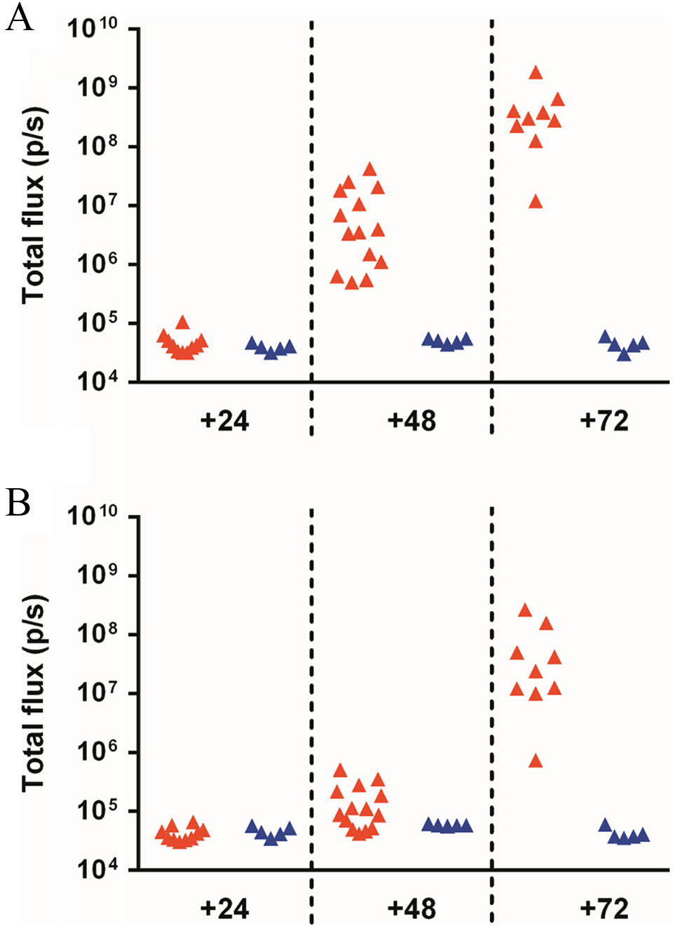
Luminescence of the (A) thorax and (B) abdomen following intranasal infection of BALB/c mice with Yersinia pestis strain Lux9 (red triangles) or wildtype Y. pestis CO92 (blue triangles).
Summary
We have designed and established a purpose built isolator system to house an IVIS Spectrum instrument within a bespoke stainless steel isolator, connected to a procedures module and a passbox with a Rapid Transfer Port. The system has been designed to ensure the highest levels of operator protection, maintain high standards of animal welfare, and promote maximum flexibility and utility of the IVIS Spectrum. We have developed and validated operating procedures and decontamination strategies that facilitate safe access for service engineers, and we have confirmed the functionality of the IVIS Spectrum in containment through imaging of an acute infection model using luminescent Y. pestis. We anticipate that this contained imaging system will be of value for future research, ultimately reducing the number of animals that are required for evaluation of new vaccines or therapeutics and providing more comprehensive information about infection dynamics than traditional sampling methods. Work is now underway to generate additional luminescent strains of pathogens that are valuable for use in such studies.
Footnotes
Acknowledgment
The authors gratefully acknowledge Dr. E. Carniel for kindly providing the pGEN-luxCDABE plasmid.
Authors’ Note
All animal work was approved by the Dstl Animal Welfare and Ethical Review Body, and according to the requirements of the UK Animal (Scientific Procedures) Act 1986.
Declaration of Conflicting Interests
The author(s) declared no potential conflicts of interest with respect to the research, authorship, and/or publication of this article.
Funding
The author(s) disclosed receipt of the following financial support for the research, authorship, and/or publication of this article: This work was funded by the UK Ministry of Defence.