
Abstract
Background. Annona muricata L. has been reported to possess antitumor and antiproliferative properties. Not much work has been done on its effect on BPH-1 cell lines, and no in vivo studies targeting the prostate organ exist. The study determined the effect of A muricata on human BPH-1 cells and prostate organ. Methods. The MTT assay was performed on BPH-1 cells using the aqueous leaf extract of A muricata. Cells (1 × 10⁵ per well) were challenged with 0.5, 1.0, and 1.5 mg/mL extract for 24, 48, and 72 hours. Cell proliferation and morphology were examined microscopically. BPH-1 cells (1 × 10⁴ per well) were seeded into 6-well plates and incubated for 48 hours with 0.5, 1.0, and 1.5 mg/mL A muricata extract. Reverse transcriptase polymerase chain reaction was performed using mRNA extracted from the cells. Possible target genes, Bax and Bcl-2, were examined. Twenty F344 male rats (≈200 g) were gavaged 30 mg/mL (10 rats) and 300 mg/mL (10 rats) and fed ad libitum alongside 10 control rats. Rats were sacrificed after 60 days. The prostate, seminal vesicles, and testes were harvested for histological examination. Results. Annona muricata demonstrated antiproliferative effects with an IC50 of 1.36 mg/mL. Best results were obtained after 48 hours, with near cell extinction at 72 hours. Bax gene was upregulated, while Bcl-2 was downregulated. Normal histological architecture was observed for all testes. Seminal vesicle was significantly reduced in test groups (P < .05) and demonstrated marked atrophy with increased cellularity and the acinii, empty of secretion. Prostate of test groups were reduced with epithelial lining showing pyknotic nucleus, condensation, and marginalization of the nuclear material, characteristic of apoptosis of the glandular epithelium. Furthermore, scanty prostatic secretion with flattening of acinar epithelial lining occurred. Conclusion. Annona muricata has antiproliferative effects on BPH-1 cells and reduces prostate size, possibly through apoptosis.
Introduction
Prostate cancer is the most frequent cancer in men and the second highest cause of mortality by cancer for the male population. Approximately 29% and 9% of leading new cancer cases and deaths, respectively, in the United States were attributed to the prostate in 2012. 1 A 1.33-fold increasing trend of incidence rate between 1999 and 2002 was reported in Korean men. 2 Furthermore, prostate cancer may become problematic if a less than 15-year survival is predicted. 3 In Ghana, 17.35% of male cancer death is attributed to the prostate. 4 Treatment, on the other hand, has adverse effects, 5 and in some cases unneeded, as some men do not die from their cancer and may harbor tumors that are indolent even in the absence of therapy.3,6 Benign prostatic hyperplasia (BPH) affects more than 50% of men in their 60s and as much as 90% in their 70s and 80s. In the United States in 2000, there were 4.5 million visits to physicians with issues relating to BPH. Globally and nationally, more and more people are turning to complementary and alternative medicine for various ailments of which the use of medicinal plants is foremost.
Annona muricata L., commonly called soursop, is a small erect evergreen tropical fruit tree plant belonging to the family Annonaceae, growing 5 to 6 meters in height. The leaves of A muricata have been reported to contain several groups of substances collectively called annonaceous acetogenins. Monotetrahydrofuran annonaceous acetogenins, cis-corossolone (4) annocatalin (5), annonacin, annonacinone, solamin, and corossolone have been isolated from the leaves of A muricata. The first 2 isolates have significant cytotoxic activity in vitro against 2 human hepatoma cell lines, Hep G(2) and 2,2,15. Compound 5 showed a high selectivity toward the Hep 2,2,15 cell line. 7 Additionally, acetogenins 1 (annoreticuin-9-one) and 2 (cis-annoreticuin) isolated from A reticulata and A montana, respectively, have been reported to have cytotoxicity against certain cancer cell lines. Acetogenin 1 targets the human pancreatic tumor cell line (PACA-2), human prostate adenocarcinoma (PC-3), and human lung carcinoma (A-549), while acetogenin 2, targets human hepatoma carcinoma cell line (Hep G2). The dichloromethane extract of the seeds of A muricata yielded annoreticuin-9-one (1), while the flesh of the fruit yielded cis-annoreticuin (2). 8 The presence of Annonaceous acetogenins, muricoreacin (1) and murihexocin C (2) (mono-tetrahydrofuran acetogenins) in the leaves of A muricata (Annonaceae) with significant cytotoxic activities targeting human prostate adenocarcinoma (PC-3) and pancreatic carcinoma (PACA-2) cell lines has been demonstrated. 9 Leaves of A muricata in ethyl acetate showed a higher death rate to HeLa cells than the ethanol distilled water extract. Similarly, chloroform extract application to HeLa cells showed a higher death rate than ethyl acetate extract. The chloroform extracts appear to be a better option for cancer causing viruses. 10 The aqueous extract is said to contain general glycosides, saponins, and flavonoids. 11 In an acute toxicity study (LD50 < 5000 mg/kg body wt), the aqueous extract did not show any toxicity on systemic organs. 11 However, the use of an aqueous infusion of about 140 µg/cup is said to have caused neurotoxicity related to atypical parkinsonism in Guadeloupe. 12 The ethanolic extract of the leaves of A muricata is said to have hypoglycemic and antidiabetic effects. 13 Furthermore, its protective effect on lipid profile has been documented. 14
The aim of the study therefore was to investigate the effect of A muricata on human benign prostate cells (BPH-1) and whole prostate organ in male F344 rats.
Materials and Methods
Plant Material Extraction
The leaves of A muricata were collected from the outskirts of the capital city Accra from July to August 2013 and authenticated by the national herbarium. Specimens were deposited with voucher number UG 00178.AM.215/13. Leaves were hand-washed by rubbing the surface gently under running water. They were later sun-dried for 3 days. Leaves were milled and soaked by the proportion of 1 kg of the milled substance soaked in 4000 mL of water for 24 hours. The mixture was then boiled for 1 hour and filtered through fine linen gauze. The marc was then soaked with another 3000 mL of water for another 24 hours and filtered. The 2 filtered solutions were then pooled and freeze-dried. The yield from 1 kg of ground substance was 25.2 g.
High-Performance Liquid Chromatography (HPLC) Analysis
Different batches of the extract were monitored by chromatographic fingerprint. Samples were analyzed on a Shimadzu HPLC system (Kyoto, Japan), Ultimate XB-C18 column (150 × 4.6 mm, 5 µm), and the absorbance was measured at 208 nm. The mobile phase solvent A was water and solvent B acetonitrile (ACE) at a flow rate of 1 mL/min and an injection volume of 1 µL. The gradient run ACE–H2O was as follows: from 10%:90% to 10%:90% (0-10 minutes); from 10%:90% to 85%:15% (10-30 minutes); from 85%:15% to 85%:15% (30-40 minutes). An optimum easily controlled and reproducible procedure of extraction described previously was established from the fingerprint results.
Effect of A muricata on BPH-1 Cell Viability
Cell viability assays were performed on BPH-1 cells. In brief, cells were seeded into 96-well plates at a density of 1 × 10⁵ cells/well in 0.1 mL RPMI 1640, 10% fetal bovine serum (FBS) medium. Cells were treated with 0.5, 1.0, and 1.5 mg/mL extract (in phosphate-buffered saline [PBS]) and incubated at 37°C for 24, 48, and 72 hours. At the end of treatment time for various plates, the medium was replaced by 100 µL MTT (Sigma, St Louis, MO) per well and incubated for an additional 4 hours at 37°C. The reaction was stopped by adding 100 µL DMSO, AR grade (Sigma) to each well to dissolve the purple-blue MTT formazan precipitate. The absorbance was read at 570 nm on an ELISA microplate reader (BioTek, Elx800, VT). The inhibition of growth was assessed as percent viability where vehicle treated cells were considered as 100% viable.
RNA Extraction and Reverse Transcriptase Polymerase Chain Reaction (RT-PCR) Analysis
BPH-1 cells were seeded into 6-well plates at a density of 1 × 10⁴ per well in 2 mL medium (in 10% FBS) and treated with 0.5, 1.0, and 1.5 mg/mL plant extract (in PBS) for 48 hours. Total RNA was isolated using TriZol reagent (Invitrogen, Carlsbad, CA). Oligo(dT)-primed RNA (1 µg) was reverse-transcribed using the SuperScript II transcriptase kit (RR047A, Takara, Shiga, Japan) according to the manufacturer’s instructions. cDNA obtained was amplified by PCR with TaqDNA polymerase (Fermentas, Burlington, Canada). The presence of possible target genes, Bax and Bcl-2, was determined using the obtained cDNA and glyceraldehyde-3-phosphate dehydrogenase (GAPDH) as the internal control. The sequence of primers used for amplification were as follows: Bcl-2—forward 5′-GG TGGTGGAGG AACTCT TCA-3′ and reverse 5′-GAGCAGCGTCT TCAGAGACA-3′; Bax—forward 5′-CCAAGAAGCTG AGCGAG TGT-3′ and reverse 5′-TC ACGGAG GAAGTCCAG TGT-3′; GAPDH—forward 5′ TGCTGAGTATGTCGTGGAG-3′ and reverse 5′-GTGTTCTGAGTGGCAGTGAT-3′ (bcl2—268 bp; bax—248 bp; GAPDH—240 bp). The PCR reaction was performed under the following conditions: Bcl-2, denaturation at 94°C for 30 seconds, annealing at 58°C for 60 seconds, and extension at 72°C for 60 seconds. For Bax, denaturation at 94°C for 30 seconds, annealing at 55°C for 30 seconds, and extension at 72°C for 45 seconds; for GAPDH, denaturation at 94°C for 30 seconds, annealing at 58°C for 60 seconds, and extension at 72°C for 60 seconds. The samples were analyzed by running 1.5% agarose gel electrophoresis and DNA bands examined using a Bio-Rad 2000 gel documentation system.
Animal Study
The protocol adopted followed the OECD 15 document on the use of laboratory animals and was approved by the ethics committee of the Noguchi Memorial Institute for Medical Research. Thirty male F344 rats were divided into 3 groups of 10 rats each and housed in stainless steel cages. Group I (normal control) was fed the standard diet and water. Rats in group II (low dose [LD]) were orally administered A muricata extract at a dose of 30 mg/kg body wt of A muricata, while rats in groups III (high dose [HD]) were orally administered extract at 300 mg/kg body wt. Plant extract administration was repeated for 60 days. After 60 days of extract administration, all animals were sacrificed and the prostate, seminal vesicles, and testes were harvested.
Histopathological Analysis
Fat- and connective tissue-free prostate, seminal vesicles, and testes were harvested, blotted with clean tissue, examined, and weighed to obtain organ to body weight ratios. Thereafter, prostate and seminal vesicle were immediately fixed in 10% buffered formaldehyde solution. Testes were fixed in Bouin’s solution. Three-micrometer sectioned slides of prostate were hematoxylin and eosin (H&E) stained and evaluated microscopically for histological changes using Olympus BX 51TF (Olympus Corporation, Tokyo, Japan) light microscope connected to a digital camera. Images of selected sections were captured at 100×, 200×, and 400× magnifications.
Statistical Analysis and Data Evaluation
Statistical analysis of the data was done using Graph Pad Software, Version 5.0, for Windows (Graph Pad software, San Diego, CA). Results were expressed as mean ± SEM, n = 10. Significance of difference between controls and dose groups were evaluated by performing a 1-way ANOVA. Post hoc analysis was performed with Bonferroni multiple comparison test where ANOVA showed significant differences. P values ≤.05 were considered statistically significant.
Results
In Vitro Assays
From Figure 1 and Table 1, it can be seen that the areas of peaks 3, 4, 6, 5, 2, 1, 8, 7 were in the ratio of 27.1:18.8:15.6:12.5:11.4:7.8:3.5:3.4. Results from the study are therefore reproducible if the fingerprint and its ratios are the same.
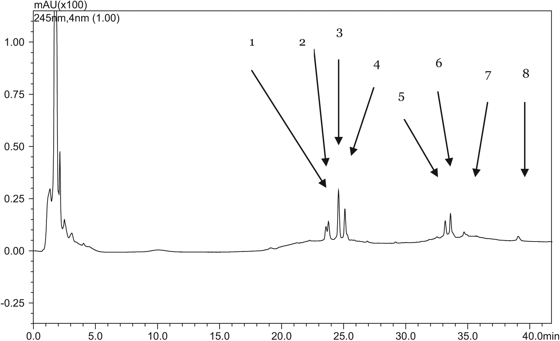
A Shimadzu HPLC system with DAD detector and an Ultimate XB-C18 column was used.
HPLC quantitative results showing area under the curve (AUC) and peak ratios of the 8 peaks isolated.
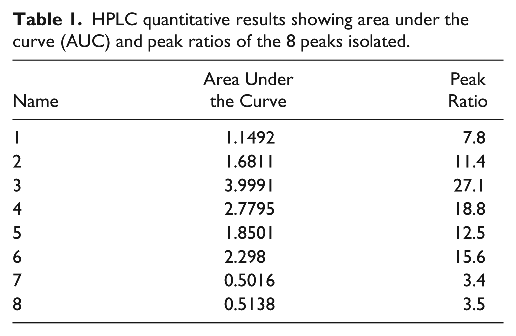
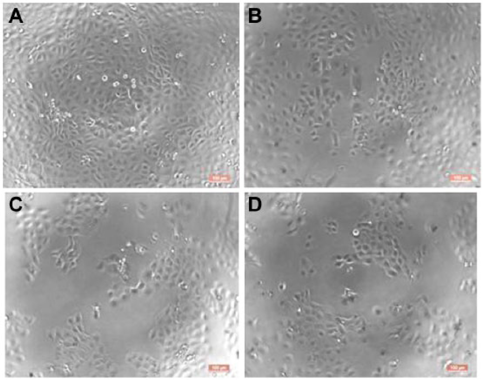
Cell morphology showed diminishing cells as the concentration of the plant extract increased from 0 mg/mL to 1.5 mg/dL (Figure 2). BPH-1 cell viability using MTT assay demonstrated significant dose-dependent decrease. Cell viability reduced from 100% to 80.1%, 65%, and 47% as the dose increased from 0, 0.5, 1.0 to 1.5 mg/mL, respectively (Figure 3). Statistical differences of each dose (0.5, 1, and 1.5 mg/mL) compared to the control was significant (P < .05, P < .05, P < .001, respectively). IC50 recorded was 1.36 mg/mL.
BPH-1 cell proliferation at various concentrations.
BPH-1 cell viability test after 48-hour treatment with A muricata.
PCR results demonstrated a decrease in band intensity for Bcl-2 gene downregulation as the concentration of A muricata increased from 0 mg/mL to 1.5 mg/mL. Conversely, there was an upregulation of Bax in a dose-dependent manner. The internal control GAPDH gave strong positive bands (see Figure 4).
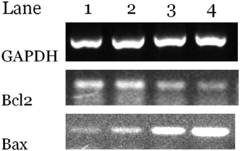
Shows a downregulation of BCl-2 and upregulation of Bax mRNA extracted from BPH-1 cells after 48 hours of treatment with A muricata at doses of 0 mg/mL (lane 1), 0.5 mg/mL (lane 2), 1.0 mg/mL (lane 3), and 1.5 mg/mL (lane 4). GAPDH was used as a positive control.
Effect of A muricata on Prostate Organ and Seminal Vesicle
The extract reduced the size of the prostate in the test groups. Prostatic index (PI) for the Control, LD, and HD groups were 0.178 ± 0.086, 0.152 ± 0.075, and 0.157 ± 0.061, respectively (Figure 5). Although there was a slight reduction in PI differences they were not statistically significant. The “seminal vesicle index” (wet wt. of seminal vesicle/total body wt. × 100) for the control, LD, and HD were 0.437 ± 0.069, 0.184 ± 0.041, and 0.227 ± 0.052, respectively. Statistical differences between control and LD, and control and HD were significant (P = .004 and .009, respectively; Figure 6).
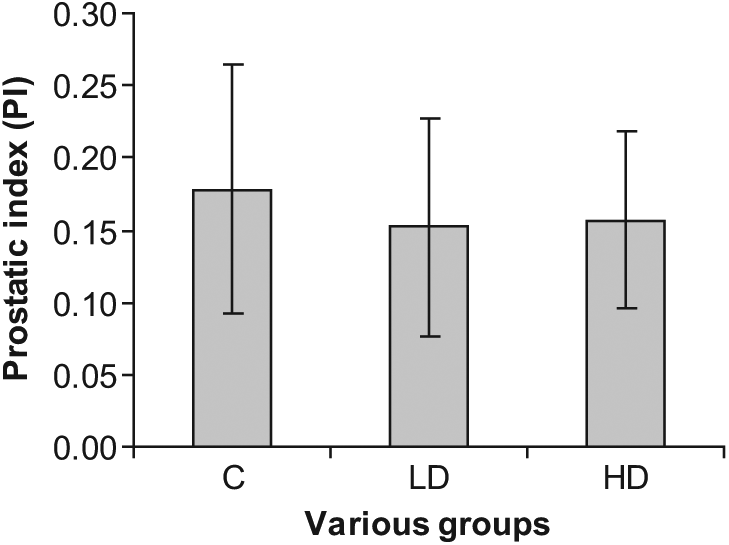
The figure demonstrates the reduction in prostatic index (PI) at day 60 of A muricata administration. There was a reduction in PI both with the LD (30 mg/kg body wt.) and HD (300 mg/kg body wt.). However, decreases were not statistically significant.
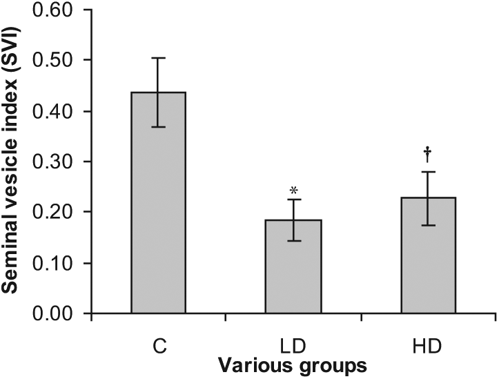
“Seminal vesicle index” (SVI) was significantly reduced from 0.44 ± 0.07 (Control group) to 0.18 ± 0.04 (LD) and 0.23 ± 0.05 (HD). Thus, A muricata caused about 50% relative reduction in the seminal vesicle size. Differences between control and LD as well as control and HD were statistically significant (*P = .004 and †P = .009, respectively).
Microscopic Changes
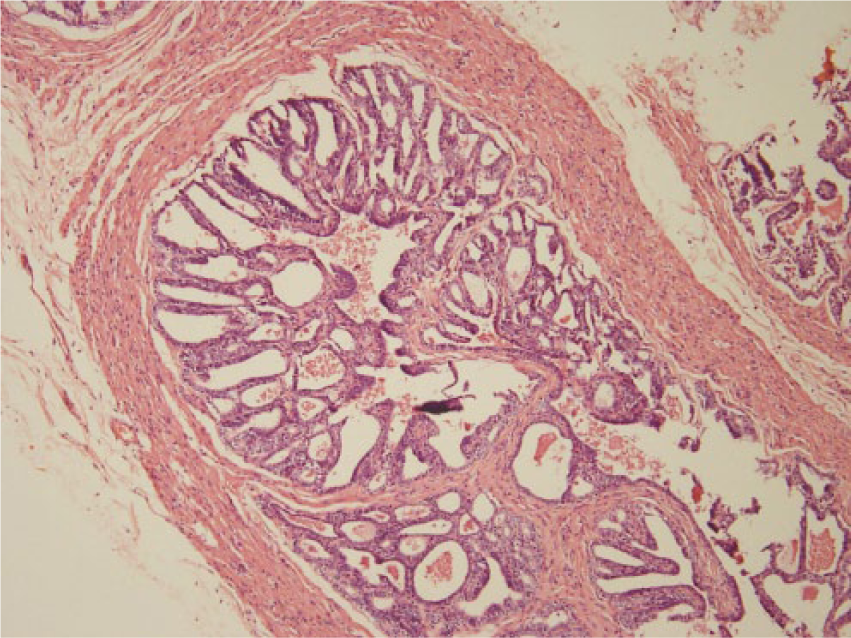
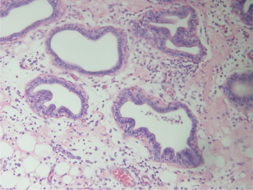
Microscopically, the acinii of the seminal vesicle of the control rats had the normal structure where nuclei are basal and the cytoplasm appeared eosinophic (Figure 7A). Many of the cells showed vacuolation in the cytoplasm, which is the indication of maturity. However, both low- and high-dose levels of leaf extract resulted in atrophy and loss of secretion in the seminal vesicle (Figure 7B and C). The nuclei of the acinar cells appeared to be smaller, and structureless eosinophilic substances were found in the acinii. The central lumen of the gland showed occasional pyknotic nuclei as seen in the lining epithelium, characteristic of apoptosis. Nonetheless, rats from either low- and/or high-dose groups showed no evidence of inflammatory changes in the seminal vesicle (Figure 7D and E).
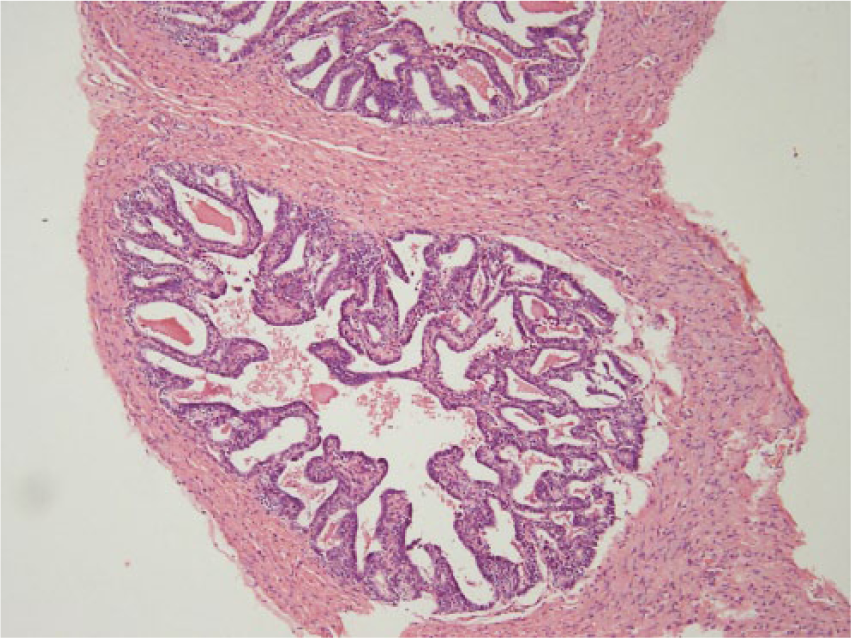
Section through the seminal vesicle of a rat fed on control rat chow for 60 days. Note normal histology, with presence of pseudostratified epithelium of low cylindrical cells that were identified in the base line. H&E, 100×.
Shows thickening of the basal epithelium of connective tissue and reduction of secretion in the acinii. Representative section of seminal vesicle from a rat fed 30 mg of aqueous leaf extract of A muricata per day for 60 days (low-dose group). H&E, 100×.
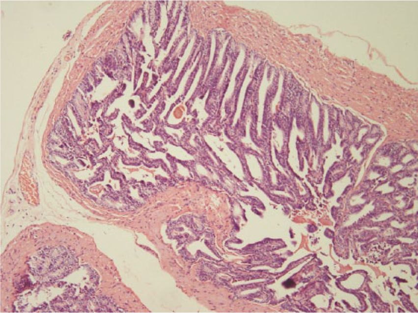
Section through the seminal vesicle of a rat from high-dose group for a period of 60 days. Marked atrophy of the seminal tissues were noticed. Note increased cellularity and the acinii are empty of secretion. H&E, 100×.
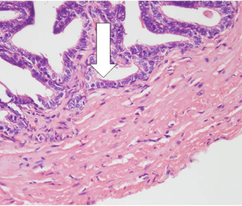
Section through seminal vesicle of a rat fed 30 mg of leaf extract for 60 days (low-dose group). Note fluid separates the epithelium from the submucosa (arrow). The nuclei are densely colored and packed. H&E, 400×.
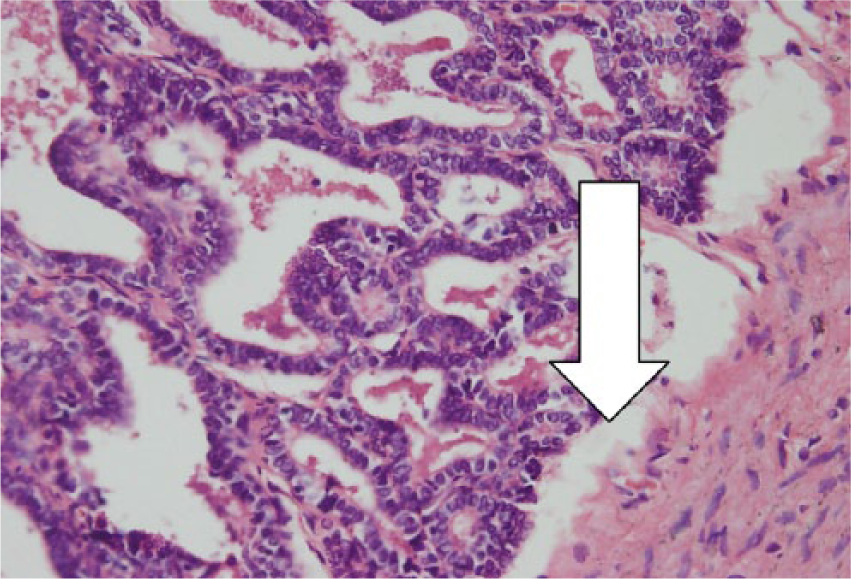
Section through the seminal vesicle of a rat fed 300 mg of leaf extract (high dose) for 60 days. Note epithelial lining showed pyknotic nucleus (arrow). H&E, 400×.
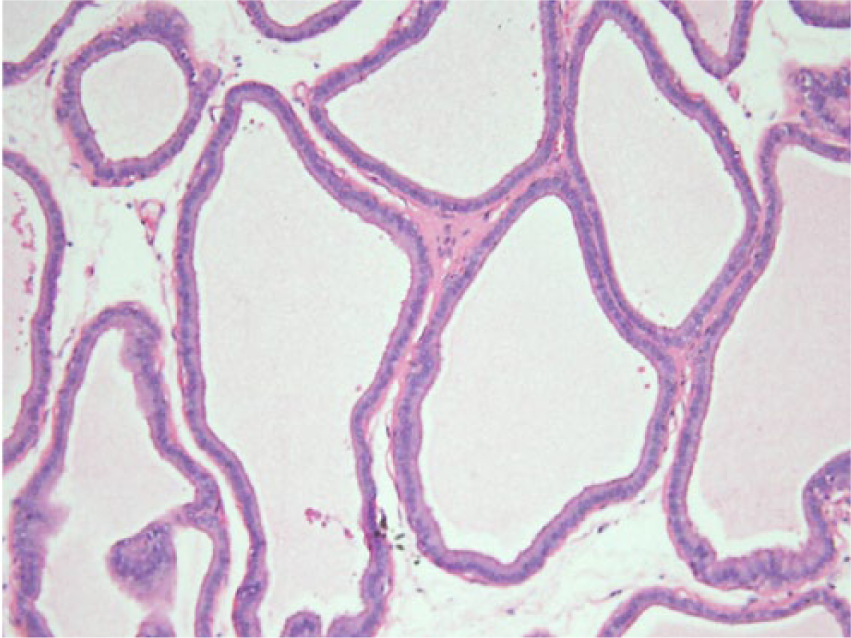
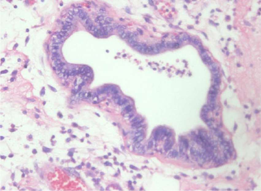
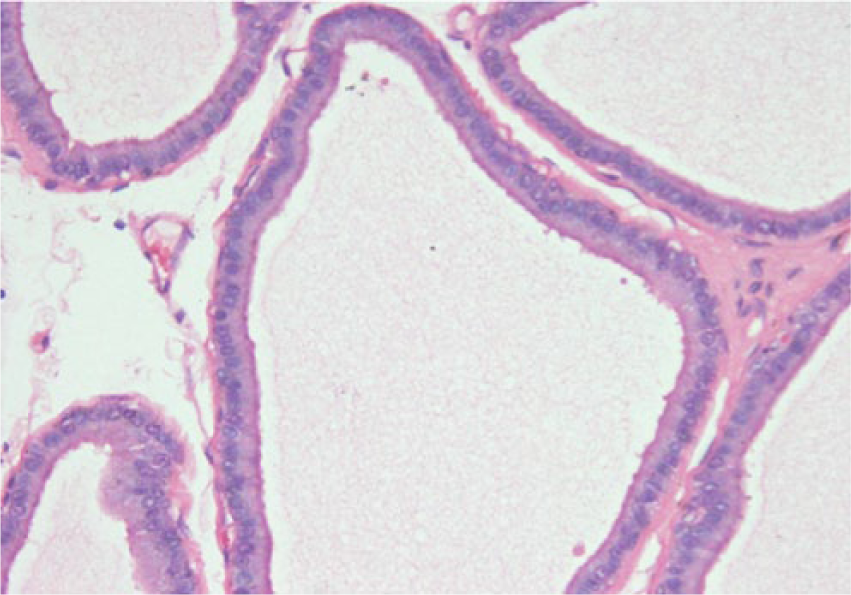
The prostate of rats fed on controlled chow showed normal histological structures (Figure 8A and B). The alveoli showed tall columnar epithelial cells with an apparent high ratio of cytoplasm to nucleus. However, representative sections of tissues of prostate obtained from rats that received low dose of leaf extract showed apoptosis in the epithelium of the glandular acinii (Figure 8C and D). They include discrete condensation of the chromatin, to sharply delineated granular masses along the nuclear envelope, shrinking of the cells, twisting of the cellular and nuclear outlines, and fragmentation of the nucleus. The affected cell disintegrated into membrane-bound apoptotic bodies that remained as a ghost space along with its neighboring cells. Furthermore, the cell membrane and the membrane encasing the apoptotic fragments retained their integrity. However, there was no associated inflammation in the apoptotic areas. Rats that received low dose of the leaf extract showed marked reduction in cytoplasm and secretory activity of the acinii.
Normal glandular structure of the prostate from rats fed on control rat cow. Note conspicuous microvilli. H&E, 200×.
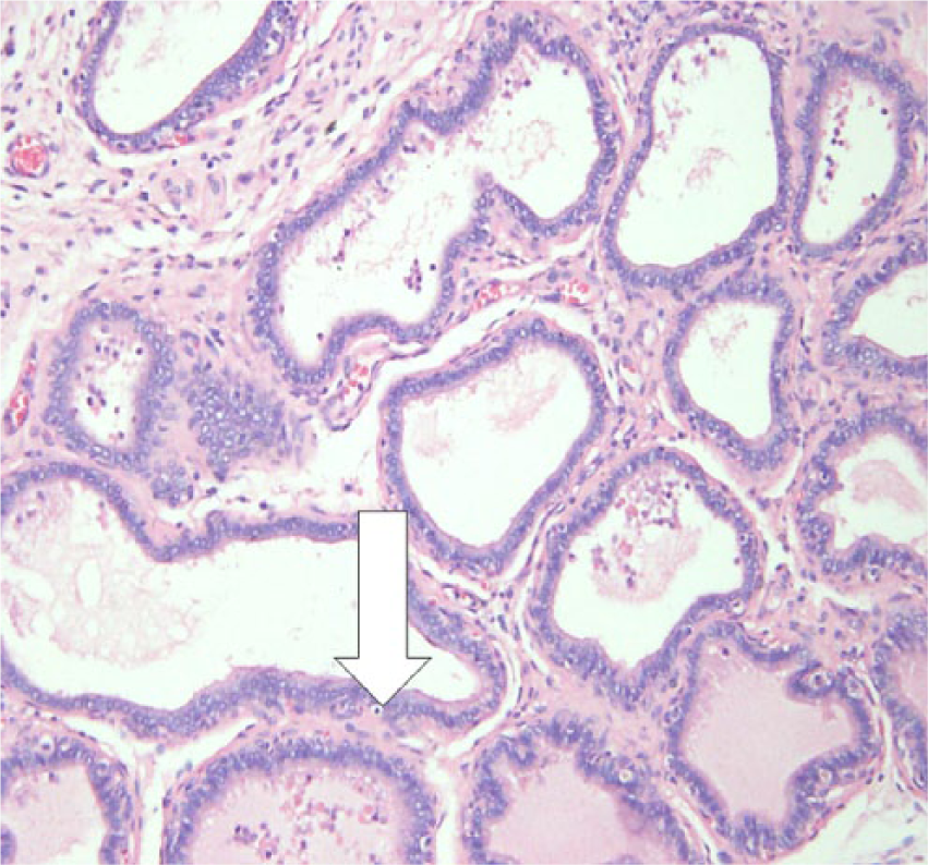
Section through prostate from a rat fed 30 mg of leaf extract/day (low-dose prostate) for 60 days. Note condensation of nuclear material (apoptosis) of the glandular epithelium. H&E, 200×.
Section through prostate of rat that received 300 mg of aqueous leaf extract of A muricata per day for a period of 60 days (high-dose group). Note scanty prostatic secretion with flattening of the acinar epithelial lining. There is a significant reduction in the epithelial thickness. H&E, 200×.
Section through prostate from a rat fed on control rat chow. Note conspicuous microvilli projection with an apparent high ratio of cytoplasm to nucleus. H&E, 400×.
Compared to those of control prostate glands, representative sections of prostrate obtained from the high-dose group showed dramatic decrease in prostatic fluid in the acinar lumens. The lumens were empty and there was flattening of the internal lining of the acinar lumen. This led to an apparent decrease in the average cell number per unit area, and this decreased the thickness of the prostate epithelium. Treatment with high dose of leaf extract for 60 days caused reduction of the stroma, acini size, and shrinking of the epithelium, but it appeared flat with a decrease in the thickness of the fibromuscular layer (Figure 8E and F). In comparison with the control, representative sections of testes examined from rats fed on both low and high levels of leaf extracts showed active spermatogenesis (not shown in the figure). Representative sections of tissues showed normal histological structures of the testes, expressed seminiferous tubules containing different kinds of germ cells: spermatogonia, spermatocytes, spermatids, spermatozoa, and somatic Sertoli cells. The interstitial tissues found between seminiferous tubules showed interstitial cells and Leydig cells. There was no evidence of adverse effects of any kind including degenerative, inflammatory, and/or atrophic alterations in the testicular tissues.
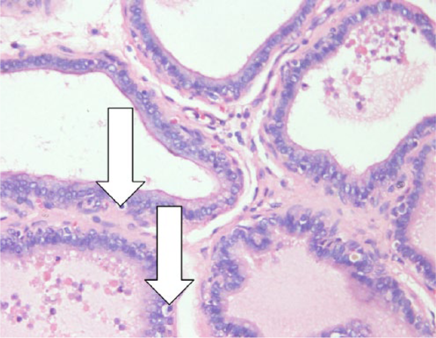
Prostate section from a rat fed 30 mg of aqueous leaf extract of A muricata per day for a period of 60 days (low-dose group). Note condensation and marginalization of the nuclear material. H&E, 400×.
Microphotograph of prostate from a rat that received 300 mg aqueous leaf extract of A muricata per day for a period of 60 days (high-dose group) showing pyknotic nuclei and decreased secretion in acinii with flattening of the epithelial linings. H&E, 400×.
Discussion
In this study, a yield of 2.52% (w/w) was obtained. A similar yield of 2.62% of the aqueous extract of A muricata was obtained by Adewole and Ojewole. 16 However, 50 g of leaf powder extracted with n-butanol yielded 5.4 g of the crude extract. 17 It appears the yield is higher with polar solvents among other factors.
The leaf, stem, bark, and seeds of A muricata contain varying amounts of biologically active chemicals called Annonaceous acetogenins.18,19 So far 14 different tested acetogenins have demonstrated ATP-blocking properties and were found to be more potent against multidrug resistant cancer cells. 20
Profiling of medicinal plant extract is important and critical in order to establish the authenticity and quality of the extract. The process has been used in many studies in order to ensure that results produced from such extracts are a true reflection of the said plant. 21 HPLC fingerprinting of the aqueous extract of leaf revealed 8 peaks with Rf values in the range of 0.1 to 0.3 under conditions previously described. This was reevaluated anytime a new batch of extract was received. The practice was adopted for correct identification of the plant, as a marker of the active phytochemicals and also a reliable indicator of genetic variability in plant populations. 22 Similar practices were done for A squamosa L. 23 including DNA fingerprinting of 4 Annona species.24,25
The antiproliferative potential of A muricata on BPH-1 cells was seen in a dose-dependent manner. Cell viability decreased as the dose increased from 0.5 mg/mL to 1.5 mg/mL. The effect of A muricata on various cell lines have been reported before from hydrodistilled oils. Regarding breast carcinoma cell line (MCF-7), the IC50 was 4.34 µg/mL for the ethanol extract, whereas colon carcinoma cell line (CACO) displayed IC50 values 2.82 µg/mL, and liver carcinoma cell line (HEPG2) showed values of 3.43 µg/mL. 25 Acetogenins from A muricata have been reported to exhibit cytotoxic activities against the human pancreatic tumor cell line (PACA-2), human lung carcinoma, and human prostate adenocarcinoma (PC-3) (A-549). 8 In this study, proliferation of benign prostatic hyperplasia cell line (BPH-1) treated with A muricata showed a reduction in the number of cells in a dose-dependent manner that was not due to direct cytotoxicity but growth inhibition (0.0 mg/mL > 0.5 mg/mL > 1.0 mg/mL > 1.5 mg/mL), indicating that the aqueous plant extract was capable of exerting inhibition of human benign prostate cell proliferation and, possibly, its tumor.
Other aqueous plant extracts such as Artemisia vulgaris inflorescence have been reported to exert an inhibitory effect on cell growth and colony formation of prostate cancer PC-3 cells. A vulgaris demonstrated 8% to 65% inhibition at similar doses for 24 hours. Furthermore, an increased cell growth inhibition was observed after incubation of these cells from 24 to 72 hours at various doses. 26 Such dose-dependent increases with corresponding morphological changes were also observed in this study. Near cell extinction was observed at the highest dose of 1.5 mg/mL after 72 hours
Some studies on human breast carcinoma cells (MDA-MB-435S) and human immortalized keratinocyte cells (HaCaT) have confirmed the presence of therapeutically active antineoplastic compounds in the n-butanolic leaf extract of A muricata. Isolation of the active metabolites from the extract is a prospect 17 as they could possibly suppress proliferation of prostate cancer cells as observed in BPH-cells in this study. The antitumor effects of A muricata leaves may be due to the presence of acetogenins. Previous studies revealed that most of the acetogenins act as a DNA topoisomerase I toxin, causing cancer cell arrest at the G1 phase and inducing apoptotic cell death in a Bax- and caspase-3-related pathway. Furthermore, the inhibition of the enzyme NADH-ubiquinone oxidoreductase (complex I) in mitochondria was suggested.27-29
Imbalance of molecular mechanisms of proliferation and apoptosis has been found to be associated with the development of BPH and its cancer, with evidence of reduction of apoptosis as a major underpinning factor. Several studies have revealed that there is upregulation of pro-apoptotic proteins Bax and Bak and downregulation of anti-apoptotic protein Bcl2 in cell lines undergoing apoptosis. In this study, administration of the aqueous leaf extract of A muricata induced Bax upregulation and bcl-2 downregulation in a dose-dependent manner in BPH-1 cells using RT-PCR.
The histological changes observed in the prostate of rats given 30 and 300 mg of leaf extract of A muricata appear to agree with the findings of Olamide et al 30 and Dhanotiya et al. 31 These authors used methanolic and/or ether extracts of Citrullus lantatus and C colocynthis Schrad, respectively. Cell apoptosis from prostate and seminal vesicle do not appear to have been described for rats feeding plant extracts. However, the present experiment showed apoptosis in the glandular epithelium in both prostate and seminal vesicles. It would seem that the onset of apoptotic effect in the seminal vesicle may not necessarily correlate with the appearance of prostatic cell apoptosis, although the nature of induction of the effect may well be changed in the latter organ, a matter that needs careful investigation. In addition, the absence of ill effects toward spermatogenesis in the rats so gavaged shows that A muricata would have an advantage over other plant extracts so far used to prevent prostate hyperplasia. 32
The changes in the seminal vesicle showed accumulation of fluid in the submucous layer. The authors regard such changes to be due to different cause or causes. In the case of the seminal vesicle, the immediate cause of apoptosis observed is likely due to accumulation of fluid deep into the submucous layer and thus isolated epithelial cells must have been deprived of nutrients. This effect has the resemblance to those produced in the vas deferens of mouse by the administration of oestrogen. 33 Although, in the present study, the androgen level in the rats were not measured, it is obvious that it had great influence in the development of the reproductive organs. Estrogen may act closely with those of androgens to develop accessory sex organs, whereas androgens stimulate the epithelia, and estrogens the connective tissue. 34
It may be mentioned here that apoptosis or programmed cell death involves a sequential cascade of cellular event, resulting from chromatin condensation, DNA fragmentation, cytoplasmic membrane blebbing, and cell shrinkage. 35 Various initiator and executor caspases are involved. Anticancer agents that inhibit the poly (ADP-ribose) polymerase (PARP) and other substances have been reported to be susceptible to the cleaving action of caspase-3 in response to DNA strand breaks leading to apoptosis.36,37 Results from a previous study indicated that Artemisia vulgaris extracts caused caspase-3 activation that resulted in PARP cleavage. 26
In rats, increased secretory activity of the seminal vesicle is associated with increased testosterone production followed by increased seminal vesicle weight.38,39 Indeed, 5-α-reductase, the enzyme that converts testosterone to dihydrotestosterone (DHT), is active in the seminal vesicle. DHT is important for the development of the prostate. However, it is also responsible for the pathologic growth of the prostate. DHT binds to androgen receptors with subsequent modulation of target genes causing BPH and its related cancer. 40 To arrest BPH and the further development of cancer, 5-α-reductase inhibitors are administered to act as pathologic substrates of the disease, thereby arresting the disease, reducing the prostate volume, and improving symptoms. 41 The significant decrease in SVI in the treatment group therefore implies a possible reduction in DHT and the shrinkage of the prostate as seen in the reduced PI of the treated groups.
The evidence presented here on the aqueous leaf extract of A muricata and its possible anti-BPH properties provides the basis for further studies using BPH animal models.
Footnotes
Declaration of Conflicting Interests
The author(s) declared no potential conflicts of interest with respect to the research, authorship, and/or publication of this article.
Funding
The author(s) received no financial support for the research, authorship, and/or publication of this article.