
Abstract
Purpose. The study was designed to screen Sphaeranthus indicus, Ganoderma lucidum, and Urtica dioica for their anticancer activity against human cancer cell lines. Phytochemical screening of active extracts was also planned. Methods. Petroleum ether, ethanolic, and aqueous extracts of S indicus Linn, G lucidum P Karst, and U dioica Linn were subjected to cytotoxicity studies using 7 different cancer cell lines. Potent cytotoxicity was noted in petroleum ether extract of S indicus (SIP), which inhibited proliferation of various cancer cell lines. Growth inhibition was determined by sulforhodamine B assay. Two biochemical markers, namely β-sitosterol and 7-hydroxyfrullanolide were isolated and characterized using high-performance thin layer chromatography, melting point, Fourier transform infrared spectroscopy, nuclear magnetic resonance spectroscopy, and mass analysis. Cytotoxicity of isolated β-sitosterol and 7-hydroxyfrullanolide were also determined. The IC50 of SIP was calculated in the HL-60 cells and was found to be 53 µg/mL. Furthermore, SIP induced apoptosis in human leukemia HL-60 cells as measured by several biological end points. Cell cycle analysis and change in mitochondrial membrane potential was quantified by flow cytometry. Subsequently, using annexin V/PI assay, proportion of cells actively undergoing apoptosis was determined. Changes in DNA were observed by DNA ladder assay. Results. SIP induced apoptotic bodies formation, induced DNA laddering, enhanced annexin-V-FITC binding of the cells, increased sub-G0 DNA fraction, and induced loss of mitochondrial membrane potential (ΔΨm) in HL-60 cells. SIP also elevated the caspase 3 and caspase 9 levels in the HL-60 cells, which clearly indicates the involvement of the intrinsic proteins in inducing apoptosis. Discussion. All the above parameters revealed that SIP induced apoptosis through the mitochondrial-dependent pathway in HL-60 cells. The criterion for anticancer activity in cytotoxicity assay was ≥70% growth inhibition at 100 µg/mL against at least 4 cell lines. As G lucidum and U dioica did not exhibit appreciable inhibitory activity against human cancer cell lines (less than 50%), they were not included in the study thereafter. The results established that SIP has apoptosis-inducing effect against HL-60 cells in vitro and is a promising candidate for further anticancer study. β-Sitosterol and 7-hydroxyfrullanolide can be considered to be potent anticancer compounds isolated from SIP on the basis of present studies.
Introduction
The uses of plants as medicine are as old as human civilization. About 60% of anticancer drugs are derived from plant sources, for example, taxol from Taxus brevifolia, camptothecin from Camptotheca acuminata, and so on. It is noted that the management of cancer is still not up to the mark and there are always needs to search new drugs for the management of cancer. In this context, plants still hold the hope for the treatment and prevention of cancer. 1
Sphaeranthus indicus (Compositae) is an herb found mostly in southern India. It is bitter stomachic, stimulant, alterative, pectoral, demulcent, and external emollient. 2 It is a multibranched herb with round purple flowers that grows plentifully in rice fields and is distributed throughout India, Ceylon, Malay, China, and Africa.3,4 It is used indigenously in the Indian system of traditional medicine as a remedy for various ailments, as a tonic, laxative, digestive, anthelmintic, and in the treatment of insanity, tuberculosis, diseases of the spleen, anemia, bronchitis, elephantiasis, pain of the uterus and vagina, piles, asthma, leucoderma, and hemicrania. The herb is an ingredient of certain proprietary marketed preparations in India, namely, “Prostabliss” used for the management of benign prostatic hyperplasia. There is paucity of reports about the use of this herb as an anticancer agent. Previous studies have reported the presence of stigmasterol, β-sitosterol, 5 hentriacontane, sesquiterpene lactone, 6 sesquiterpene glycoside, sphaeranthanolide, 7 flavone, and isoflavone glycosides 8 in the powdered caputula of S indicus. When our studies were underway, we came across a reference by Mitra et al, 9 who obtained a patent on the anticancer activity of S indicus and reported the sesquiterpene lactones present in the extracts to be responsible for the activity. The anticancer activity in the plant extract were attributable primarily to β-sitosterol, which is a well-established steroidal compound having prominent activity against benign prostatic hyperplasia and prostatic cancer cell lines.10,11
We screened S indicus, Ganoderma lucidum (a mushroom), and Urtica dioica for their cytotoxicity against human cancer cell lines and found S indicus to be the most effective in inhibiting the proliferation of prostate cancer cell lines, that is, PC-3 and DU-145. The petroleum ether, ethanolic, and aqueous extracts of the test drugs were screened for their in vitro cytotoxicity. S indicus proved to be the best in these studies and its petroleum ether extract exhibited inhibitory activity against most of the human cancer cell lines, namely, lung (A549), prostate (PC-3 and DU-145), colon (Colo-205), neuroblastoma (IMR-32), and breast cancer (MCF-7). The other 2 plants have a history of use in the management of benign prostatic hyperplasia and they are being used widely for its prevention.12-15 But they did not exhibit appreciable inhibitory activity against human cancer cell lines, including prostate. Hence only S indicus petroleum ether extract (SIP) was subjected to mechanistic studies to have an insight into its mechanism of anticancer activity.
Deregulation of apoptosis is the hallmark of all cancer cells, and agents that activate programmed cell death in cancer cells could be valuable anticancer therapeutics. Anticancer drugs act through different pathways converging ultimately into activation of apoptosis in cancer cells leading to cell cytotoxicity. We thus became interested to know the mechanism of cancer cell killing by SIP so that the lead might be developed into a novel antineoplastic therapeutic. In this article, we report the cell cytotoxicity of SIP in several cancer cell lines and mechanism of action of SIP involved in the induction of apoptosis in human leukemia HL-60 cells. The observed apoptotic activity of SIP is associated with early loss of mitochondrial membrane potential, DNA fragmentation, enhanced annexin-V-FITC binding of the cells, and increased sub-G0 DNA fraction, which altogether account for apoptotic cell death.
Materials and Methods
Chemicals
RPMI-1640, trypsin, EDTA, gentamycin, penicillin, 5-fluorouracil (5-FU), adriamycin, mitomycin-c, paclitaxel, propidium iodide (PI), sulforhodamine B (SRB), phosphate-buffered saline (PBS), agarose, ethidium bromide, sodium dodecyl sulfate (SDS), proteinase-K, DNase-free RNase A, dimethyl sulfoxide (DMSO), rhodamine-123 (Rh-123), and fetal bovine serum (FBS) were purchased from Sigma Chemical Co (St Louis, MO). Annexin V-FITC kit was purchased from Axxora (San Diego, CA). Other reagents and chemicals used were of analytical grade and purchased locally.
Plant Material and Extraction
Flower heads of S indicus were collected from the outskirts of Sagar city. U dioica roots were procured from Nainital, a city amid the Himalaya mountains. G lucidum powder was supplied by Gano Excel industries, Malaysia. All of them were identified in the Botany Department of Dr Hari Singh Gour Vishwavidyalaya, Sagar. The flower heads of S indicus and roots of U dioica were shade dried and coarsely powdered. All the 3 drugs were subjected to Soxhlet extraction successively with petroleum ether and ethanol. After ethanolic extraction, aqueous extract was obtained by maceration for 24 hours. Thus petroleum ether, ethanolic, and aqueous extracts were obtained for all the drugs.
Characterization of Extract and Selection of Marker
Chromatographic profile of SIP in toluene:ethyl acetate (8:2) revealed the presence of 10 spots. One of the spots showed identical Rf value with the standard compound β-sitosterol at Rf 0.95 when visualized under ultraviolet at 254 nm.
HPTLC Analysis and Isolation of Marker Compounds
Precoated and preactivated thin layer chromatography (TLC) plates (E. Merck No. 5548) of silica gel 60 F254 + 366 with the support of aluminum sheets 0.1 mm thick and 20 × 20 cm were used. SIP (10 mg) was weighed accurately and dissolved in 10 mL of petroleum ether. The extract sample was applied in the form of a band using CAMAG LINOMAT V, an automatic sample application device, maintaining a bandwidth 6 mm, space 10 mm, 250 nL/s. The quantity of sample applied was 10 µL. The mobile phase used was toluene:ethyl acetate (8:2). Standard β-sitosterol (10 mg) and isolated compound (isolation procedure described below) were dissolved in petroleum ether (10 mL). The solution was analyzed in the same manner as described above.
For isolation of compound showing identical TLC profile as standard β-sitosterol, column chromatography was performed. Column was packed in methanol and was eluted with methanol. A white crystalline material melting at 140°C was isolated. It gave a positive Libermann Burchard test indicating the presence of sterol. It was characterized as β-sitosterol after co-chromatography with the standard using TLC and high-performance TLC (HPTLC). The percentage of isolated compound in the extract was found to be 46.76% (Figure 1). The melting point of the isolated compound was determined using the Superfit melting point determination apparatus and found to be 140°C, which was identical to the standard sample (140°C). Furthermore, superimposable Fourier transform infrared (FTIR) analysis with the standard confirmed the identity of isolated compound as β-sitosterol (Figure 2). The nuclear magnetic resonance (NMR) analysis of the isolate further confirmed its identity as β-sitosterol. The 1H-NMR spectrum of the isolated compound exhibited a one-proton doublet at δ 5.35 (J = 1.0 Hz) assigned to H-6 proton. A broad one-proton multiplet at δ 3.51 (J = 1.15 Hz) is ascribed to α-oriented H-3 methine proton (axial) interacting with C-2 equatorial, C-2 axial, C-4 axial, and C-4 equatorial protons. Three doublets, integrating 3 protons each, at δ 0.94 (J = 6.34 Hz), 0.84 (J = 4.49 Hz), and 0.82 (J = 3.83 Hz) are because of C-21, C-26, and C-27 secondary methyls, respectively. A 3-proton triplet at δ 0.81 (J = 2.74 Hz) is ascribed to C-29 primary methyl protons. The remaining 2 tertiary C-18 and C-19 methyl signals appeared as singlets at δ 0.67 (J = 3.08 Hz) and δ 1.00 (J = 4.13 Hz), respectively. The presence of all the methyls in the region δ 0.67 to δ 1.00 suggests that these functionalities are attached to saturated carbons. The remaining methylene and methine protons resonated in the region δ 2.28 to δ 1.02.

High-performance thin layer chromatography profile of β-sitosterol (standard), isolated compound, and petroleum ether extract of Sphaeranthus indicus (SIP)

Overlaid Fourier transform infrared spectra of β-sitosterol and the isolated compound
Furthermore, we also isolated another compound, namely, 7-hydroxyfrullanolide (7-HF) from SIP using the aforementioned procedure. The FTIR spectra of the compound showed bands at 3604, 3430, 3200, 2918, 1760, 1714, 1652, 1604, 1462, 1433, 1376, 1260, 1155, 1135, 970, 887, 813, and 723 cm−1. The peak at 3604 cm−1 indicates the presence of an –OH group. Stretching at 2918 cm−1 indicates the C–H stretching. The peaks at 1760 and 1714 cm−1 are indicative of a ketonic group, which is a part of a lactone ring (5-membered lactone). The band at 1670 cm−1 is indicative of C=C stretch and that at 1604 cm−1 indicates an exocyclic methylene group. Hence the IR spectra pointed toward the presence of a lactone ring in the isolated compound.
The comparison of FTIR and mass spectra of the compound with the reported literature led us to conclude that the compound isolated was 7-hydroxyfrullanolide. 6 The compound had a mass of 233 with molecular ion peak m/z at 248. The peaks obtained by mass spectral analysis confirmed the structure of the compound. The mass spectrum was significant in that it did not show an M − 18 peak, but showed M − 15 and M − 33 peaks, the latter arising because of the loss of a methyl and a water molecule.
In conformity with its structure, the compound exhibited 1H-NMR signals due to tertiary methyl (0.89, 3H, s) and a methyl on a tetrasubstituted double bond (1.25, 3H, s), in addition to sharp signals (3.35 and 8.14, 1H each, s) attributable to exomethylene protons. The presence of these, and the absence of any signal due to vinylic proton, indicated a δ4-5 eudesmane skeleton for frullanolide.
The HPTLC studies of the compound revealed it to be present at Rf 0.31 occupying 33.53% of the total proportion of the extract. Hence the major proportion of the extract with β-sitosterol and 7-hydroxy frullanolide taken together (46.76 + 33.53= 80.29%) account for about 80% of the extract.
Cell Lines and Cell Cultures
The human cancer cell lines were obtained either from National Center for Cell Science, Pune, India or National Cancer Institute, Frederick, MD. The human colon (Colo-205), prostate (PC-3 and DU-145), lung (A-549), breast (MCF-7), ovary (IGR-OV-1), and acute lymphoblastic leukemia (HL-60) cell lines were grown and maintained in RPMI-1640 medium, pH 7.4, whereas DMEM was used for neuroblastoma (IMR-32). The media were supplemented with FBS (10%), penicillin (100 U/mL), streptomycin (100 µg/mL), glutamine (2 mM), and cells were grown in CO2 incubator (Heraeus GmbH, Hanau, Germany) at 37°C with 90% humidity and 5% CO2. Cells were treated with SIP dissolved in DMSO whereas the untreated control cultures received only the vehicle (DMSO, <0.2%).
Cytotoxicity Assay
Stock solution of 100 µg/mL of extracts was prepared in 2% DMSO in complete medium. The stock solution was serially diluted with complete medium supplemented with 50 µg/mL gentamycin to obtain solutions of desired concentrations. In vitro cytotoxicity against human cancer cell lines was determined using sulforhodamine B assay as described previously. 16
Isolated β-sitosterol and 7-hydroxyfrullanolide were also tested for their cytotoxicity profile against human cancer cell lines using the aforementioned protocol. Lung (A-549), leukemia (THP-1), liver (HEP-2), colon (HCT-15, Caco-2), prostate (PC3 and DU145), and cervix (Hela) cell lines were used.
Assay of Cell Proliferation in HL-60 by MTT
HL-60 cells were plated in 96-well plate at a density of 2 × 10⁴ cells/well/100 µL of RPMI medium containing 10% FBS. Cultures were treated with different concentrations of SIP and were incubated for 48 hours. The concentrations used in the MTT study were 0, 1, 10, 30, and 100 µg/mL. The stocks of SIP were initially prepared in DMSO and from these stocks, 2× concentration stocks were prepared in media, such that the concentration of DMSO is uniform in all stocks and not more than 0.5% in the final well of the 96-well plate. The 2× concentration stocks are prepared in media because the volume introduced in the well is 100 µL while 100 µL media is already present in the well that contains cells. Cell proliferation using MTT was assayed as described earlier.17,18 The experiment was repeated thrice. Control cultures were simultaneously treated with culture medium containing 0.5% DMSO v/v.
DNA Ladder Assay
DNA fragmentation, which is a typical hallmark of apoptosis, was assessed by electrophoresis of extracted genomic DNA from HL-60 cells as described previously with some modifications. 19 HL-60 cells in the log phase were cultured (37°C, 5% CO2) in RPMI medium (pH 7.2) in a CO2 incubator. Overall, 2 × 10⁶ cells after various treatments (10, 30, and 100 µg/mL) were incubated for 24 hours and then washed in PBS containing 10 mM EDTA. The pellet was lysed in 250 µL of lysis buffer (100 mM NaCl, 5 mM EDTA, 10 mM Tris–HCl, pH 8.0, 5% Triton X-100, 0.25% SDS) containing 400 µg/mL DNase-free RNase and incubated at 37°C for 90 minutes followed by 1-hour incubation with proteinase-K (200 µg/mL) at 50°C for 1 hour. The DNA was extracted with 200 µL of phenol:chloroform:isoamyl alcohol (25:24:1) mixture for 1 minute and centrifuged at 13 000 × g for 3 minutes. The aqueous phase was further extracted with chloroform and centrifuged. DNA was precipitated from aqueous phase with 3 volumes of chilled alcohol containing 0.3 M sodium acetate at 4°C overnight. The precipitate was centrifuged at 13 000 × g for 10 minutes. The DNA pellet was washed in 80% alcohol, dried, dissolved in 50 µL TE buffer, and electrophoresed in 1.6% agarose gel at 50 V, stained with ethidium bromide, and visualized in Bio-Rad gel documentation system.
Cell Cycle Phase Distribution
Cell cycle phase distribution was done as described earlier. 20 Briefly, HL-60 cells (1 × 10⁶/mL) in exponential phase of growth were seeded in 6-well plates and allowed to adhere for 24 hours. The old medium was replaced by fresh medium without and with SIP in different concentrations (10, 30, and 100 µg/mL) and further incubated for 24 hours. Cells were trypsinised, centrifuged, and washed with PBS. Cells were fixed in 70% ethanol, washed with PBS, subjected to proteinase and RNase digestion (37°C for 30 minutes), followed by staining of clean nuclear materials (nuclei) with PI (25 µg/mL) using procedures and reagents as described by manufacturer in the Cycle Test plus DNA reagent kit (Becton-Dickinson, Franklin Lakes, NJ). The preparations were analyzed for DNA content using BD-FACS Caliber flow cytometer. Data were collected in list mode on 10 000 events for FL2-A versus FL2.
Annexin V Binding: Flow Cytometric Analysis of Apoptosis and Necrosis
The mode of cell death can be clearly distinguished by annexin FITC assay. During the early events of apoptosis, the plasma membrane phospholipid phosphatidylserine is translocated from the inner side of membrane leaflet to the outer side, which has a very high affinity for annexin V antibody. Thus, annexin V-FITC–positive and PI-negative cell populations are indicative of apoptosis and annexin V-FITC–positive and PI-positive cells indicate necrosis. The extent of apoptosis was determined using FITC-labeled annexin V by flow cytometry. 21 Cells (1 × 10⁶/mL) were incubated with the designated doses of SIP (10, 30, and 100 µM) for 24 hours, washed, and stained with annexin V-FITC antibody and PI as per the instructions given by the manufacturer and scanned for fluorescence intensity in FL-1 (FITC) and FL-2 (PI) channels. The fraction of cell population in different quadrants was analyzed using quadrant statistics. Cells in the lower right quadrant represented apoptosis and in the upper right quadrant represented necrosis or postapoptotic necrosis.
Loss of Mitochondrial Membrane Potential
Changes in mitochondrial transmembrane potential (ΔΨm) as a result of mitochondrial perturbation were measured after staining with Rh-123. Cells (1 × 10⁶/1.5 mL/12-well plates) were treated with the test compound and incubated for 24 hours. Rh-123 (5 µM) was added 1 hour before the termination of the experiment. Cells were collected and washed in PBS. The decrease in fluorescence intensity because of mitochondrial membrane potential loss was analyzed in FL-1 channel on flow cytometer. Intensity of Rh-123 is directly related to mitochondrial membrane potential. 22
Caspase Assays
Cells (2 × 10⁶/3 mL) were incubated with SIP for the indicated time periods. At the end of treatment, cells were washed in PBS, and cell pellets lysed in cell lysis buffer. Activities of caspase-3, -8, and -9 in the cell lysates were determined fluorimetrically using BD Apoalert caspase fluorescent assay kits as per the instructions given by the manufacturer. Procedure was followed according to the work carried out by Malik et al. 17
Statistical Analysis
All in vitro experiments were done in triplicates and each data point represents the average of at least 3 independent experiments. The comparisons were made between control and treated cultures using unpaired Student’s t test and the difference was considered to be statistically significant if P < .001.
Results
In Vitro Cytotoxicity Against Human Cancer Cell Lines
SIP showed maximum growth inhibition against all the human cancer cell lines used in the present study except ovarian cancer cell line IGR-OV-1. G lucidum and U dioica were ineffective in inhibiting the proliferation of human cancer cell lines. Both these species exhibited no significant cytotoxic activity and hence they were not carried over further in the studies (Table 1). 5-Flurouracil (20 µM), adriamycin (1 µM), paclitaxel (10 µM), and mitomycin C (1 µM), which were used as positive control showed 54%, 75%, 69%, and 54% growth inhibition, respectively, after 48-hour treatment. Data are representative of 1 of 3 similar experiments. Isolated β-sitosterol and 7-HF were also subjected to in vitro cytotoxicity studies and they also exhibited potent cytotoxic effects against the cell lines used in the study. Results are shown in Tables 2 and 3. Hence, SIP was forwarded to the later phase of anticancer screening as described in sections below.
Preliminary Cytotoxic Activity of Test Extracts Against Various Human Cancer Cell Lines a

Abbreviations: 5-FU, 5-fluorouracil; GLE, Ganoderma lucidum ethanolic extract; GLP, Ganoderma lucidum petroleum ether extract; GLA, Ganoderma lucidum aqueous extract; UDP, Urtica dioica petroleum ether extract; UDE, Urtica dioica ethanolic extract; UDA, Urtica dioica aqueous extract; SIP, Sphaeranthus indicus petroleum ether extract; SIE, Sphaeranthus indicus ethanolic extract; SIA, Sphaeranthus indicus aqueous extract.
Data are representative of 1 of 3 similar experiments.
Preliminary Cytotoxic Activity of Isolated β-Sitosterol Against Various Human Cancer Cell Lines a

Data are representative of 1 of 3 similar experiments.
Preliminary Cytotoxic Activity of Isolated 7-Hydroxyfrullanolide Against Various Human Cancer Cell Lines

Data are representative of 1 of 3 similar experiments.
MTT Assay
The IC50 (half maximal inhibitory concentration) of SIP was calculated in HL-60 cells and was found out to be 53 µg/mL. The cells were treated with different concentrations of SIP and incubated for 48 hours. The inhibition of cell growth was found to be occurring in a concentration-dependent manner (Figure 3). Other parameters and details are described in the Materials and Methods section.
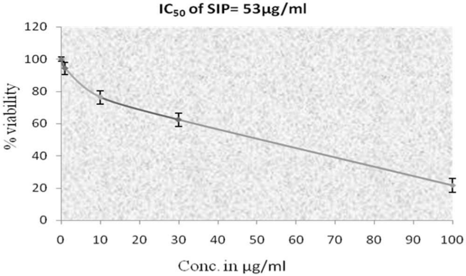
Percentage viability of HL-60 cells using different concentrations of petroleum ether extract of Sphaeranthus indicus (SIP; MTT assay)
SIP Induces DNA Laddering, a Hallmark of Apoptosis
To elucidate whether SIP decreases cell survival by the induction of DNA fragmentation, genomic DNA was isolated from cells exposed to different concentrations of SIP from HL-60 cells and electrophoresed. The compound induced DNA laddering of 180 to 200 bp in HL-60 cells treated for 24 hours. The minimal concentration inducing DNA fragmentation was evident at 100 µg/mL. The ladder pattern indicated that the test material induces apoptosis. DNA isolated from untreated control did not show DNA ladder (Figure 4).

SIP (petroleum ether extract of Sphaeranthus indicus)-induced DNA fragmentation in the HL-60 cells
SIP Increases Sub-G0 DNA Fraction of Cell Cycle Phase Distribution
To further determine and confirm whether or not the growth inhibitory activity of SIP was related to induction of apoptosis, the subdiploid (character of apoptosis) fraction was measured by flow cytometry. After the cells were exposed to 10, 30, and 100 µg/mL SIP for 24 hours exhibited continuous increase in sub-G0 fraction comprising both apoptotic and debris population, implying together the extent of cell death (Figure 5). The damage was more apparent with higher SIP concentration over the period of study. The results indicated that, compared with the control, 10, 30, and 100 µg/mL SIP treatment increased the population of sub-G0 phase from 4% to 10% and 80%, respectively. There was hardly any significant effect after 24 hours of treatment on G2/M fraction, which indicated that SIP does not cause mitotic arrest.
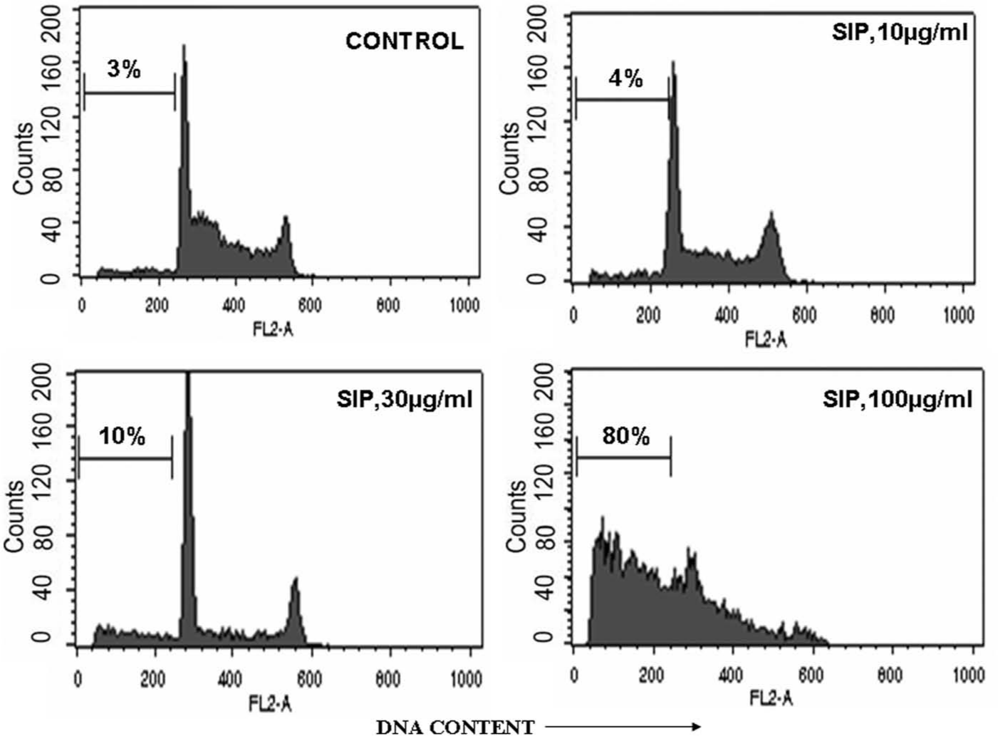
Cell cycle analysis of SIP (petroleum ether extract of Sphaeranthus indicus)-treated HL-60 cells
The cell cycle arrest at sub-G0/G1 denotes the inhibition of cell proliferation. This arrest is because of the effect of SIP, which does not allow the cancer cells to proceed on with the next phase of cell cycle, that is, the G1 phase. This arrest occurs at 100 µg/mL concentration, which means that at 100 µg/mL concentration, the cells die.
Flow Cytometric Estimation of SIP-Induced Apoptosis and Necrosis
Exposure of phosphatidylserine (PS) on the surface of cells is an early event in the onset of apoptosis, which has strong binding affinity for annexin V in the presence of calcium. HL-60 cells were incubated with different concentration of SIP and cells were stained with annexin V-FITC and PI to assess the apoptotic and necrotic cell population (Figure 6). SIP produced dose-dependent increase in the apoptotic cell population. The basal apoptotic population in the untreated culture was 3%, which increased to 85% at 100 µg/mL. The annexin antibody binding revealed that there is very high number of apoptotic cells at the 100 µg/mL concentration. Apoptosis thus appeared to be the primary mode of cell death induced by SIP.

Flow cytometric analysis of SIP (petroleum ether extract of Sphaeranthus indicus)-induced apoptosis and necrosis in HL-60 cells using annexin V-FITC/PI double staining
SIP Induces Loss of Mitochondrial Membrane Potential
The loss of mitochondrial membrane potential (ΔΨm) is largely because of the opening of mitochondrial permeability transition pores (PTP), which conduit the leakage of cytochrome c and proapoptotic proteins from mitochondria to the cytosol. SIP caused concentration-dependent mitochondrial damage and loss of mitochondrial membrane potential in HL-60 cells (Figure 7) that showed the role of PTP in loss of ΔΨm. SIP caused concentration-dependent mitochondrial damage and hence the decrease of mitochondrial membrane potential. There was a significant decrease of ΔΨm from 7% in control to 14%, 40%, and 80%, in treatments ranging from 10, 30, to 100 µg/mL SIP, respectively, after 24 hours. This shows that SIP primarily targets mitochondrial cell death. Thus, it can be observed that apoptosis is induced by mediating mitochondrial outer membrane potential loss.

Analysis of SIP (petroleum ether extract of Sphaeranthus indicus)-induced alterations in mitochondrial membrane potential (ΔΨm)
Caspase Activation by SIP
We next evaluated caspase activities, namely, caspase-3, -8, and -9 in HL-60 cells exposed to SIP for 24 hours, because activation of these enzymes might suggest whether the cell death involved mitochondrial-dependent and/or -independent pathways. HL-60 cells incubated with SIP for 24 hours activated caspase-3 activity by approximately 6-fold whereas caspase-9 was upregulated by approximately 5.9-fold. Caspase-8 was moderately affected (Figure 8).

SIP (petroleum ether extract of Sphaeranthus indicus)-induced activation of caspases in HL-60 cells
Discussion
In recent years, considerable attention has been focused on identifying naturally occurring substances capable of inhibiting, retarding, or reversing the process of multistage carcinogenesis. 23 Anticancer drugs having minimum side effects, inducing apoptosis, and targeting specific cytotoxicity to the cancer cells are the drugs of choice. 16 Keeping this in mind, we investigated the cytotoxic potential of petroleum ether, ethanolic, and aqueous extracts of 3 drugs having a history of use in benign prostatic hyperplasia, namely, G lucidum, S indicus, and U dioica in various human cancer cell lines. The in vitro cytotoxicity induced by G lucidum and U dioica was not significant. Hence both of them were eliminated from further stages of the present study. Only petroleum ether extract of S indicus, or SIP, showed significant inhibitory activity and it was therefore selected for mechanistic studies to identify the exact mechanism of its action on cancerous cells. The results of the present study describe the cytoxicity and proapoptotic activity of SIP on human leukemia HL-60 cells. SIP was able to inhibit the cell proliferation of 7 cell lines of 6 different tissue origins. Considering the potential that SIP offers in its development as anticancer agent, we further sought to understand the mechanism of apoptotic cell death.
Apoptosis is a major form of cell death, characterized by a series of stereotypic morphological changes such as formation of apoptotic bodies, chromatin condensation, shrinkage of cells and bleb formation, internucleosomal DNA fragmentation, and sub-G0 DNA accumulation. 24 DNA fragmentation is a relatively late event and hallmark of apoptosis. To confirm apoptosis induced by SIP, we have further employed flow cytometry to determine the extent and causes of apoptosis. SIP significantly induced the DNA fragmentation, apoptotic body’s formation and annexin V binding, increased the sub-G0 DNA fraction of HL-60 cells, and triggered a significant loss of mitochondrial membrane potential.
One of the most commonly used techniques for confirmation of apoptosis is identification of DNA ladders. 25 Apoptosis typically involves intranucleosomal chromatin cleavage by endonucleases in multiples of 180 bp leading to DNA fragments, producing thereby a typical DNA laddering. 26 To elucidate whether SIP decreases cell survival by the induction of DNA fragmentation, genomic DNA was isolated from cells exposed to different concentrations of SIP from HL-60 cells and electrophoresed. The SIP treatment of HL-60 cancer cells indicated internucleosomal DNA breakdown, leading to DNA fragmentation as expected for apoptotic cells.
The development of cytofluorimetric approaches to monitor these multiple cell alterations permits a precise and reliable quantification of apoptosis. 27 Apoptotic cells exhibit some morphological modifications that are readily detected by flow cytometry according to their light scatter properties (FSC/SCC).25,26 To further determine and confirm whether or not the growth inhibitory activity of SIP was related to induction of apoptosis, subdiploid (character of apoptosis) fraction was detected by flow cytometry. The presence of the sub-G0 peak in the histogram of the DNA distribution indicated that a population of cells was dying by apoptosis.
Phosphatidylserine occurs exclusively on the inner leaflet of the plasma membrane in viable cells but becomes exposed on the outer leaflet usually during the early stages of apoptosis and before the loss of membrane integrity. The loss of lipid asymmetry and exposure of phosphatidylserine can be detected using fluorochrome-conjugated annexin V (a Ca2+-dependent phospholipid binding protein), but because phosphatidylserine exposure also occurs during necrosis, simultaneous staining for membrane permeability (eg, with PI) enables the 2 states to be distinguished.24,28 So the flow cytometric analysis of annexin V-/PI-labeled cells was done. Healthy cells were negative for both PI and annexin V. Apoptotic cells were PI negative and annexin V positive. Late apoptotic and necrotic cells were positive both for PI and annexin V. HL-60 cells treated with the SIP showed concentration-dependent increase in apoptotic cells.
Mitochondrial function was indirectly assessed by variation in mitochondrial transmembrane potential using Rh-123, a cell permeable cationic dye, which preferentially partitions into mitochondria because of the highly negative mitochondrial membrane potential (ΔΨm). Depolarization of ΔΨm results in the loss of Rh-123 from the mitochondria and a decrease in intracellular fluorescence. 29 Mitochondrial depolarization was assessed by flow cytometry using the Rh-123 dye, showing that this parameter was also altered by treatment with SIP, which indicated apoptosis induction by mitochondrial pathway. SIP also elevated the caspase-3 and caspase-9 levels in the HL-60 cells, which clearly indicate the involvement of the intrinsic proteins in inducing apoptosis or an involvement of mitochondrial-dependent pathway.
The cell lines used in our experiments have been tested on this plant for the first time and it was found that S indicus has potent cytotoxicity against lung, prostate, neuroblastoma, breast, and colon cancer cell lines. S indicus contains primarily sterols, β-sitosterol being the major one. 5 As sterols are easily soluble in petroleum ether and the presence of β-sitosterol was confirmed in TLC analysis, HPTLC analysis, melting point determination, and superimposable FTIR analysis with the standard, it may not be unreasonable to attribute the observed anticancer activity to the presence of β-sitosterol. It is interesting to note that β-sitosterol has been shown to cause mitotic cell cycle arrest in G2/M phase of the cell cycle in PC-3 cells, 11 whereas loss of mitochondrial membrane potential has been found to cause apoptotic cell death in our experiments with HL-60 cells. The variation in mechanism of apoptosis suggests that β-sitosterol is not the sole contributor of activity or the activity is modified by the presence of other constituents of the extract such as 7-hydroxyfrullanolide, which constitutes about 33.53% of SIP. This may also be because of different molecular characteristics of the cells of PC-3 and HL-60. Our studies differ from the previous reports about the anticancer activity of S indicus wherein only sesquiterpene lactones were held responsible for the activity. We found promising activity in petroleum ether extract and the constituent responsible was found to be β-sitosterol, although not being the sole contributor of activity. But still it has appreciable anticancer activity as compared with the parent extract, that is, SIP. β-Sitosterol has shown more than 50% cytotoxicity against prostate, colon, liver, and leukemia cell lines. These studies nevertheless provide important information about the proapoptotic nature of SIP, projecting this candidate for development into a potential anticancer therapeutic and also demonstrate potential avenues for targeting the mitochondria in apoptosis.
Footnotes
Declaration of Conflicting Interests
The authors declared no potential conflicts of interest with respect to the research, authorship, and/or publication of this article.
Funding
The authors disclosed receipt of the following financial support for the research, authorship, and/or publication of this article: Funding from All India Council for Technical Education, New Delhi (F. no. 1-10 RID/ NDF-PG / (20) 2008-09) in the form of National Doctoral Fellowship to Mr. Alok Nahata is duly acknowledged.