
Abstract
Previous cytotoxic (anticancer) evaluations of Elephantopus mollis were mainly focused on its elephantopin derivatives neglecting the combined effect of the phytochemicals in its traditionally used extracts. In this study, the cytotoxic mechanism of its extracts was investigated using methylene blue assay. The cytotoxic screening results revealed the ethyl acetate extract as the most potent extract by displaying prominent dose-dependent and time-dependent growth inhibitions in human liver carcinoma HepG2 cells with the lowest EC50 value of 9.38 ± 0.43 µg/mL after 72 hours of treatment. Acute exposure of the HepG2 cells to the ethyl acetate extract produced a significant regulation of caspase-3 with the peak expression at 8 hours of treatment (P < .05). DNA fragmentation indicated by DeadEnd Apoptosis Detection System–labeled nuclei cells confirmed that the extract induced apoptotic cell death through caspase-3-dependent pathway in HepG2 cells.
Introduction
Hepatocellular carcinoma is one of the most invasive malignant tumors and the fourth leading cause of cancer-related mortality worldwide, with the highest incidence in Asia.1-3 According to Malaysian cancer statistics, 4 liver cancer ranked fifth among males and ninth among females in Peninsular Malaysia. Many of the currently used chemotherapeutic drugs for hepatocellular carcinoma are not highly effective and may lose their efficacy due to the development of drug resistance.5,6 Hence, cytotoxic screenings of medicinal plants are carried out continuously worldwide to discover new naturally occurring anticancer agents from plants. Some of the phytochemicals in dietary plants are believed to be nontoxic and have been used as natural remedies since ancient times. 7
A medicinal plant, Elephantopus mollis Kunth. (synonyms: E scaber L., E tomentosus L., and E carolinianus Raeusch.), 8 which belongs to the Asteraceae family, is reputed for curing many diseases, including different types of cancers and liver infections.9,10 The scientific validations of its hepatoprotective and anticancer properties were limited to a single study for each activity, which were by Lin et al 11 and Rajkapoor et al, 12 respectively. Several sesquiterpene lactones isolated from its extracts were discovered to be the main contributors to its in vitro cytotoxic activity.13-16 However, to date, cytotoxic assessment to determine the combined effect of various compounds in its therapeutic decoction, infusion, and herbal soup 17 has yet to be performed. Therefore, in this study, the cytotoxic activity of its decoction was compared with the extracts obtained through quantitative recovery procedure (using sequential extraction with 3 different polarities of solvents).
Induction of apoptosis is well recognized as a target for mechanism-based drug discovery in which malignant cells are selectively eliminated from multicellular organisms in a nonharmful way. 18 Cancer cells were found to exhibit more apoptosis than normal cells, thus the effectiveness of the cytotoxic agents in increasing apoptosis of cancer cells can certainly be measured.19,20 Apoptosis is a genetically regulated programmed cell death that is characterized by a discrete set of biochemical and morphological changes, including the activation of caspases, externalization of phosphatidylserine, membrane blebbing, chromatin condensation, and fragmentation of the cell into subcellular fragments called apoptotic bodies. 21
To date, despite the apoptosis-based cytotoxicity of its sesquiterpene lactones (elephantopins) that were reported mostly based on the morphology changes and DNA fragmentation of human cervical carcinoma HeLa and neuroblastoma B104 cells,16,22 there is still no scientific evidence on the apoptotic gene expression analysis, especially caspase-3 that is responsible to these apoptotic alterations elicited by this plant extract. The activation of caspase-3 has received great attention because the gene is a main executor for apoptosis and is constantly induced by various plant extracts and compounds in human hepatocellular carcinoma.23-25 It is expressed as an inactive proenzyme in living cells and becomes activated when cells receive an apoptosis-inducing signal by cleaving a number of key cellular proteins, leading to typical hallmarks of apoptosis. The deficiency of this gene was also found to cause chemotherapeutic resistance in human cancer cells.26,27 Therefore, the main objectives of the present study were (a) to conduct a preliminary screen for the cytotoxic activities of the different extracts of E mollis against human hepatocellular carcinoma HepG2 cells, (b) to further determine the cytotoxic effect of the most active extract in 3 different cell lines and treatment times, (c) to investigate the mechanism of apoptosis triggered by the best extract, and (d) to detect the major classes of phytochemicals in the best extract.
Materials and Methods
Chemicals
Methylene blue, dimethylsulfoxide (DMSO), ethidium bromide, Mayer’s, Liebermann–Buchard, vanillin, and Folin–Ciocalteu reagents were obtained from Sigma-Aldrich Chemicals (St Louis, MO). Tri-Reagent LS was obtained from Molecular Research Center (Mobile, AL). All culture media and additives (ie, fetal calf serum [FCS], DMEM,
Preparation of Crude Extracts
Elephantopus mollis was collected from Penang Agriculture Department, Relau, Malaysia. The plant was identified and verified by Mr V. Shunmugam of Universiti Sains Malaysia. The voucher specimen (No. 11003) was preserved and deposited in the herbarium of School of Biological Sciences, Universiti Sains Malaysia. The whole plant materials (3 kg) were washed, dried, and finely chopped using a grinder. The dried material was then successively extracted using nonpolar (petroleum ether) followed by polar (ethyl acetate) and most polar (methanol) solvents in a Soxhlet extractor. The extracts were filtered and concentrated using a rotary evaporator and then evaporated to dryness. The water extract was obtained by refluxing the dried material at 100°C for 3 hours, and the extract was dried using freeze drier. All the dried extracts were then weighed using a microbalance (Sartorius; Goettingen, Germany) and reconstituted with 99.9% (v/v) DMSO to prepare a stock solution at concentration of 10 mg/mL and serially diluted into 7 different concentrations ranging from 10 to 1.563 mg/mL.
Cell Line and Culture Medium
All cell lines were obtained from American Type Cell Culture (ATCC; Manassas, VA). The human liver carcinoma HepG2 (ATCC HB-8065) and human ovarian carcinoma Caov-3 (ATCC HTB-75) cells were, respectively, cultured in MEM/EBSS and DMEM medium. Both human lung carcinoma NCI-H23 (ATCC CRL-5800) and human breast carcinoma T-47D (ATCC HTB-133) cells were cultured in RPMI 1640 medium. All the cell lines were supplemented with 2 mM
In vitro Cytotoxicity Assay
Nearly confluent cultures of cells were harvested with 0.05% (w/v) trypsin–EDTA. The cells were then centrifuged and pellet resuspended with complete medium with 10% (v/v) FCS. Then, 100 µL of cells were plated into each well of a 96-well plate at a density of approximately 6000 cells/well. Cell viability was routinely determined using trypan blue exclusion test to ensure cell viability was always in excess of 95%. The cells were then allowed to attach and were incubated at 37°C in a CO2 incubator for a further 24 to 48 hours. After the cells reached 80% to 90% confluency, the medium was removed and replaced with medium containing only 0.5% (v/v) FCS, and the cells were incubated for 4 hours. Subsequently, the cells were treated with different concentrations of extract by adding 1 µL of serial diluted extract into each well. In all cases, the final concentration of DMSO in all wells was kept less than 1% (v/v). The cells were incubated over a period of 72 hours. Cell survival was determined by the procedure using methylene blue staining as described by Yamazaki et al 28 and Li and Hwang. 29 After 72 hours of incubation with plant extracts, the surviving cells were fixed with 2.5% (v/v) glutaraldehyde for 15 minutes and were then washed with 0.15 M NaCl solution to remove the dead cells. The fixed cells were subsequently stained with 100 µL of 0.05% (w/v) methylene blue solution for 15 minutes. After washing off the excess dye with NaCl, dye elution was carried out using 200 µL 0.33 M HCl. Absorbance was read at 650 nm using Vmax Kinetic Microplate Reader (Molecular Devices, Sunnyvale, CA). The number of surviving cells was determined from the absorbance value. The most potent extract was subjected to further cytotoxic investigations on different cell lines and time points of treatment by using the similar screening method.
Determination of Apoptotic-Related Genes Expression
To investigate the mRNA expression level of caspase-3, which is responsible for triggering the apoptosis mechanism, semiquantitative reverse transcription polymerase chain reaction (RT-PCR) was carried out. The selected cell line was cultured in T25 flasks and starved with medium containing only 0.5% (v/v) FCS for 4 hours. The cells were then treated at different incubation periods with the most potent extract using concentration of EC50 at 72 hours (9.38 ± 0.43 µg/mL). After the treatment, the total cellular RNA was isolated using the Tri-Reagent LS according to the manufacturer’s instructions (Molecular Research Center, Mobile, AL). One microgram of isolated total cellular RNA sample was treated with DNaseI, reverse-transcribed into cDNA, and subjected to PCR amplification. The optimized PCR amplification program comprised an initial denaturation step at 94°C for 2 minutes, followed by denaturation at 94°C for 45 seconds, annealing at 56°C for 1 minute, extension at 72°C for 2 minutes, and final extension step at 72°C for 10 minutes. The PCR optimization process including the optimal cDNA concentration and the number of amplification cycles used to amplify caspase-3 and β-actin genes was in the exponential phase of PCR amplification (ie, provide a linear relationship between the amount of amplification and the concentration of the original cDNA templates; data not shown), indicating that the conditions were optimized for semiquantitative studies. The sequences of primers for analysis were synthesized according to Tan et al. 30 The amplification products were separated on 1.2% (w/v) agarose gel and stained with ethidium bromide. Gene expressions signaling at each point of time were quantified using GeneTools analysis software on GENEGENIUS gel documentation system (Syngene; Cambridge, UK). The signals of caspase-3 were normalized against β-actin, and the ratio in untreated cells (negative control) was assigned as 1. The housekeeping gene β-actin was used as an internal standard control.
Detection of DNA Fragmentation
Nuclear DNA fragmentation was detected using a DeadEnd Colorimetric Apoptosis Detection System as suggested by the manufacturer (Promega; Madison, WI) and described by Ooi et al. 31 The staining was immediately viewed under the light microscope (Olympus BH2; Tokyo, Japan).
Phytochemical Analysis
Detection of the chemical constituents in the ethyl acetate extract was carried out using phytochemical screening methods and thin layer chromatographic systems as suggested by Harborne. 32 For the alkaloid test, 5 mg of the ethyl acetate extract was dissolved in 5 mL of chloroform and 25 µL of ammonia (NH4OH) was added into it. The extract was then treated with a few drops of 2 M sulfuric acid. Two layers were formed and the upper layer was transferred into a new test tube and a few drops of Mayer’s reagent were added into it. The formation of white precipitate indicated the presence of alkaloids. For the terpenoid test, the ethyl acetate extract (5 mg) was re-extracted with chloroform (5 mL) and then tested with a few drops of Liebermann–Buchard reagent. The formation of blue-violet color indicated the presence of terpenoids. For the saponin test, the dry extract (5 mg) that was re-extracted in water (5 mL) was shaken vigorously, and the formation of more than 1 cm foam indicated the presence of saponins. The tannin test was carried out according to the proanthocyanidin detection procedure. An aliquot (1 mL) of ethyl acetate extract (that was redissolved in methanol) was hydrolyzed in 2 mL 2 M HCl for 40 minutes in boiling water. The formation of red color indicated the presence of tannin. Polyphenolics were detected using Folin–Ciocalteu reagent. In brief, 0.5 mL of the extract (1.0 mg/mL in concentration in DMSO) was mixed with 1 mL of 10% (v/v) Folin–Ciocalteu reagent. After 3 minutes, 3 mL of 2% (w/v) sodium carbonate was added to the mixture. The formation of blue color indicated the presence of polyphenolics.
Calculation and Statistics
Cytotoxicity experiments were performed in 6 replicates, and the results were expressed as percentage growth inhibition of control. EC50 values for growth inhibition were derived from a nonlinear model (curvfit) based on sigmoidal dose–response curve (variable) and computed using GraphPadPrism (San Diego, CA) software. Data are given as mean ± standard deviation (SD). The criterion of cytotoxic or noncytotoxic was adapted from the guidelines set by the National Cancer Institute (NCI), which indicated that plant extracts with EC50 ≤ 20 µg/mL are considered to be cytotoxic and noncytotoxic if otherwise. 33
The significance of differences in the ratio to β-actin (between each time point and negative control) was determined using 1-way analysis of variance (ANOVA) and Dunnett’s multiple comparison test for postcomparison tests, computed using GraphPadPrism. Differences were considered to be significant if P < .05.
Results
Cytotoxic Activities of Extracts of Elephantopus mollis Against HepG2 Cells
The EC50 values of different extracts of E mollis against HepG2 carcinoma cells that were obtained from the preliminary cytotoxicity screening (Table 1) revealed the highest effect of ethyl acetate extract with the lowest EC50 value of 9.38 ± 0.43 µg/mL. This was followed by the petroleum ether and methanol extracts with EC50 obtained at 49.31 ± 10.14 µg/mL and 75.32 ± 7.47 µg/mL, respectively. Therefore, the relative sensitivity of the plant extracts toward HepG2 cells was as follows: ethyl acetate > petroleum ether > methanol. Meanwhile, the EC50 of aqueous/water extract against this cell line cannot be determined due to the less than 50% growth inhibition at the range of concentrations used in this study. This clearly indicated that the successive extraction using different polarities of organic solvents is a much more efficient method in extracting anticancer agents from this plant than using water extraction.
EC50 Values of Elephantopus mollis Extracts and Vincristine Sulfate on HepG2 Cells. a
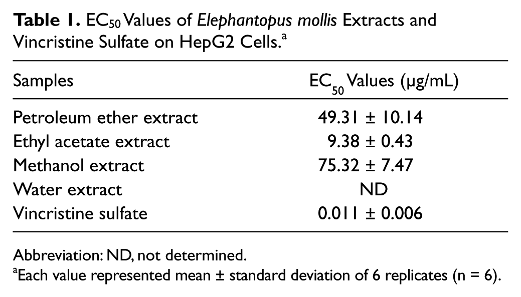
Abbreviation: ND, not determined.
Each value represented mean ± standard deviation of 6 replicates (n = 6).
Cytotoxic Effects of Ethyl Acetate Extract of Elephantopus mollis Against Different Cancer Cells
As the most potent extract, the ethyl acetate extract was selected for further cytotoxicity analysis against different cell lines. A dose-dependent inhibition of cell growth was observed in all cancer cell lines treated with the ethyl acetate extract (Figure 1). Based on the EC50 values, the cytotoxic effects of the ethyl acetate extract against 3 other cell lines, namely, human lung NCI-H23, breast T-47D, and ovarian Caov-3 carcinoma cells, were compared with that of HepG2 (Table 2). The highest cytotoxic activity of the extract was still detected in HepG2 with the lowest EC50 value (9.38 ± 0.43 µg/mL; Table 2). The extract also showed a marked cytotoxic effect on T-47D (EC50 12.57 ± 3.25 µg/mL) and NCI-H23 (EC50 13.17 ± 3.79 µg/mL). Caov-3 (EC50 42.11 ± 9.81 µg/mL) cells were found to be the most resistant cell toward the extract. As indicated in Figure 1, the range of concentrations that significantly inhibited more than 50% of cell growth was found much wider in HepG2 cells (12.5-100 µg/mL) when compared with other cell lines (P < .001). The relative sensitivity of the cell lines against the ethyl acetate extract was in the following order: HepG2 > T-47D > NCI-H23 > Caov-3.

Effects of Elephantopus mollis ethyl acetate extract on different cell lines.
EC50 Values of Elephantopus mollis Ethyl Acetate Extract Against Different Cell Lines.

Each value represented mean ± standard deviation of 6 replicates (n = 6).
Cytotoxic Effects of Ethyl Acetate Extract of Elephantopus mollis at Different Incubation Periods
The cytotoxicity of the ethyl acetate extract against HepG2 cells was further evaluated at 24, 48, and 72 hours of incubation times. Concentration-dependent inhibitions were observed in HepG2 cells when treated with the extract at all incubation intervals (Figure 2). Interestingly, for all time points, this extract displayed more than 75% of growth inhibition, which was relatively unchanged at higher concentrations (50-100 µg/mL). After 24 hours of treatment, the extract significantly inhibited the growth of HepG2 cells at concentrations of 25 µg/mL and above (P < .001). The range of concentrations with significant growth inhibition was increased from 12.5-100 µg/mL at 48 hours to 6.25-100 µg/mL at 72 hours (P < .001). The EC50 values were decreased as the incubation periods were increased from 24 (25.98 ± 3.85 µg/mL) to 48 (19.33 ± 2.20 µg/mL) and 72 hours (9.38 ± 0.43 µg/mL; Table 3).

Effects of Elephantopus mollis ethyl acetate extract on the proliferation of HepG2 cells at 24, 48, and 72 hours of incubation.
EC50 Values of Elephantopus mollis Ethyl Acetate Extract Against HepG2 Cells at 24, 48, and 72 Hours of Incubation.

Each value represented mean ± standard deviation of 6 replicates (n = 6).
Induction of Caspase-3 Gene Expression in HepG2 Cells by Ethyl Acetate Extract of Elephantopus mollis
Semiquantitative RT-PCR was used in this study to determine the expression levels of an apoptosis-related gene, caspase-3, in the ethyl acetate extract-treated HepG2 cells. As shown in Figure 3, the caspase-3 mRNA was induced at the earlier phase of treatment (4 hours) before reaching a peak at 8 hours (nearly 3-fold increase). The level was then gradually decreased to the lower point at 16 hours (1.7-fold increase) and nearly unchanged for the next 8 hours of incubation. When compared with control cells, the increase of caspase-3 mRNA expression was proven to be statistically significant from 4 hours to 24 hours poststimulation (P < .05). Thus, there was a clear indication that the cell death induced by the ethyl acetate extract of E mollis in HepG2 cells was mediated via the activation of caspase-3 gene expression.

(A) Time course mRNA expression of β-actin and caspase-3 in HepG2 cells incubated in the absence or presence of Elephantopus mollis ethyl acetate extract. β-actin was used as an internal standard control for each PCR reaction. (B) Semiquantitative analysis of the caspase-3 mRNA level in HepG2 treated with ethyl acetate extract of Elephantopus mollis using densitometric scanning.
Induction of DNA Fragmentation in HepG2 Cells by Ethyl Acetate Extract of Elephantopus mollis
To confirm the apoptotic death mechanism elicited by the plant ethyl acetate extract, the treated HepG2 cells were further subjected to the DeadEnd Apoptosis Detection System. As depicted in Figure 4B, the extract-treated cells exhibited similar dark brown stained nuclei as in the positive control treated with DNase I (data not shown). Most of the stained nuclei were globular in shape and were equally distributed in the slides. Therefore, DNA fragmentation was clearly observed in the extract-treated cells as compared with the negative control cells (DMSO), which displayed almost all nonstained nuclei cells (Figure 4A). These results strongly denoted that the ethyl acetate extract triggered apoptosis as a main death mechanism in HepG2 cells, with the induction of caspase-3 gene expression and apoptotic DNA fragmentation.

The effect (A) dimethyl sulfoxide 1%(v/v) and (B) ethyl acetate extract of Elephantopus mollis (9.38 µg/mL) on HepG2 cells after 24 hours of treatment and subjected to the DeadEnd Colorimetric Apoptosis Detection System.
Phytochemicals in Ethyl Acetate Extract of Elephantopus mollis
The ethyl acetate extract that was subjected to preliminary phytochemical screening methods revealed the presence of polyphenolics and terpenoids. The tests for alkaloids, tannins, and saponins, however, demonstrated negative results. Two-dimensional chromatographic analysis using cellulose TLC plate (in solvent systems: n-butanol–acetic acid–water; 4:1:5 and 15% acetic acid) indicated the presence of 3 glycosidic flavonols in the extracts. This preliminary identification was made based on the Rf values and colors of the spots under long-wave ultraviolet light. 32 Best separation of the extract with 7 positive spots of terpenoids (detected using vanillin and antimony chloride reagents) was achieved using a mobile phase of hexane–ethyl acetate (7:3) on silica gel TLC plate. Furthermore, 2 red spots on silica gel plate (run in chloroform–ether; 4:1) that was sprayed with concentrated sulfuric acid and heated for 5 minutes at 110°C were detected as sesquiterpene lactones. Based on a visual comparison of the size and intensity of each and every separated spots on the cellulose and silica gel TLC plates, these sesquiterpene lactones could possibly be considered the major constituents in the extract. However, for confirmation, further quantitative analysis of the extract is needed.
Discussion
Elucidation of the molecular basis of the cytotoxic mechanism of plant extract should be considered as a promising approach in improving the cancer treatment by targeting the drug-resistance cells.34,35 In the present study, the cytotoxicity of E mollis extracts and the apoptotic gene expression profile of the most potent extract were assessed by methylene blue assay and RT-PCR analysis, respectively. Among all extracts, the ethyl acetate extract displayed the highest reproducible cytotoxic activity with the lowest EC50 of 9.38 ± 0.43 µg/mL. Based on our preliminary phytochemical analysis, the activity is suggested to be attributed to either the combined effect of polyphenolics and terpenoids or potentiation of sesquiterpene lactones in the extract. Although its EC50 in HepG2 cells was found to be much higher than the positive control, vincristine sulfate (EC50 0.011 ± 0.006 µg/mL), with this EC50, the ethyl acetate extract was still considered cytotoxic based on the criterion set by the NCI. 33 Meanwhile, both petroleum ether and methanol extracts showed low cytotoxic effects on the cell line with higher EC50 (>30 µg/mL). According to Tabopda et al, 16 some sesquiterpene lactones isolated from the methanol extract of this plant were cytotoxic against neuroblastoma B104 cells. However, in this study, the EC50 of the methanol extract was nearly 7-fold higher than the ethyl acetate extract, suggesting the lower susceptibility of the HepG2 cells to the methanol extract. In addition, the water extract was found to be noncytotoxic on this cell line as it displayed low growth inhibition at all concentrations used. Although the water extract is traditionally used for preparation of decoction or infusion of this plant and has been reported to possess anti-inflammatory and antidiabetic effects,36,37 it was found to have a low efficiency in extracting anticancer agents from this plant. Thus, the ethyl acetate extract was selected for further cytotoxicity analysis in different cell lines and incubation times.
Recent surveys have reported that most lung, breast, and ovarian cancer patients are keen to use herbal remedies as alternative medicines.38,39 This is apparently the first study on the effect of the E mollis against lung NCI-H23, breast T-47D, and ovarian Caov-3 cancers. At the highest concentration of the extract (100 µg/mL), the maximum growth inhibition of HepG2 was found to be nearly 90%. This was followed by the NCI-H23 and T-47D cells with 80% of growth inhibition. Caov-3 cells were found to be the most resistant cell toward the extract with the lowest inhibition at all tested concentrations. This clearly indicated the different sensitivity of the extract toward the cell lines with obvious selective toxicity against the HepG2 cells. This might be due to the high affinity of HepG2 cell membrane (lipophilic) to react with the active constituents in the ethyl acetate extract. Likewise, Than et al 15 also reported the different sensitivities of a panel of human cell lines toward elephantopin derivatives and found the melanoma MEXF 394NL cells as the most susceptible to all treated compounds.
Further cytotoxic evaluations not only unveiled the dose-dependent inhibition of the ethyl acetate extract on HepG2 cells but also exerted time-dependent cytotoxicity against this cell line. The extract exhibited early reaction (24 hours time point) on HepG2 cells with pronounced cytotoxic activities. The inhibitory activities were further increased when incubation times were increased to 48 and 72 hours, which reduced nearly 2.0-fold and 2.5-fold of EC50 values than 24 hours, respectively. Therefore, the longer duration of the treatment may prolong the exposure of HepG2 cells to the ethyl acetate extract and thus may require lower dose to abrogate 50% of the cell growth, suggesting the occurrence of a cumulative cytotoxic effect of the extract. The lowest EC50 concentration that was obtained after 72 hours of treatment was used for further investigation on the molecular mechanism of cell death elicited by the extract.
At molecular level, the RT-PCR analysis exhibited a differential mRNA expression levels of caspase-3 in HepG2 cells exposed with the ethyl acetate extract of E mollis over 24 hours. Among the treatment periods, the peak expression of this gene was significantly observed at 8 hours (P < .05), indicating caspase-3 was highly induced at early phase of the treatment of this plant extract. The activation of caspase-3 was required to activate an endonuclease caspase–activated DNase (CAD) that is responsible for specifically cleaving DNA at internucleosomal chromatin regions during apoptosis. 40 This has subsequently led to the apoptotic morphological changes and the fragmentation of DNA. 41 Further DNA fragmentation analysis that was performed in this study verified the occurrence of apoptotic morphological and biochemical alterations of the ethyl acetate extract–treated HepG2 cells. This clearly indicated the role of caspase-3 in executing DNA fragmentation. The fragmented DNA was catalyzed by TdT enzyme to be incorporated with biotinylated dUTP and was then detected by chromogen and peroxidase substrates. 42 Xu et al 22 has identified these apoptotic features in the human HeLa cervical carcinoma cells treated with the deoxyelephantopin derived from this plant. Furthermore, the consistent induction of caspase-3 (around 1.7-fold increase) at the late periods of treatment (16-24 hours) could be due to its involvement in the late phase of the apoptotic pathway as a downstream executor, which is activated after a series of upstream cellular processes during apoptosis. 43 The results strongly postulated the induction of caspase-3-dependent apoptosis pathway by the ethyl acetate extract.
Conclusions
In conclusion, the anticancer property of the ethyl acetate extract of E mollis has been partially validated by verifying its remarkable cytotoxic activities against the HepG2 cells in concentration-dependent and time-dependent manner via an apoptotic cell death. The apoptotic death mechanism was mediated by the caspase-3-dependent pathway. This clearly speculated that the induction caspase-3 may contribute to the cytotoxic action of the plant extract on HepG2 cells. Our findings provide preliminary experimental evidence for further in-depth study on E mollis.
Footnotes
Declaration of Conflicting Interests
The author(s) declared no potential conflicts of interest with respect to the research, authorship, and/or publication of this article.
Funding
The author(s) disclosed receipt of the following financial support for the research, authorship, and/or publication of this article:
The authors would like to acknowledge the Ministry of Agriculture and Agro-based Industry Malaysia (EScience Fund: 05-01-05-SF1001) and Universiti Sains Malaysia (Research University Grant: 1001/PBIOLOGI/811123) for the financial support.