
Abstract
Objective. To observe the effects of Qingyihuaji formula (QYHJ) on the progression of liver metastases from human pancreatic cancer and to detect the expression changes of some biological factors associated with angiogenesis and metastasis during the development of advanced pancreatic cancer. Methods. Nude mice were inoculated intrasplenically with human pancreatic cancer cell line SW1990 and then randomly assigned into 4 groups: a control group and groups QYHJ-A, QYHJ-B, and QYHJ-C. Following this, the mice were treated with or without QYHJ formula for 4 weeks and were sacrificed at the end of the sixth week. The changes in body weight were observed, followed by the livers being excised and weighed. Then, both the numbers and the volume of metastatic nodules per liver were evaluated. Subsequently, the expressions of MMPs, VEGF, and Cyr61 in the tissue of liver metastases were detected by reverse transcription polymerase chain reaction, immunohistochemistry, or Western blot. Finally, the correlation was evaluated between the expressions of the factors associated with metastasis and the growth of liver metastasis. Results. Liver metastases were identified in 11 of 15 mice (73%) in the control group, 9 of 15 mice (60%) in group QYHJ-A, 6 of 14 mice (43%) in group QYHJ-B, and 8 of 14 mice (57%) in group QYHJ-C both the number and the volume of metastatic nodules per liver same as the ratio of liver-body weight in QYHJ groups were significantly less than the controlled group (P < 0.05). The expressions of Cyr61, MMP-2, and VEGF at the levels of mRNA and protein were decreased in the QYHJ groups when compared with the control, as confirmed by immunohistochemistry detection (P < .05). However, no significant difference was observed in the mRNA expression of MMP-1 and MMP-9 between the QYHJ groups and the control group (P > .05). Regression analysis indicated that QYHJ possessed an evident inhibition against the progression of liver metastasis by downregulating the expression of VEGF and Cyr61 rather than MMP-2. Conclusions. The QYHJ formula exerted an inhibitory effect on the growth of liver metastasis from pancreatic cancer, perhaps by targeting VEGF and Cyr61 to some extent.
Introduction
Pancreatic cancer, a common malignant tumor in the digestive system, has shown a significant increasing trend in incidence in recent years. The prognosis of advanced pancreatic cancer is poor, with a 5-year survival rate less than 5%, despite advances in medical therapy and surgical techniques. 1 One of the major features of pancreatic cancer is its early systemic dissemination, which leads to the fact that 80% of patients diagnosed with this disease, among whom the incidence of liver metastasis is more than 50%, cannot receive curative treatment. 2 At present, effective medication and drugs are lacking for the treatment of pancreatic cancer with liver metastasis because the metastases resist radiation and most chemotherapeutic agents. Hence, there is an urgent need to find better modalities for the treatment and prevention of pancreatic cancer, especially for those with liver metastasis. In addition, the molecular mechanisms contributing to distant metastasis of pancreatic cancer are not well understood, but it is well known that the hematogenous remote metastasis involves a multistep process. 3 In the metastatic process, extracellular matrix degradation and angiogenesis are thought to play important roles in the metastasis of cancer cells and their growth in distant locations. The formation of tumor is closely related to angiogenesis, which is devoted to vasculature supplying oxygen and nutrients, and vascular endothelial growth factor (VEGF) is particularly important among the factors involved in angiogenesis, as it clearly acts on endothelial cells in a direct manner. 4 VEGF maintains not only its position as the most critical driver of vascular formation but also enhances the invasion and metastasis of tumor cells.5,6 Matrix metalloproteinases (MMPs), a type of proteolytic enzyme, appear to be implicated in the vascularization and metastasis of malignant tumors including pancreatic cancer.7,8 Furthermore, elevated expression of MMPs, including MMP-2 and MMP-9, was found in pancreatic cancer. 9 Some studies suggested that MMPs also enhanced the angiogenesis of tumor by triggering the angiogenic switch. 10 Cysteine-rich protein 61 (Cyr61), belonging to growth factor–inducible, immediate-early genes, stimulated the progression of tumor in breast cancer, pancreatic cancer, and glioma.11,12 It was reported that the expression of Cyr61 increased 6-fold in the peritoneal metastases, as high as that in primary pancreatic cancer. 13 In addition, Cyr61 was overexpressed in invasive and metastatic human breast cancer cells and tumor biopsies. 14 It has been reported that Cyr61 served to mediate cell adhesion, stimulate chemostasis, augment growth factor–induced DNA synthesis, foster cell survival, and enhance angiogenesis.15-18
Qingyihuaji formula (QYHJ), consisting of traditional Chinese herbs, has been used as an integrative agent in the clinical treatment of advanced pancreatic cancers, especially those with liver metastasis, for many years in the Cancer Hospital, Fudan University, Shanghai, China. Prior clinical research indicated that QYHJ treatment combined with conventional Western medicine had some advantages such as median survival prolongation in treating patients with pancreatic cancer,19,20 and animal studies showed that QYHJ could inhibit the growth of subcutaneously transplanted pancreatic tumors in nude mice. 21 Additionally, a study of advanced pancreatic cancer with liver metastases showed a response advantage of QYHJ treatment. 22
However, it has not been confirmed in animal models whether QYHJ could execute a suppressive effect on the development of liver metastasis from human pancreatic cancer, and it has not been established how QYHJ functions on the inhibition of liver metastasis either. Therefore, in this primary study, we established an intrasplenically inoculated xenograft animal model in nude mice with human pancreatic cancer cell line SW1990 to explore the impact of QYHJ treatment on liver metastasis from advanced pancreatic cancer. In addition, the expressions of MMPs, VEGF, and Cyr61 related to the invasion and metastasis of pancreatic cancer were detected. Also, an evaluation was performed of correlation between the progression of liver metastasis and the expression of those factors related with metastasis after QYHJ treatment.
Materials and Methods
Cell Lines and Mice
Human pancreatic cancer cell line SW1990 was obtained from the American Type Culture Collection and grown in complete growth medium as recommended by the manufacturer. The cultured cells were maintained in a humidified 5% CO2 atmosphere at 37°C. Female BALB/c-nu/nu nude mice (18-22 g) were obtained from Shanghai Laboratory Animal Center, Chinese Academy of Sciences, Shanghai, China, and housed in laminar flow cabinets under specific pathogen-free conditions with food and water ad libitum. The study protocol was approved by the Shanghai Medical Experimental Animal Care Committee.
Drugs and Reagents
QYHJ was made from Amorphophallus konjac as the main material and Hedyotis diffusa, Scutellaria barbata, Semen coicis, Fiveleaf gynostemma herb, and Amomum kravanh as the minor material, in the ratio of 12:6:6:6:4:1, according to the theories of traditional Chinese medicine. Granules for each herb were produced by Jiangyin Tianjiang Pharmaceutical Co, China. The final decoction of QYHJ was prepared by dissolving herb granules into distilled water together to the required concentration and treating the herb mixture by high-temperature sterilization in the Pharmaceutical Department of Cancer Hospital, Fudan University, Shanghai, China. The daily dosage of nude mice was dependent on routine dosage(4 g/kg/d) in our department and was calculated according to the following human–mouse transfer formula: Db = Da × Rab, where Da, Db, and Rab represent human dosage (g/kg), mouse dosage (g/kg), and conversion coefficient, respectively. RPMI-1640 and fetal bovine serum were purchased from Gibco (Carlsbad, CA), VEGF monoclonal antibody from BD Pharmingen (San Jose, CA), and Cyr61 polyclonal antibody and MMP-2 monoclonal antibody from Santa Cruz Biotechnology, Inc (Santa Cruz, CA).
Establishment of Xenograft Model in Nude Mice
SW1990 cells (6 × 10 6 in 0.2 mL) in logarithmic phase were inoculated into mouse right axillae. Mice were sacrificed and then the subcutaneous tumor was removed after about 3 weeks, when the tumor grew approximately 1000 mm3 in volume. The tumor tissue was sheared and then ground into cell suspensions of 3 × 10 7 /mL final concentration in serum-free phosphate buffered saline, with cell viability greater than 95% determined by trypan blue exclusion. The mice were anesthetized with 10% chloral hydrate and the spleen was exteriorized through a left flank incision. A total of 3 × 10 6 cells in 100 µL of suspension were slowly injected into the splenic pulp by an inoculator needle. After hemostasis by compression, the spleen was relocated into the abdominal cavity, and the peritoneum and skin were closed. All animals tolerated the procedure well.
Administration
A total of 58 mice with intrasplenic inoculation were randomly divided into 4 groups: control group (15 mice) administered with normal saline and QYHJ-A group (15 mice), QYHJ-B group (14 mice), and QYHJ-C group (14 mice) administered with QYHJ formula at a dose of 18 g/kg body weight, 36 g/kg body weight, and 72 g/kg body weight daily, respectively. One week after randomization, all mice received oral gavages of 0.4 mL each day for 4 weeks (days 7-35) and were weighed each week. All experiments on mice were conducted in accordance with the guidelines of the National Institutes of Health for the Care and Use of Laboratory Animals.
Assessment of Liver Metastasis In Vivo
Mice were sacrificed 6 weeks after inoculation. The livers were excised and weighed for assessment of the ratio of liver to body weight (g/100g), and the metastases on the surface of liver were enumerated by a dissecting microscope. To estimate the volume of liver tumors, the diameter of each metastatic nodule was measured to the nearest millimeter by Vernier caliper, and the volume of each nodule was calculated by assuming it to be a sphere. 23 The sum of the volumes of all tumors in each liver was determined. Then, a portion of liver nodules was placed in liquid nitrogen for polymerase chain reaction (PCR) and Western blot; other liver metastases were preserved in 10% neutral buffered formalin for immunohistochemistry.
Reverse Transcription Polymerase Chain Reaction (RT-PCR) Analysis
Total RNA was extracted from liver nodules in liquid nitrogen with TRIzol Reagent (Invitrogen, San Diego, CA), and a total of 2 µg of the purified total RNA was used for RT-PCR, performed according to the manufacturer’s instructions (MBI Fermentas, Vilnius, Lithuania). The glyceraldehyde-3-phosphate dehydrogenase (GAPDH) gene was chosen as an internal control. We used Quantity-One Gel Imaging software (BIO-RAD) to perform quantitative RT-PCR analyses of mRNA levels relative to GAPDH. All primers were synthesized at Shanghai Sangon Biological Engineering & Technology Services Co, Shanghai, China (Table 1).
Primer Sequences Used for PCR Analysis

Western Blot Analysis
A 200-mg sample of the liver nodules from each group was dissected and immediately lysed in ice-cold NP40 lysis buffer with protease inhibitor (0.5 mM phenylmethylsulfonyl fluoride). Insoluble debris was removed by centrifugation at top speed for 15 minutes. Samples were vortexed, incubated on ice for 30 minutes, centrifuged again, and the supernatants were stored at −80°C. Equal amounts of supernatant protein (50 µg) of liver metastasis were denatured by boiling for 5 minutes in sodium dodecyl sulfate (SDS) sample buffer, separated by 10% SDS-polyacrylate gel electrophoresis, and transferred to a polyvinylidene fluoride membrane for Western blot analysis. Blot quantitative analysis was done with ImageJ software supported by the NIH. Protein levels were calculated relative to β-actin.
Immunohistochemical Analysis
A portion of liver metastases were placed in 10% buffered formalin, dehydrated, and embedded in paraffin. Dewaxed paraffin-embedded sections measuring 4 µm were immunostained according to the protocol of a streptavidin–peroxidase (SP) kit (ZYMED, Carlsbad, CA). Immunoreactions were visualized with HRP conjugated goat antirabbit/mouse IgG and developed in diaminobenzidine. The sections were counterstained with hematoxylin. The positive sections known were used as positive controls, and the negative control was obtained by replacing the primary antibody with phosphate-buffered saline, pH 7.4, of the same species. The presence of brown staining, more apparent in cytoplasm than in nonspecific dyeing background, was considered as a positive identification. The slides in 10 randomly selected high-power fields (400×) were examined in a blinded fashion with light microscopy by 2 independent researchers.24,25 ImageJ software (NIH) was used to analyze the optical density of positively stained cells on the slides.
Statistical Analysis
The data shown here were representative of the identical results obtained from 2 independent experiments and were expressed as mean ± standard deviation. The data of small samples were evaluated by logarithmic transformation. Statistical analyses were performed with Student’s t test for unpaired values, Mann–Whitney’s U test, or multiple linear regression. The level of significance was set at P < .05. The statistical analysis was performed with GraphPad Prism software, version 5.01 (San Diego, CA), except for the multiple linear regression analysis performed with SPSS software, version 13.0.
Results
Effect of QYHJ on the Body Weight in Liver Metastasis–Bearing Mice
Two weeks after treatment, the body weights were more improved in the treated mice than in the controls. At the end of the sixth week, some mice in each group appeared angular and had obviously lost weight similar to cachexia caused by progressive tumor. At that time, mice were weighed and then sacrificed humanely. We found that all the mice gained some weight after treatment with QYHJ when compared with pretreatment and that the mean bodyweight was more improved in the treated mice than in the controls (P < .05, Figure1A). This showed that QYHJ could help slow down the loss of body weight in the experimental animals, as was coincident with the clinical outcomes of QYHJ.

The effect of QYHJ on the weight of body and liver in each group
Effects of QYHJ on the Progression of Liver Metastases
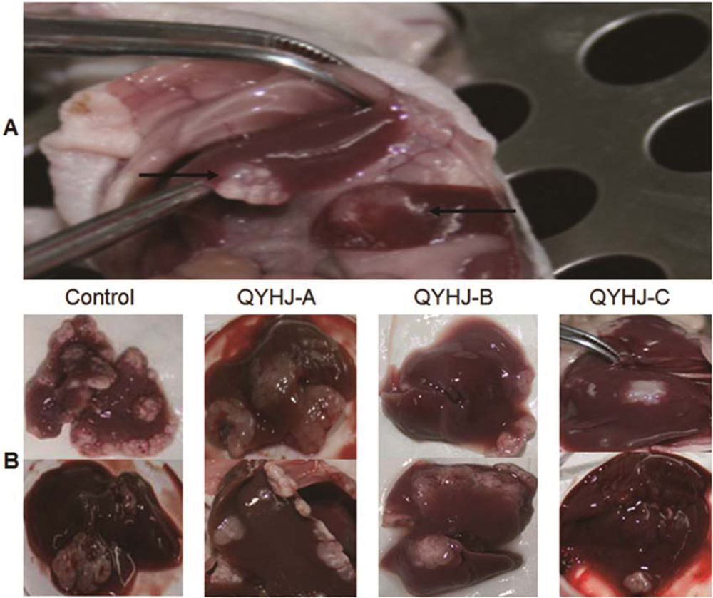
In the present investigation, liver metastases were identified in 11 of 15 mice (73%) in the control group, 9 of 15 (60%) in group QYHJ-A, 6 of 14 (43%) in group QYHJ-B, and 8 of 14 (57%) in group QYHJ-C, which indicated a downward trend in incidence of liver metastasis after QYHJ treatment, though of no significant difference statistically. To observe the impact of QYHJ on liver metastasis, we chose to use the ratio of liver to body weight (g/100 g) and both the numbers and the total volume of metastatic nodules in liver as assessment standards. The ratio of liver to body weight (g/100 g) was greater in the control group versus the treated groups (4.72 ± 0.65 vs 3.71 ± 0.32, 3.51 ± 0.35, and 3.53 ± 0.20 for groups QYHJ-A, -B, and -C, respectively; P < .05; Figure 1B). The appearance of livers of representative animals is shown in Figure 2. The number of metastatic nodules per liver in each group ranged from 4 to 64, 4 to 35, 1 to 22, and 3 to 27 in the control group and groups QYHJ-A, -B, and -C, respectively. The mean volume of metastatic nodules per liver was obviously different (2.746 ± 0.720 cm3, 2.070 ± 0.680 cm3, 1.493 ± 0.501 cm3, 1.495 ± 0.487 cm3 in the control group and groups QYHJ-A, -B, and -C, respectively; data were obtained after logarithmic transformation). When the numbers and volume of liver metastases were analyzed, there was a dramatic reduction in the QYHJ groups, compared with the control, which made the influence of QYHJ treatment even more evident (P < .05; see Table 2 and Figure 3). The reduction in the numbers and size of liver nodules as well as the ratio of liver to body weight showed the suppression of QYHJ on progression of liver metastasis.
The liver metastases that occurred were identified in 11 of 15 mice (73%) in the control group, 9 of 15 mice (60%) in group QYHJ-A, 6 of 14 mice (43%) in group QYHJ-B, and 8 of 14 mice (57%) in group QYHJ-C, respectively
The Effect of QYHJ Treatment on Athymic Mice Bearing Experimental Hepatic Metastases From Human Pancreatic Carcinoma

P value by Mann–Whitney U test.
Mean ± SD. Tumor volume approximately accorded with normal distribution by logarithmic transformation. P value, estimated between QYHJ groups and control group, <.05, was significant.
P value by Student’s t test.

In vivo the antimetastasis effect of different doses of QYHJ compared with the control (untreated) in the xenograft model of SW1990 pancreatic tumor. A, The number of metastasis nodules per liver. B, The tumor volume per liver in each group measured on the 42nd day (tumor volumes, mean ± SD)
mRNA Expression of MMPs, VEGF, and Cyr61 of Liver Metastasis After QYHJ Treatment
In this experiment, the expression of key genes closely related to invasion and metastasis was first determined by RT-PCR analysis. As shown in Figure 4, RT-PCR analysis revealed that the mRNA expression of VEGF, Cyr61, and MMP-2 in liver metastasis decreased significantly in the QYHJ groups when compared with the control (Figure 4A, C, D, F; P < .05), whereas there were no distinct changes of MMP-1 and MMP-9 (Figure 4A, B, E; P > .05). This suggests that QYHJ could inhibit the expression of VEGF, Cyr61, and MMP-2 in liver metastasis. In addition, we observed that the dose of QYHJ-B and QYHI-C showed more inhibitory effect than QYHJ-A (P < .05, data not shown).

RT-PCR analysis to detect the mRNA expression of VEGF, Cyr61, MMP-1, MMP-2, and MMP-9 in the liver metastasis from pancreatic cancer cell line SW1990
Protein Expression of MMP-2, VEGF, and Cyr61 of Liver Metastasis After QYHJ Treatment
Subsequently, to further examine the protein expression of VEGF, Cyr61, and MMP-2 in liver metastasis, we performed Western blotting. Here, we applied the ImageJ software, version 1.42, to assess the densitometry of protein bands. The analysis showed that the protein expression of VEGF, Cyr61, and MMP-2 was significantly downregulated in the QYHJ groups, compared with the control (Figure 5A-D; P < .05) and this reduction was more remarkable in groups QYHJ-B and QYHJ-C than in group QYHJ-A (data not shown), which showed the same tendency as the results of the RT-PCR analysis.

Western blotting analysis of the lysates of liver metastasis to determine the expressions of metastasis-related proteins
QYHJ Could Reduce the Positive Immunostaining of MMP-2, VEGF, and Cyr61 of Liver Metastasis Tissue
To investigate whether the expressions of VEGF, Cyr61, and MMP-2 at the tissue level were in accordance with those at the level of mRNA and protein, immunohistochemical analysis was performed (Figure 6A and B). ImageJ software, version 1.42, was used to measure the optical density of the staining specimens as a quantitative analysis. Subtracting the nonwhite background color, the hematoxylin component was separated from the DAB component using color deconvolution. 26 For each specimen, the lowest and the highest mean optical density (MOD) values measured from 8 manually identified positively stained cells selected from the DAB component (with visual comparison with the original image) were used to set the threshold manually for whole sample staining density quantification (Figure 6C). The data of the measured MOD were compared statistically with the expression of VEGF, Cyr61, and MMP-2 between QYHJ groups and the control. The analysis revealed that liver metastases in treated mice showed less immunohistochemical staining of VEGF, Cyr61, and MMP-2 expression when compared with the control (Figure 6D; P < .05). IHC analysis further confirmed the outcomes of RT-PCR and Western blotting analysis that QYHJ could reduce the expression of VEGF, Cyr61, and MMP-2 in liver metastasis.

Immunohistochemical staining for the expression of VEGF, Cyr61, and MMP-2 in liver metastases tissues
Immunostaining Expression of MMP-2, VEGF, and Cyr61 Correlate With Development of Liver Metastasis
To assess whether the decreased expression of VEGF, Cyr61, and MMP-2 led to the reduction of the metastasis volume after QYHJ treatment, the correlation was analyzed between variables. We observed a significant positive correlation between the expression of VEGF, Cyr61, and MMP-2 and the volumes of liver metastases (Figure 7, P < .001). We then performed further regression analysis of these data and found that VEGF and Cyr61 played a direct role in the growth of liver metastases in this experiment (P < .005), whereas MMP-2 exhibited little effect (B = .111, P = .88), as shown in Table 3. This result suggested that QYHJ formula may have targeted VEGF and Cyr61 to inhibit the growth of liver metastases through some pharmacological effects, both complex and extensive.

Correlation analysis between the volume of liver metastases and the positive staining of VEGF, Cyr61, and MMP-2 (n = 34)
The Volume of Liver Metastases Responding to the Expression of VEGF, Cyr61, and MMP-2 in Liver Metastases Tissue With QYHJ Treatment (n = 34) a
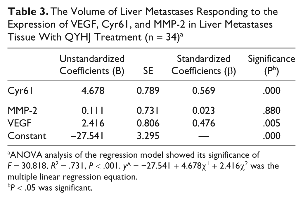
ANOVA analysis of the regression model showed its significance of F = 30.818, R2 = .731, P < .001. y^ = −27.541 + 4.678χ1 + 2.416χ2 was the multiple linear regression equation.
P < .05 was significant.
Discussion
The poor prognosis of pancreatic cancer is attributable to its local invasion, early metastases, and poor response to chemotherapy. 27 Gemcitabine remains the best chemotherapeutic agent available for the treatment of advanced pancreatic cancer, though its treatment results in an objective tumor response rate of <10%. 28 Surgical resection remains the only definitive approach for long-term survival, but only 10% to 20% of patients with resectable pancreatic adenocarcinoma benefit from it. 1 Despite recent advances in diagnostic and surgical procedures, pancreatic cancer is still one of the worst prognoses among cancers of the digestive organs because of its high rate of liver metastasis and local recurrence. 29 This reflects the inability of current systemic therapy to prevent or treat pancreatic cancer metastases and underscores the need for novel therapeutic strategies and more research about metastatic mechanisms.
Traditional Chinese medicine (TCM) as a medicine not only empirical but also clinical, widely used for cancer treatment for thousands of years, has been applied in treatment of advanced cancers in combination with modern Western medicine in recent decades in China. Our retrospective clinical studies have shown the efficiency of QYHJ as a TCM formula in the treatment of advanced pancreatic cancer with liver metastasis. Based on our clinical evidence, we speculated that QYHJ might inhibit the progression of advanced pancreatic cancer with liver metastasis through interfering in the metastatic process. In an effort to better observe the inhibitory effect of QYHJ treatment on liver metastasis from pancreatic cancer, in this study we chose to use a model of experimental liver metastasis in the athymic mouse. In addition, human pancreatic cancer cell line SW1990, well known for its highly metastatic potential, 30 was used to fit much with this experiment.
Emaciation is one of the features of patients with advanced pancreatic cancer and is commonly caused by this disease-derived obstructive jaundice, metastasis, and malnutrition. In this experiment, we found that the weight of QYHJ-treated mice improved relative to the control. This result indicated that QYHJ may contribute to ameliorating the survival quality of patients with advanced pancreatic cancer, which was in accordance with our clinical observation that QYHJ formula greatly improved appetite. In particular, QYHJ treatment displayed its inhibition against the growth and progression of liver metastasis, as the data obtained clearly showed that QYHJ could reduce the number of metastatic lesions in liver and diminish the size of metastases in liver.
To further explore the mechanism of the antimetastasis effect of QYHJ, we carried out a preliminary detection of mRNA expression of MMPs, VEGF, and Cyr61, closely associated to metastasis, by RT-PCR analysis. The results showed that QYHJ distinctly downregulated the mRNA expression of MMP-2, VEGF, and Cyr61, yet it exerted insignificant effect on MMP-1 and MMP-9. Then, we further examined the expression of VEGF, Cyr61, and MMP-2 by Western blot and IHC detection to confirm the results of RT-PCR analysis. The downregulated expression of these factors showed the wide pharmacological effects of this formula after the intervention of QYHJ. Ultimately, simple correlation and multiple linear regression analysis based on the liver metastasis volume and the expression of VEGF, Cyr61, and MMP-2 were carried out. These analyses revealed that the expression of MMP-2 was positively correlated to the volume of liver metastases, yet without depending on each other; however, we observed the dependent relationship between the volume of liver metastasis and the expression of VEGF and Cyr61. This experiment indicated that QYHJ might inhibit the growth and progression of pancreatic cancer with liver metastasis by targeting VEGF and Cyr61.
It is known that there is an unclarified and complex network of interaction among many factors such as VEGF, MMPs, bFGF, and TGF-β1 in the process of tumorigenesis, progression, invasion, and metastasis of malignant tumor. It was reported that there was a positive link between VEGF and MMP-2 in some cancers.31,32 In the intervening process, QYHJ may have indirectly reduced the expression of MMP-2 through regulating other factors such as VEGF and Cyr61.
Recent evidence hinted that tumors might develop resistance to VEGF-targeted therapy by switching to other signaling pathways.33,34 This information suggested that it might be crucial to develop potential multitargeted anticancer therapy. The expression changes of MMPs, VEGF, and Cyr61 after QYHJ treatment displayed the anticancer complexity of the Chinese medicinal preparation. This experiment also indicated that QYHJ had the potential effect of multitargeted therapy through inhibiting the expression of VEGF and Cyr61. However, this was only a preliminary study, leaving more space for further research in later experiments.
In conclusion, QYHJ displayed its preventive effect against the progression of pancreatic cancer with liver metastasis by targeting VEGF and Cyr61. The present findings support the clinical effectiveness of QYHJ in the therapy of advanced pancreatic cancer. Therefore, this study may help better understand the clinical effects of QYHJ, and the further exploration of QYHJ may offer a new insight to develop novel targets for therapeutic intervention in advanced pancreatic cancer.
Footnotes
The author(s) declared no potential conflicts of interests with respect to the authorship and/or publication of this article.
The author(s) disclosed receipt of the following financial support for the research and/or authorship of this article:
This study was supported by the ‘Climbing Up’ Project of the Shanghai Municipal Commission for Science and Technology, Shanghai, China (GJ-KW0601), and Shanghai Nature Science Fund, Shanghai, China (09ZR1406800).