
Abstract
Early diagnosis of Alzheimer’s disease (AD) allows individuals and their health managers to manage healthier medication. We proposed an approach for classification of AD stages, with respect to principal component analysis (PCA)-based algorithm. The PCA has been extensively applied as the most auspicious face-recognition algorithm. For the proposed algorithm, 100 images of 10 children were transformed for feature extraction and covariance matrix was constructed to obtain eigenvalues. The eigenvector provided a useful framework for face recognition. For the classification of AD stages, magnetic resonance imaging (MRI) and functional magnetic resonance imaging (fMRI) data were obtained from Alzheimer’s Disease Neuroimaging Initiative database. Hippocampus is one of the most affected regions by AD. Thus, we selected clusters of voxels from the “hippocampus” of AD screening stage (mild cognitive impairment), AD stage 1, AD stage 2, and AD stage 3. By using eigenvectors corresponding to maximum eigenvalues of fMRI data, the purposed algorithm classified the voxels of AD stages effectively.
Introduction
Faces are complex and multidimensional and have meaningful visual stimuli. Developing a system for face recognition is fairly problematic because of related shape of faces collective with several variations between images of the identical face. 1 Nawaf et al developed a method for face recognition based on modular principal component analysis (PCA). 2 Algorithms based on PCA are standard because of simplicity in implementation and have turned into the benchmark of algorithms. 3 Moon and Phillips established PCA-based algorithm comprises of normalization, PCA projection, and recognition modules. 3 The combination of optic flow and PCA can be used for the face recognition. 4 An algorithm was developed based on Hidden Markov model and optic flow for face recognition. 5 Cottrell and Metcalfe applied backpropagation neural network and PCA for recognition of facial expressions from still images. 6
Alzheimer’s disease (AD) is utmost numerous neurodegenerative disorder and emergent health problem. 7 Primary and accurate diagnosis/identification of AD is challenging but is decisive for the future treatments. 7 A method was purposed to classify AD (or mild cognitive impairment [MCI]) and healthy controls using kernel combination method by combining the magnetic resonance imaging (MRI), fluorodeoxyglucose positron emission tomography, and cerebrospinal fluid biomarkers. 8 The predetection of AD stages is perceived as significant because treatment might be more effectual if detected as timely as possible. 9 The MRI’s high-dimensional mode classification identifies patterns of brain structure that characterize MCI, which is usually the precursor to AD. 10 For the AD, a molecular test can lead for the better cure and rehabilitations, and 18 signaling proteins founded that could be used for the discrimination of AD and healthy controls blinded samples. 11
Many MRI studies in AD stages have relied on measurements of volumes of particular brain regions, specifically the hippocampus and entorhinal cortex which shows histopathological changes at initial stages of AD. 9
Hippocampus is one of the primary regions of brain that is affected in AD, leading to loss of memory and confusion. 12 The core function of hippocampus is primarily related with memory, and other major functions are spatial navigation, spatial memory, and behavioral inhibition. 12 Impairment to the hippocampus can lead to loss of memory and difficulty in building new memories. This study used 2 data sets, first had 100 images of 10 children (10 × 10 = 100) with different facial expressions for face recognition while other had functional magnetic resonance imaging (fMRI) data of AD. The objective of this study was classification of faces (face recognition) and classification of AD stages with PCA-based algorithm. For classification of AD stages, brain’s region “hippocampus,” located in medial temporal, was analyzed.
Methodology
Principal component analysis is a well-known multivariate technique and well-recognized method for dimensionality reduction and feature extraction. 13 In PCA, we transform high-dimensional data into low-dimensional space. By reducing the degree of freedom and the complexity of space and time, PCA signifies data into a space that best expresses the variation. The PCA calculates the PCs that are base vectors in decreasing order of variability. 14
The method involves covariance matrix of input data, its eigenvectors and corresponding eigenvalues, and the projected data in new reductional space, defined by PCs. The first eigenvalue gives the direction of largest variance of data. Algebraic definition and process of PCs is given below.
Step 1: Given a sample of n observations on a vector of p variables. 14

Calculate the covariance matrix of data such as: 13 -15
where X is the data matrix and m represents the mean vector. S is symmetric matrix.
Step 2: Define the first PC by the linear combination, 14

where the vector
Here, the vector
Step 3: To find

So, λ1 is the largest eigenvalue of S. The first PC,

The kth largest eigenvalue of S is the variance of kth PC. The kth PC
Step 4: Arrange the eigenvectors

where each row of
Analysis and Results
The PCA has been frequently used for problems related to faces recognition. 15 Ease and rapidity of PCA algorithm in face recognition is one advantage over other face-recognition algorithms. 15 In computation of PCA, there were 100 images of 10 children (10 images of each child). The face database is shown in Figure 1.

Face database of children images.
Each grayscale image is converted into

Mean face image of all images, except the test image.
Next, subtract the mean image from the data matrix and calculate the covariance/correlation matrix to apply PCA algorithm. As PCA used the eigenvalues and eigenvectors for recoginition, calculate the eigenvalues and eigenvectors/eigenfaces of covarinace matrix. Based on 10 largest eigenvalues, pick the eigenvector and calculate signature for each image. For the recognition of test image, subtract the mean image from test image and compare it with feature data matrix, by using PCA. The proposed algorithm will exactly match the test image, as shown in Figure 3.
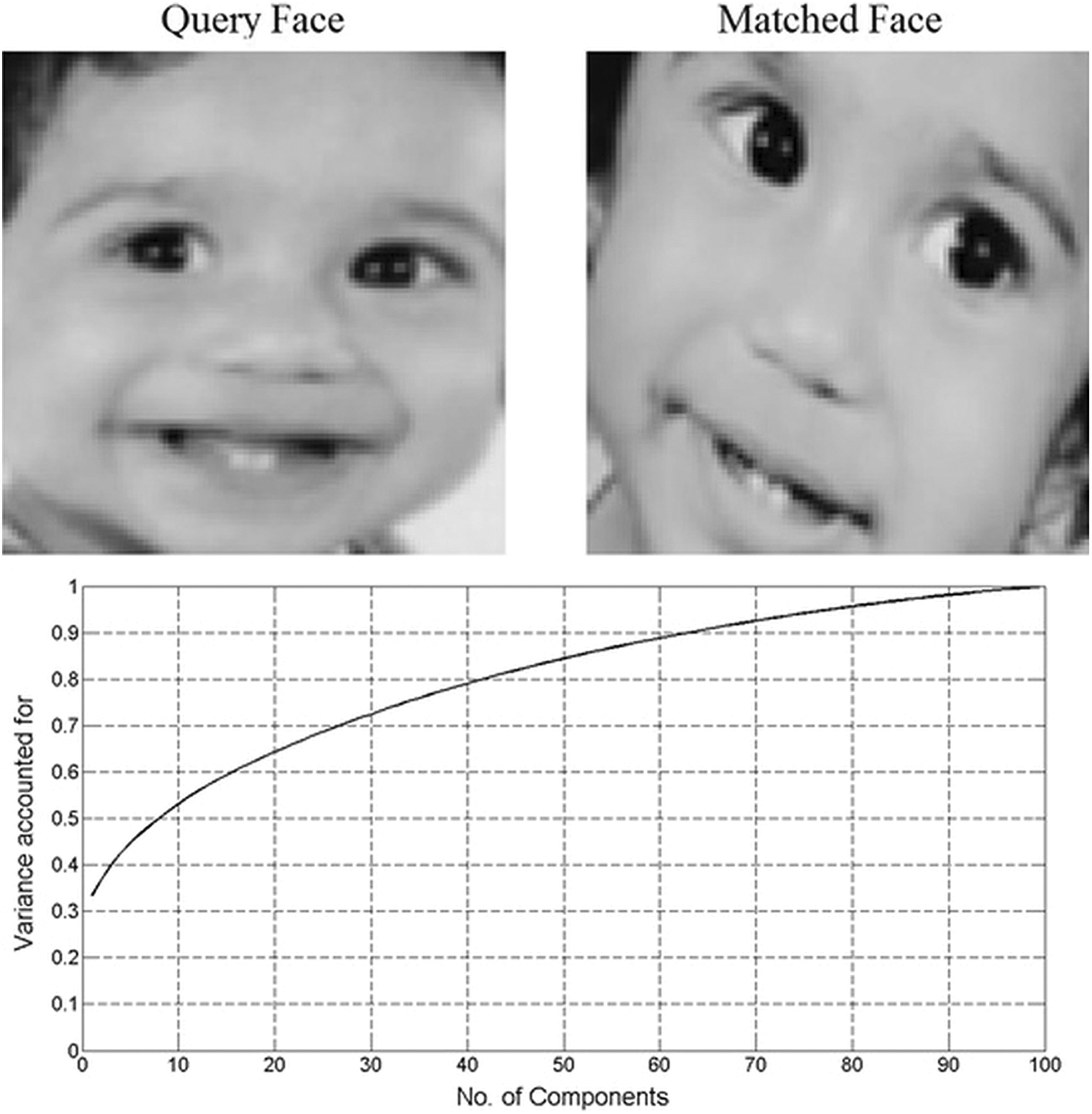
Test face (query face) and matched face from the database and number of PCAs used for best classification. PCA indicates principal component analysis.
We analyzed real fMRI data that were obtained from the Alzheimer’s Disease Neuroimaging Initiative (ADNI) database (http://adni.loni.usc.edu). Investigators within ADNI contribute to the design and implementation of ADNI and provide the data but did not participate in analysis or write-up of this article. A complete list of ADNI investigators can be found at http://adni.loni.usc.edu/wp-content/uploads/how_to_apply/ADNI_Acknowledgement_List.pdf.
For preprocessing and analysis of fMRI AD data, MATLAB (version 2015, Statistics Toolbox: MathWorks, Massachusetts), Statistical Paramteric Mapping (SPM), version 2016 and MRIcron, version 2016, were used. The SPM involves 2 steps, preprocessing and model specification. During preprocessing of functional data, a low-pass filter option was applied to remove respiratory and cardiac noise effects, and generalized linear model was applied to analyze signal of subject. After preprocessing, to check the activation of fMRI data in SPM, 100 smoothed, realigned, and normalized images in model specification were used. Model specification involved some specifications or conditions about fMRI experiment to estimate the βs of fMRI brain data. In the next step, that is, results, because we used the resting-state fMRI data and there was no specific task-related condition in fMRI experiment, so for checking the activation of brain, we assumed dummy contrast, that is, [1 0]. Activations in brain regions during resting-state experiment are shown in Figure 4.
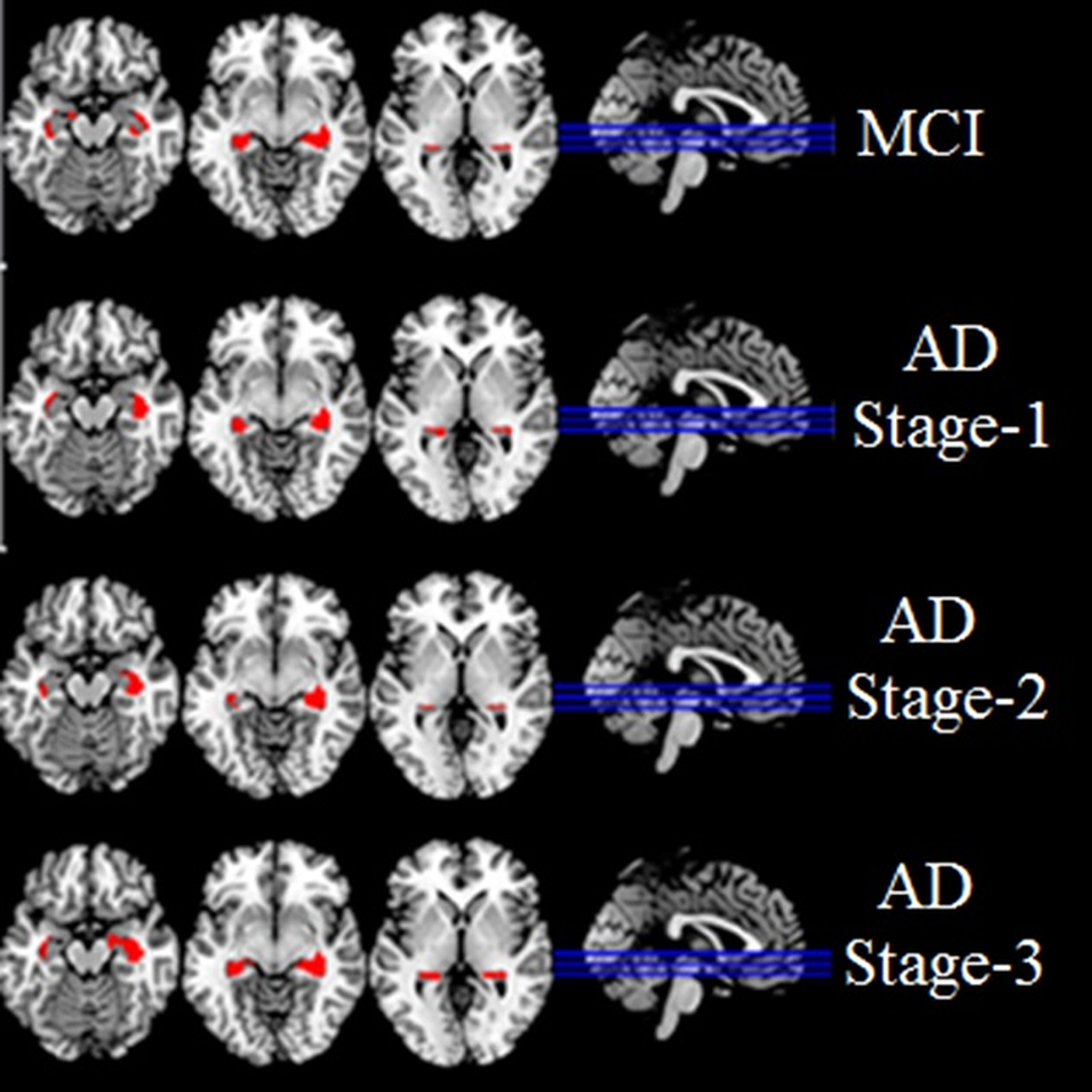
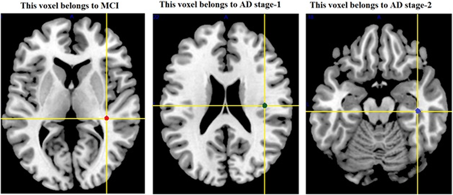
Activation of brain’s region “hippocampus” in multislice view at MCI stage, AD stage 1, AD stage 2, and AD stage 3. AD indicates Alzheimer’s disease; MCI, mild cognitive impairment.
On the basis of activated voxels of the brain, region of interest, that is, hippocampus, was selected for the classsifaction of AD stages, as shown in Figure 5. While classifying the stages of AD, we selected the voxel clusters of hippocampus and used that voxels as vectors to apply the PCA algorithm. For classification of AD stages, we applied purposed alogorithm on that voxels. The objective of applying PCA was to develop an approach for classification of AD stages. The PCA-based algorithm classified the voxels of 4 stages of AD similar to classifcation of face images. This method involves the same steps as mentioned above for face recognition, that is, selecting a voxel for testing and calculation of covariance matrix of data, calcluation of eigenvalues and eigenvectors corresponding to largest eigenvalues of covarinace matrix, and the testing voxel matched with feature data matrix (which involves the voxels of 4 stages of AD). The PCA effectively matched the testing voxel with its actual stage of AD, as shown in Figure 6. Figure 7 showed the location of voxels recognized by proposed PCA-based algorithm, where red-, green-, and blue-colored voxels belongs to MCI, AD stage 1, and AD stage 2, respectively. The Scree plot of PCA shows the number of PCAs (approximately 10 PCAs) used for best classification, as shown in Figure 8.
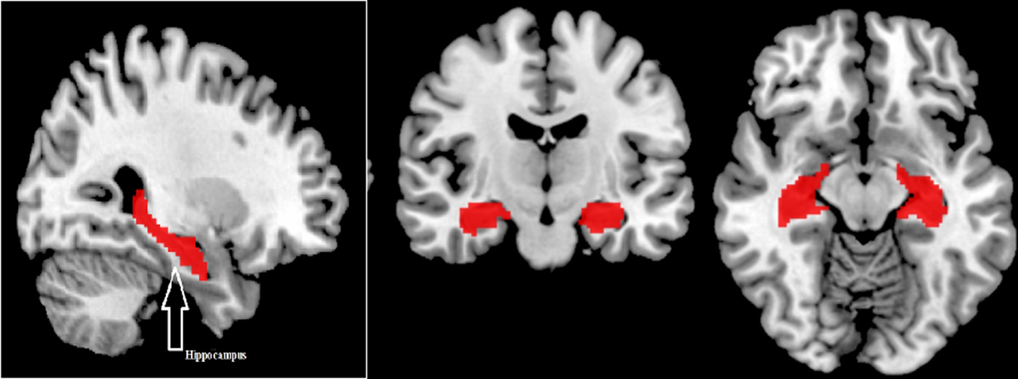
Red-colored area showing the region of interest (hippocampus) in coronal, axial, and sagittal view for classification of Alzheimer’s disease stages.
Location of voxels in Montreal Neurological Institute coordinate system with detected stage of Alzheimer’s disease.
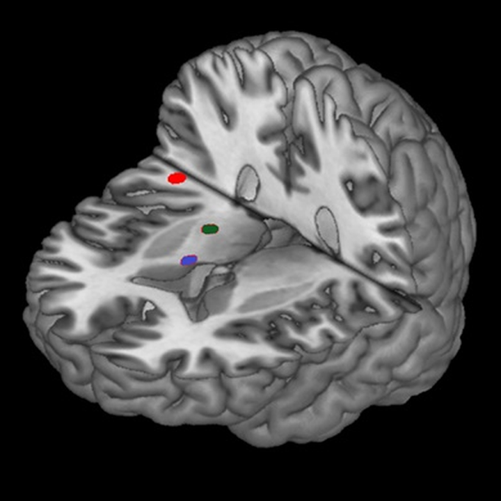
Cutout brain showing the location of voxels recognized by proposed PCA-based algorithm where red-, green-, and blue-colored voxels belong to MCI, AD stage 1, and AD stage 2, respectively. AD indicates Alzheimer’s disease; MCI, mild cognitive impairment; PCA, principal component analysis.
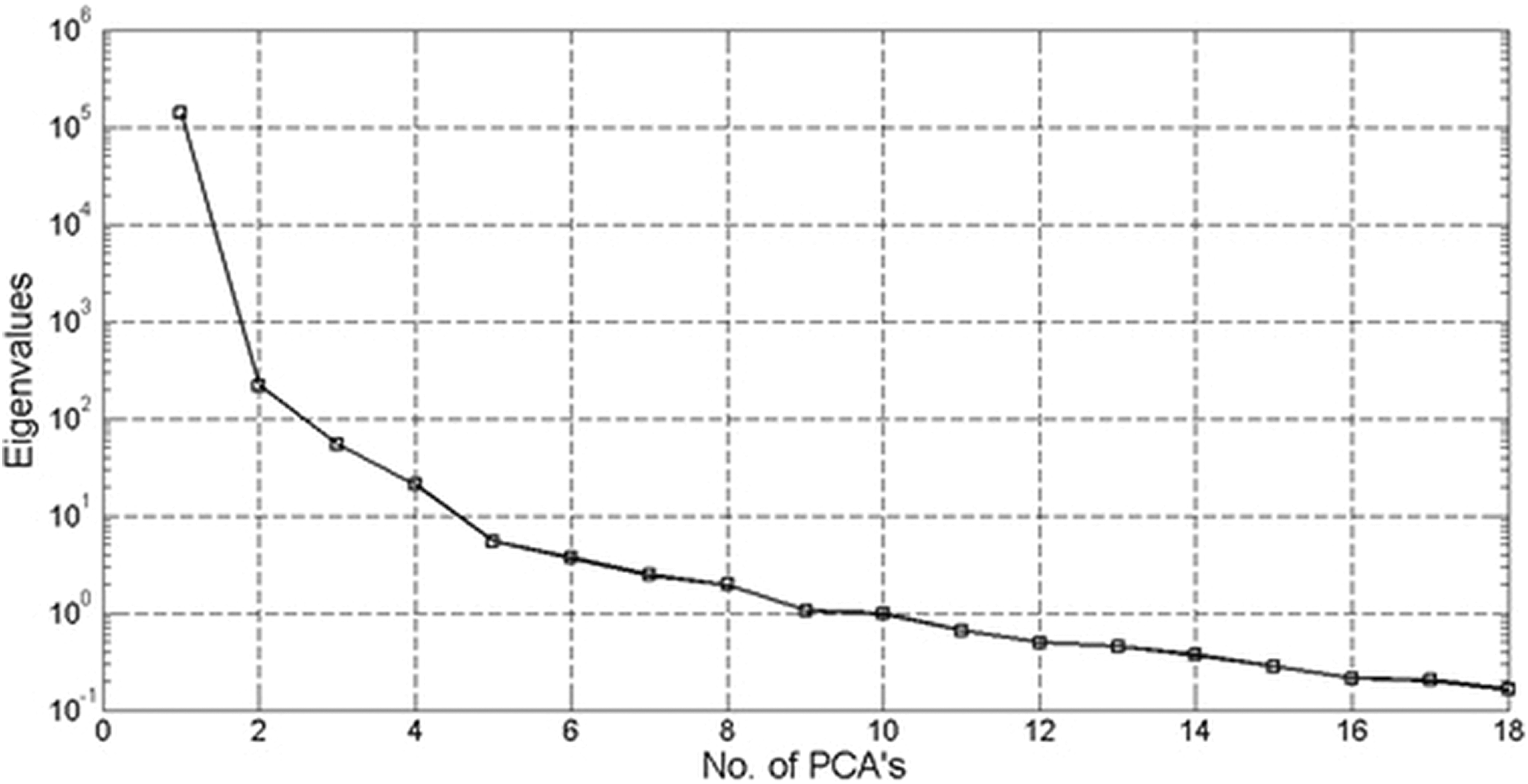
Scree plot showing that almost 10 PCAs used for best classification of Alzheimer’s disease stages. PCA indicates principal component analysis.
Discussion
The PCA-based algorithm has been widely used in problems related to face recognition. As PCA performs well in classification of faces, we made an approach by using PCA-based algorithm for classification of images and AD stages. Previously, we identified and classified the voxels of brain, used for rewardless-related decision-making using artificial neural network technique. 16 In our present approach, we used images as vectors of covariance matrix, and eigenvectors were obtained corresponding to largest eigenvalues for the classification and recognition of images. The AD has become common cause of dementia in all age groups. 17 The nature of AD is progressive, 18 so early diagnosis of AD is important and challenging for future treatments. 7 As we have mentioned earlier that hippocampus is one of the first regions of brain that is affected by AD, we analyzed the fMRI data using SPM and extracted the voxels of hippocampus region of 4 stages (MCI, AD stage 1, AD stage 2, and AD stage 3) of AD and obtained 95% classification rate. In our approach, we used that voxels as independent variables (vectors of covariance matrix). Eigenvectors based on largest eigenvalues were extracted and used for classification of AD stages. Our PCA-based algorithm for face-recognition approach classified the AD stages effectively, as shown Figure 7. This approach is simple and helpful for early diagnosis of AD. Early diagnosis of AD can help doctors healthier manage an individual’s conditions and sidestep medications that may deteriorate cognition or function. 18 Premature recognition of AD enables the best medical care and health outcomes for people with the disease, it also helps to doctors to identify and treat revocable conditions that mimic cognitive impairment and dementia, such as depression or insufficient vitamin. 18 -20
Conclusion
In this article, an efficient human face-recognition technique based on PCA is proposed. On the basis of eigenvectors corresponding to largest eigenvalues, PCA provided a structure for face recognition. In addition, the technique presented here promises to distinguish the stages of AD. Rather than MRI, with the usage of fMRI data, we were able to identify and classify the stages of AD using activated voxels of hippocampus as input vectors of covariance matrix and through eigenvalues and eigenvectors which covers maximum variability of data, PCA provided an effective framework for the classification of AD. The PCA also provided best classification rate as compared to other methods. As hippocampus is one of the first regions of brain that is affected by AD, we purposed that, using fMRI data of a patient based on the voxels from hippocampus, PCA-based algorithm can identify and accurately classify the early diagnosis of AD in simple way using significant number of PCAs.
Footnotes
Authors’ Note
The grantee organization is the Northern California Institute for Research and Education, and the study is contributed by the Alzheimer’s Therapeutic Research Institute at the University of Southern California. ADNI data are disseminated by the Laboratory for Neuroimaging at the University of Southern California.
Acknowledgments
The authors would like to appreciate the financial support provided by National Research Program for Universities (NRPU) of Higher Education Commission (HEC) Pakistan.
Declaration of Conflicting Interests
The authors declared no potential conflicts of interest with respect to the research, authorship, and/or publication of this article.
Funding
The authors disclosed receipt of the following financial support for the research, authorship, and/or publication of this article: Data collection and sharing of this project was funded by Alzheimer’s Disease Neuroimaging Initiative (ADNI; National Institute of Health grant U01 AG024904) and DOD ADNI (Department of Defense award number W81XWH-12-2-0012). ADNI is funded by the National Institute on Aging, the National Institute of Biomedical Imaging and Bioengineering, and through generous contributions from the following: AbbVie, Alzheimer’s Association; Alzheimer’s Drug Discovery Foundation; Araclon Biotech; BioClinica, Inc; Biogen; Bristol-Myers Squibb Company; CereSpir, Inc; Cogstate; Eisai Inc; Elan Pharmaceuticals, Inc; Eli Lilly and Company; EuroImman; F. Hoffmann-La Roche Ltd and its affiliated company Genetech, Inc; Fujirebio; GE Healthcare; IXICO Ltd; Janssen Alzheimer’s Immunotherapy Reseach and Development, LLC; Johnson and Johnson Pharmaceutical Research & Development, LLC; Lumosity; Lundbeck; Merck & Co, Inc; Meso Scale Diagnostics, LLC; NeuroRX Research; Neurotrack Technologies; Novartis Pharmaceuticals Corporation; Pfizer, Inc; Piramal Imaging; Servier; and Takeda Pharmaceutical Company and Transition Therapeutics. The Canadian Institutes of Health Research is providing funds to support ADNI clinical sites in Canada. Private sector contributions are facilitated by the Foundation for the National Institutes of health (![]() ). This work was supported by the National Research Program for Universities (NRPU), Higher Education Commission Pakistan (grant number: 7776/Punjab/NRPU/R&D/HEC/2017).
). This work was supported by the National Research Program for Universities (NRPU), Higher Education Commission Pakistan (grant number: 7776/Punjab/NRPU/R&D/HEC/2017).