
Abstract
The Polycomb group genes are a general class of regulators that are responsible for maintaining homeotic gene expression throughout cell division. Polycomb group expression plays an important role in oncogenesis of several types of human cancer. Melanoma nuclear protein 18 and B-cell-specific Moloney leukemia virus insert site 1 are key Polycomb group proteins. Studies have shown that melanoma nuclear protein 18 is a potential tumor suppression, and B-cell-specific Moloney leukemia virus insert site 1 is overexpressed in several human malignancies. However, the roles of melanoma nuclear protein 18 and B-cell-specific Moloney leukemia virus insert site 1 in esophageal squamous cell carcinoma are still unclear. In this study, we analyzed the expression levels of melanoma nuclear protein 18 and B-cell-specific Moloney leukemia virus insert site 1 in 89 esophageal cancer tissues and paired normal mucosal tissues using immunohistochemistry, Western blotting, and quantitative real-time polymerase chain reaction analyses. We found that the expression of melanoma nuclear protein 18 in the carcinoma tissues was significantly lower than that in the noncancerous mucosal tissues (P < .05), and B-cell-specific Moloney leukemia virus insert site 1 expression in the carcinoma tissues was significantly higher than that in the noncancerous mucosal tissues (P < .05). In addition, the expression of melanoma nuclear protein 18 was correlated with clinical stage, depth of invasion, and lymph node metastasis (P < .05) but was not correlated with gender, age, degree of differentiation, or disease-free survival (P > .05). B-cell-specific Moloney leukemia virus insert site 1 expression was strongly correlated with the degree of differentiation, clinical stage, and lymph node metastasis (P <.05) but was not correlated with the gender, age, depth of invasion or disease-free survival (P > .05). Moreover, there was a negative correlation between melanoma nuclear protein 18 and B-cell-specific Moloney leukemia virus insert site 1 expressions in esophageal squamous cell carcinoma (P < .05). Our study suggests that melanoma nuclear protein 18 and B-cell-specific Moloney leukemia virus insert site 1 may play a crucial role in esophageal squamous cell carcinoma. Melanoma nuclear protein 18 or B-cell-specific Moloney leukemia virus insert site 1 may be a potential biomarker for diagnosis and prognosis of esophageal squamous cell carcinoma.
Keywords
Introduction
Esophageal malignancy is one of the most lethal cancers worldwide. It comprises approximately 7% in all gastrointestinal cancers internationally. 1 Despite clinical advances in the field of oncology, esophageal cancer has the sixth highest mortality of all cancers, with 406 800 deaths per year. 2 Esophageal squamous cell carcinoma (ESCC) is the major histological type of esophageal cancer. Smoking and drinking are the major risk factors for ESCC. 3 –5 Esophageal squamous cell carcinoma is a highly aggressive malignancy due to late diagnosis, rapid progression, and poor prognosis. Thus, the rate of the mortality of esophageal cancer is similar to the rate of the incidence. 6,7 The 5-year survival rate of esophageal cancer is less than 20%. 8,9 However, the overall survival could be dramatically improved by early diagnosis, with a 5-year survival rate of up to 90%. 10 The majority of patients with early ESCC show no clinical symptoms and manifestations. Therefore, identifying ideal markers with high sensitivity and specificity will provide physicians with valuable information for diagnosis and suitable treatment options. At present, there are no suitable biomarkers for diagnosing ESCC; thus, finding specific biomarkers of ESCC is urgently needed to detect tumors and recurrence.
The Polycomb group (PcG) genes are involved in the regulation of the cell cycle, hematopoiesis, and X-inactivation. 11,12 Polycomb group gene expression plays an important role in oncogenesis of various types of human cancer. 13 Melanoma nuclear protein 18 (Mel-18) and B-cell-specific Moloney leukemia virus insert site 1 (Bmi-1) are key PcG proteins, and they are highly similar in structure. Melanoma nuclear protein 18 and Bmi-1 play important roles in oncogenesis and progression of ESCC; however, the effects of Mel-18 and Bmi-1 are different in cancer cell growth and survival. 14 Previous studies have shown that Mel-18 is overexpressed in several human tumors, including Hodgkin’s lymphomas, 15 human melanoma, 16 and medulloblastoma. 17 Recently, several reports have suggested that Mel-18 may act as a tumor suppressor restraining c-Myc expression in oncogenesis and progression of ESCC. 18,19 Thus, the function of Mel-18 in cancers is still unclear. The Bmi-1 gene was discovered in 1999, and overexpression of Bmi-1 has been found in a variety of human cancers, such as breast cancer, 20 gastric cancer, 21 and head and neck cancer, 22 and its expression was also associated with the development of tumors. 23,24 Studies have shown that the expression of Mel-18 is negatively correlated with Bmi-1 in several tumors. 24,25 However, the functions of Mel-18 and Bmi-1 in ESCC have not been fully elucidated. Therefore, we investigated the expression levels of the 2 marks in esophageal cancer tissues and paired normal mucosal tissues, and our study suggests that Mel-18 or Bmi-1 may be a biomarker for diagnosis and prognosis of ESCC.
Materials and Methods
Sample Preparation of Patient With ESCC
The samples of esophageal cancer tissue and adjacent noncancerous mucosal tissue were obtained from 29 patients who received the surgical treatment from 2015 to 2016 in our hospital. Normal tissues located 5 cm away from the tumor edge were collected during the surgery. Tissue microarrays containing 60 esophageal tissue, and adjacent normal tissue sections were commercially obtained from Chaoying Bioscience (Shanxi, China). None of the patients received prior chemotherapy or radiotherapy before the surgery. Pathology of the esophageal cancer biopsies showed they were all squamous cell carcinomas. The patient information, including gender, age, and clinicopathological characteristics, was obtained from the medical records or the manufacturer. The enrolled patients included 64 males and 25 females, and the median age was 59 years (range: 36-76 years). The median follow-up time was 13.8 months (range: 5.0-22.9 months). The duration of disease-free survival (DFS) began at the day of surgery and ended at relapse, metastasis, last follow-up, or death for any cause. Patients consented to the specimen collection, and this study was approved by the research ethics committee of our hospital (Qianfoshan Hospital affiliated with Shandong University).
Quantitative Real-Time-PCR Assays
Total RNA was extracted from frozen tissue specimens using Total RNA Kit II (Omega, Georgia). One microgram of total RNA was used to perform reverse transcription for first-strand cDNA using the Revert Aid First Strand cDNA Synthesis Kit (Thermo Scientific, Waltham, Massachusetts). Subsequently, quantitative PCR was performed in triplicate on an ABI VIIA7 Real-Time PCR System (Applied Biosystems, Foster City, California) with SuperReal PreMix Plus (SYBR Green; TIANGEN, Beijing, China). The following gene-specific primer pairs were used: Mel-18: (F) 5′-CGGACTACACGGATCAAAATCA-3′, (R) 5′-GGCGTCGATGAAGTACCCC-3′; Bmi-1: (F) 5′-CCACCTGATGTGTGTGCTTTG-3′, (R) 5′-TTCAGTAGTGGTCTGGTCTTGT-3′. β-Actin: (F) 5’-GACCACACCTTCTACAATGAG-3′, (R) 5′-GCATACCCCTCGTAGATGGG-3′. All reactions were performed in a 20 µL reaction volume. Polymerase chain reaction was carried out after predenaturing at 95°C for 15 minutes followed by 40 cycles at 95°C for 10 seconds and 62°C for 32 seconds. Gene expression was analyzed with the comparative threshold (Ct) cycle method after normalizing to β-Actin. ΔΔCt = ΔCt (esophageal cancer)-ΔCt (control) for RNA samples. The fold change for each esophageal cancer sample relative to the control sample = 2−ΔΔCt. When the expression showed a 2-fold increase or decrease compared to the normal counterpart tissue, it was considered altered expression.
Immunohistochemistry Staining
Tissue samples were soaked in 10% neutral buffered formalin, embedded in conventional paraffin, and sectioned to a thickness of approximately 3 μm. After deparaffinization, the specimens were hydrated and incubated with an epitope retrieval solution (pH 6.0) in a microwave (temperature controlled at 95°C-100°C) for 20 minutes. The slices were then incubated with 0.3% H2O2 for 10 minutes at room temperature to inactivate endogenous peroxidase and rinsed with phosphate-buffered saline. Then, the specimens were incubated with rabbit polyclonal antibody for Mel-18 (1:300 dilution, bs-9673 R, Bioss, Beijing, China) and rabbit polyclonal antibody for Bmi-1 (1:500 dilution, ab85688, Abcam) at 4°C overnight. After that, the specimens were incubated with secondary antibodies using an Elivision Plus Kit (Maixin-Bio; Lab Vision, Kalamazoo, Michigan). DAB (Diaminobenzidine) (DaKo, Carpinteria, California) chromogenic reagent was added. After the specimens were counterstained with hematoxylin, they were dehydrated, mounted, and observed with a Nikon 50i fluorescence microscope (Nikon, Tokyo, Japan).
The immunohistochemical specimens were observed under 200× optical microscope and evaluated using positive staining intensity and percentage of positive staining by 2 pathologists. The extent of the stained cells was graded as follows: 0%-5.0% = 0, 5.1%-25.0% = 1, 25.1%-50.0% = 2, 50.1%-75.0% = 3, and 75.1%-100% = 4. Positive staining intensity was rated as follows: no staining = 0, light yellow = 1, yellow = 2, and dark yellow = 3. The final score was determined by multiplying the positivity scores with the staining intensity scores of cells; a score of 0 to 2 was regarded as negative (−), 3 to 5 as weak (+), 6 to 8 as moderate (+ +), and 9 to 12 as strong (+++) .
Western Blot
Human esophageal cancer tissues and adjacent noncancerous mucosal tissue were homogenized and centrifuged at 12 000g for 30 minutes at 4°C. The protein concentrations were determined using the BCA (Bicinchonininc Acid) Protein Assay Kit (BOSTER, Wuhan, China). Thirty micrograms of total protein was separated by 10% SDS-PAGE (Sodium Dodecyl Sulfate Polyacrylamide Gel Electrophoresis), transferred to a PVDF (Polyvinylidene Fluoride) membrane, and blocked in 5% milk. The membranes were incubated with rabbit polyclonal antibody for Mel-18 (1:500 dilution, bs-9673 R, Bioss, Beijing, China) and rabbit polyclonal antibody for Bmi-1 (1:8000 dilution, ab85688, Abcam) or GAPDH (Glyceraldehyde Phosphate Dehydrogenase) (1:6000 dilution, China) at 4°C overnight. The secondary antibody (1:8000 dilution, China) was incubated with the membrane for 1 hour at room temperature. Finally, the immunoreactive protein bands were visualized with a chemiluminescence kit (Millipore, MA, USA).
Statistical Analysis
The protein expression data were statistically analyzed with χ2 tests and Fisher exact test. The correlation between Mel-18 and Bmi-1 messenger RNA (mRNA) expression levels was analyzed by Pearson coefficient test. Disease-free survival rates were plotted using the Kaplan-Meier method, and the log-rank test was used for analysis. All statistical analyses were performed using the SPSS 21.0 software package. P < .05 was considered statistically significant.
Results
Immunodetection of MEL-18 and BMI-1 in ESCC Tissues
The expression rate of Mel-18 in ESCC tissues and normal mucosa was 33.7% (30 of 89) and 68.5% (61 of 89), respectively. The expression rate of Bmi-1 in ESCC tissues and normal mucosa was 74.2% (66 of 89) and 30.3% (27 of 89), respectively. The differences in the expression of Mel-18 and Bmi-1 were statistically significant between esophageal cancer tissues and normal esophageal mucosa (χ2 = 21.606, P < .05; χ2 = 34.249, P < .05). In our cases, Mel-18 and Bmi-1 were expressed in the nuclei of tumor cells as shown by immunohistochemistry staining (Figure 1).
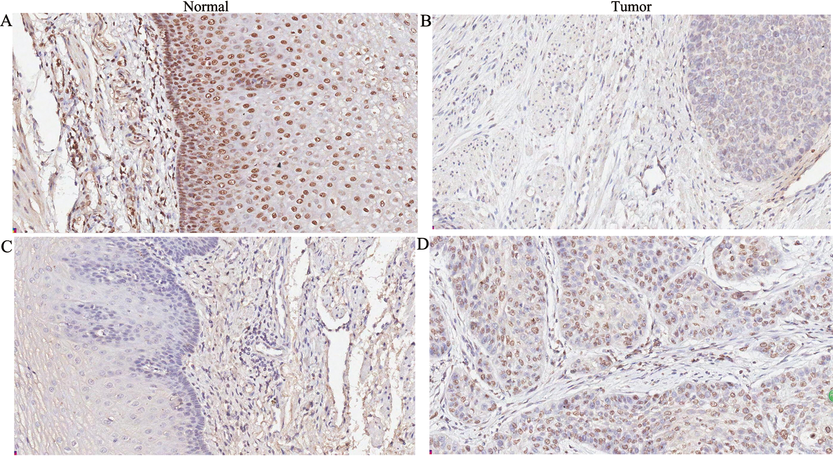
Immunohistochemical staining of esophageal squamous cell carcinoma using an antibody against Mel-18 (A and B) or Bmi-1 (C and D). Mel-18 protein expression in ESCC cells was weaker than that in adjacent noncancerous tissues. B-cell-specific Moloney leukemia virus insert site 1 (Bmi-1) protein expression in esophageal squamous cell carcinoma (ESCC) cells was stronger than that in adjacent noncancerous tissues. Original magnification was 200×.
The Association Between the Expression of MEL-18 and BMI-1 With Clinicopathological Parameters
The importance of Mel-18 and Bmi-1 in ESCC was evaluated by correlating their expression levels with clinicopathological features. Several of the analyzed characteristics showed significant associations with the expression levels (Table 1). The results showed that the expression of Mel-18 was correlated with clinical stage, depth of invasion, and lymph node metastasis but not with patient gender, age, or degree of differentiation. The expression of Bmi-1 was correlated with degree of differentiation, clinical stage, and lymph node metastasis but not with patient gender, age, or depth of invasion.
Correlations Between the Expression Level of Mel-18 or Bmi-1 and Clinical-Pathologic Variables.

Abbreviations: Bmi-1, B-cell-specific Moloney leukemia virus insert site 1; Mel-18, melanoma nuclear protein 18.
aStatistically significant. Statistically significant at 0.05 level (bilateral).
Quantifying MEL-18 and BMI-1 Expression Levels by Western Blot Analysis and Real-Time PCR
Melanoma nuclear protein 18 and Bmi-1 protein levels were quantified by Western blot analyses. The proteins were detected in ESCC and corresponding para-carcinoma tissues. Compared to the para-carcinoma tissues, the expression level of Mel-18 was significantly lower, and Bmi-1 was significantly higher in ESCC with statistically significant differences (P = .028 and P = .008, respectively, paired-sample test; Figure 2). Transcription of Mel-18 and Bmi-1 was quantified by quantitative real-time PCR. The results show that Mel-18 mRNA was downregulated in 16 (55.17%) of 29 esophageal tumor tissues (<0.5-fold), and Bmi-1 mRNA was overexpressed in 18 (62.07%) of 29 esophageal tumor tissues (>2-fold).The ESCC tissues exhibited a lower level of Mel-18 mRNA and a higher level of Bmi-1 mRNA than the para-carcinoma tissues.

Downregulation of Mel-18 and overexpression of Bmi-1 in ESCC tumors. Three paired primary esophageal tumors (T) and normal esophageal tissues (N) from the same 3 patients were analyzed for Mel-18 (A) and Bmi-1 (B) expression at the protein level by Western blotting. The expression level was normalized by GAPDH. Statistical analysis was performed using a paired-sample t test (C).
Association of MEL-18 and BMI-1 With Survival Outcome
We also analyzed the association between the expression of Mel-18 and Bmi-1 with patient survival. By the end of the study, 4 patients had relapsed and 1 patient had died from ESCC. The median DFS time was 20.4 months (95% CI: 18.5-22.4 months). Kaplan-Meier analysis shows that there was no significant correlation between the expression of Mel-18 and Bmi-1 with DFS (P > .05; Figure 3).

Kaplan-Meier analysis of the prognostic value of the expression of Mel-18(A) and Bmi-1(B) in prediction of disease-free survival.
Correlation Among MEL-18 and BMI-1 Expressions in ESCC Tissues
Correlation analysis was performed to test the relationship between Mel-18 mRNA and Bmi-1 mRNA. The Mel-18 expression was negatively associated with Bmi-1 by Pearson coefficient test (r = −.592, P = .001). Moreover, according to the result of immunohistochemical studies, there were significant negative correlations between Mel-18 and Bmi-1 (r = −.285, P =.007, χ2 test; Table 2).
Relationship Between the Expression of Mel-18 and Bmi-1 in Esophageal Cancer.
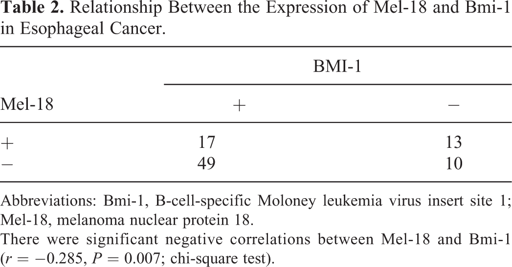
Abbreviations: Bmi-1, B-cell-specific Moloney leukemia virus insert site 1; Mel-18, melanoma nuclear protein 18.
There were significant negative correlations between Mel-18 and Bmi-1 (r = −0.285, P = 0.007; chi-square test).
Discussion
Mammalian PcG proteins are classified into Polycomb repressive complexes 1 and 2 (PRC1 and PRC2). Melanoma nuclear protein 18, a member of the PRC1, is detected in a number of tumor and normal tissues. 25 Melanoma nuclear protein 18 was reported to be overexpressed in several tumors, including human melanoma, Hodgkin lymphomas, and medulloblastoma. However, a number of previous reports have revealed that Mel-18 acts as a tumor-suppressor gene, including breast, 26,27 colorectal, prostate, 28,29 and gastric cancers. 21 Therefore, these results suggest that Mel-18 plays an oncogenic or tumor-suppressor role possibly depending upon the context of the cancer system. In our study, the expression of Mel-18 was significantly reduced in ESCC tissues compared to the corresponding noncancerous mucosal tissues. We found a negative correlation between the expression of Mel-18 and the clinical stage, depth of cancer invasion (T classification), and lymph node metastasis (N classification) with statistically significant differences (P < .05). These results suggest that Mel-18 protein is associated with carcinogenesis, progression, and metastasis. Thus, Mel-18 may play a crucial role in ESCC.
B-cell-specific Moloney leukemia virus insert site 1 is structurally highly similar to Mel-18, is another important member of PRC1, regulates proliferation and senescence in mammalian cells. It displays an inverse correlation with Mel-18 and was identified as an oncogene. 30 It has been reported to be negatively regulated by Mel-18 via repression of the c-Myc. Overexpression of Bmi-1 in esophageal cancer has been previously reported. 31,32 In this study, we found that Bmi-1 plays an important role in ESCC progression. We demonstrated that Bmi-1 expression was significantly upregulated in ESCC tissues compared to adjacent noncancerous tissues. In addition, we also observed that positive expression of Bmi-1 was correlated with the clinical stage, degree of differentiation, and lymph node metastasis (N classification), and the differences were statistically significant (P < .05). These results suggest that Bmi-1 protein may play a crucial role in the carcinogenesis, progression, and metastasis of ESCC. However, there was no correlation between Bmi-1 expression and depth of cancer invasion or DFS, which was not consistent with the results of a previous study. He et al 33 reported an association between Bmi-1 overexpression and poor OS (Overall Survival). However, Hwang et al 34 and Choy et al 35 found no association between Bmi-1 expression and OS in patients with ESCC consistent with the results of the present study. One of the reasons may the small number of samples.
Previous studies have shown that the expression of Bmi-1 was no significant associations with age, gender, tumor diameter, or nationality. 31,32,33 –35 But there is argument about the relationship between the gene expression and depth of invasion, lymph node metastasis, or clinical stage. Liu et al 32 found that high expression of Bmi-1 was significantly associated with lymph node metastasis and clinical stage but not associated with depth of invasion or distant metastasis, which was consistent with the results of our study. Zhang et al 31 reported the expression of Bmi-1 was correlated with depth of invasion and lymph node metastasis .However, Choy et al 35 shown that there were no significant associations of Bmi-1 expression with lymph node metastasis and clinical stage. For instance, esophageal carcinoma cell line including Eca109, EC 9706, TE1, and so on; however, further research is needed.
The molecular pathways by which these proteins regulate oncogenesis remain unclear. B-cell-specific Moloney leukemia virus insert site 1 can inhibit the INK4a-ARF gene and participate in the development of a variety of tumors. B-cell-specific Moloney leukemia virus insert site 1 can promote self-renewal and differentiation in liver cells, improve leukemia stem cell proliferation, and is involved in the proliferation of tumor cells under the synergy of the c-Myc gene. 23 B-cell-specific Moloney leukemia virus insert site 1 has been reported to be negatively regulated by Mel-18 via repression of the c-Myc oncogene, which is amplified and/or overexpressed in a variety of malignancies. Lee et al 36 reported that Mel-18 expression was reduced in breast cancer cells and tissues and upregulated the expression of Mel-18 and decreased the expression of c-Myc. Thus, Bmi-1 levels were decreased, reducing the activity of PKB/Akt in breast cancer cells, accelerating ageing and decreasing the life of cells, and inhibiting tumor growth. Wang et al 28 found a significant negative correlation between the expression of Mel-18 and c-Myc in prostate cancer. Zhang et al 14 found that Mel-18 also had lower expression in gastric cancer tissues and cells; overexpression of Mel-18 decreased Bmi-1 expression and upregulated p16, eventually inhibiting the growth of gastric cancer cells.
In the study, we demonstrated that neoplastic ESCC cells show abnormal expression of Mel-18 and Bmi-1. We propose that abnormal PcG expression alters the expression of target genes involved in regulation of senescence or the cell cycle in an altered composition of the PRC1 in ESCC. Our observations add to the increasing evidence that PcG genes are important contributors to carcinogenesis and progression of human tumors. We additionally found that there was a strong negative correlation between Mel-18 and Bmi-1 expressions at the mRNA (r = −0.592, P = .001) and protein level (r = −0.285, P = .007). The underlying mechanism of Mel-18 and Bmi-1 may be through the regulation of c-myc.
This study is the first to investigate the correlation between Mel-18 expression level and clinicopathological characteristics in ESCC. The expression of Mel-18 and Bmi-1 was correlated with esophageal cancer progression. Advanced-stage ESCC is more likely to express lower Mel-18 and higher Bmi-1. We examined associated gene expression, but we did not investigate the detailed mechanisms of regulation of esophageal cancers. The molecular pathways through which these genes regulate oncogenic signals need to be further studied in ESCC.
Conclusion
In conclusion, we observed an inverse correlation between the expression levels of Mel-18 and Bmi-1 in ESCC. Both Mel-18 and Bmi-1 are involved in the development and progression of ESCC. Melanoma nuclear protein 18 and Bmi-1 might be novel molecular markers for ESCC. However, the detailed mechanisms of regulation of Mel-18 and Bmi-1 remained to be elucidated.
Footnotes
Abbreviations
Acknowledgments
The authors would express our outmost gratitude to all the personnel’s in the Medical Research Center, Qianfoshan Hospital, Shandong Medical University for their excellent technical assistance.
Declaration of Conflicting Interests
The author(s) declared no potential conflicts of interest with respect to the research, authorship, and/or publication of this article.
Funding
The author(s) disclosed receipt of the following financial support for the research, authorship, and/or publication of this article: This research was supported by the Ministry of Science and Technology Research Foundation for funding support (2013GSF11836).